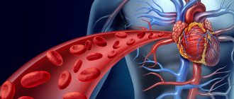
The heart has a complex structure and performs an equally complex and important job. Contracting rhythmically, it ensures blood flow through the vessels.
The heart is located behind the sternum, in the middle section of the chest cavity and is almost completely surrounded by the lungs. It may move slightly to the side as it hangs freely on the blood vessels. The heart is located asymmetrically. Its long axis is inclined and forms an angle of 40° with the axis of the body. It is directed from top to right, forward, down to the left, and the heart is rotated so that its right section is tilted more forward, and the left one – back. Two-thirds of the heart is to the left of the midline and one-third (the vena cava and right atrium) is to the right. Its base is turned towards the spine, and its apex is facing the left ribs, to be more precise, the fifth intercostal space.
Anatomy of the heart
The heart muscle is an organ that is an irregularly shaped cavity in the form of a slightly flattened cone. It takes blood from the venous system and pushes it into the arteries. The heart consists of four chambers: two atria (right and left) and two ventricles (right and left), which are separated by septa. The walls of the ventricles are thicker, the walls of the atria are relatively thin.
The left atrium contains the pulmonary veins, and the right atrium contains the hollow veins. The ascending aorta emerges from the left ventricle, and the pulmonary artery emerges from the right.
The left ventricle, together with the left atrium, makes up the left section, which contains arterial blood, which is why it is called the arterial heart. The right ventricle with the right atrium is the right section (venous heart). The right and left parts are separated by a solid partition.
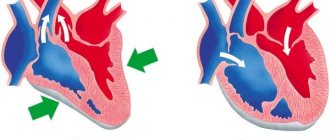
The atria are connected to the ventricles by openings with valves. On the left side the valve is bicuspid, and it is called mitral, on the right - tricuspid, or tricuspid. The valves always open towards the ventricles, so blood can only flow in one direction and cannot flow back into the atria. This is ensured by tendon threads attached at one end to the papillary muscles located on the walls of the ventricles, and at the other end to the valve leaflets. The papillary muscles contract together with the walls of the ventricles, since they are outgrowths on their walls, and this causes the tendon threads to stretch and prevent reverse blood flow. Thanks to the tendon threads, the valves do not open towards the atria when the ventricles contract.
In the places where the pulmonary artery leaves the right ventricle and the aorta leaves the left, tricuspid semilunar valves are located, similar to pockets. The valves allow blood flow from the ventricles into the pulmonary artery and aorta, then fill with blood and close, thereby preventing blood from returning.
The contraction of the walls of the heart chambers is called systole, their relaxation is called diastole.
Phases of the heart
There are several separate phases of contraction of the heart muscle:
- At the beginning, contraction of the atria occurs. Then, with some slowdown, ventricular contraction begins. During this process, blood naturally tends to fill the chambers with reduced pressure. Why doesn’t it flow back into the atria after this? The fact is that the blood is blocked by the gastric valves. Therefore, it can only move in the direction of the aorta, as well as the vessels of the pulmonary trunk.
- The second phase is relaxation of the ventricles and atria. The process is characterized by a short-term decrease in the tone of the muscle structures from which these chambers are formed. The process causes a decrease in pressure in the ventricles. Thus, the blood begins to move in the opposite direction. However, this is prevented by the closing pulmonary and arterial valves. During relaxation, the ventricles fill with blood that comes from the atria. On the contrary, the atria are filled with bodily fluid from the large and
External structure of the heart
The anatomical structure and functions of the heart are quite complex. It consists of chambers, each of which has its own characteristics. The external structure of the heart is as follows:
- apex (apex);
- basis (base);
- anterior surface, or sternocostal;
- inferior surface, or diaphragmatic;
- right edge;
- left edge.
The apex is the narrowed, rounded part of the heart formed entirely by the left ventricle. It faces forward down and to the left, abuts the fifth intercostal space to the left of the midline by 9 cm.
The base of the heart is the upper expanded part of the heart. It faces up, to the right, back and has the shape of a quadrangle. It is formed by the atria and aorta with the pulmonary trunk, located in front. In the upper right corner of the quadrangle there is the entrance of the superior vena cava, in the lower corner there is the inferior vena cava, to the right there are two right pulmonary veins, on the left side of the base there are two left pulmonary veins.
The coronary groove runs between the ventricles and atria. Above it are the atria, below it are the ventricles. Anteriorly, in the region of the coronary sulcus, the aorta and pulmonary trunk emerge from the ventricles. It also contains the coronary sinus, where venous blood flows from the veins of the heart.
The sternocostal surface of the heart is more convex. It is located behind the sternum and cartilages of the III-VI ribs and is directed forward, upward, and to the left. A transverse coronary groove runs along it, which separates the ventricles from the atria and thereby divides the heart into an upper part, formed by the atria, and a lower part, consisting of the ventricles. Another groove of the sternocostal surface - the anterior longitudinal - runs along the border between the right and left ventricles, with the right one forming the largest part of the anterior surface, the left one the smaller one.
The diaphragmatic surface is flatter and is adjacent to the tendon center of the diaphragm. A longitudinal posterior groove runs along this surface, separating the surface of the left ventricle from the surface of the right. In this case, the left one makes up the majority of the surface, and the right one makes up the smaller part.
The anterior and posterior longitudinal grooves merge at their lower ends and form the cardiac notch to the right of the cardiac apex.
There are also lateral surfaces located on the right and left and facing the lungs, which is why they are called pulmonary.
The right and left edges of the heart are not the same. The right edge is more pointed, the left is more blunt and rounded due to the thicker wall of the left ventricle.
The boundaries between the four chambers of the heart are not always clearly defined. The landmarks are the grooves in which the blood vessels of the heart are located, covered with fatty tissue and the outer layer of the heart - the epicardium. The direction of these grooves depends on how the heart is located (obliquely, vertically, transversely), which is determined by the body type and the height of the diaphragm. In mesomorphs (normosthenics), whose proportions are close to the average, it is located obliquely, in dolichomorphs (asthenics) with a thin physique - vertically, in brachymorphs (hypersthenics) with wide short forms - transversely.
The heart seems to be suspended by the base on large vessels, while the base remains motionless, and the apex is in a free state and can move.
Heart
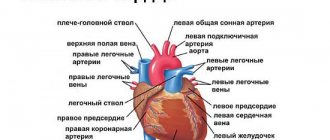
The heart (cor; Fig. 137 - 139) is a hollow cone-shaped muscular organ weighing 250 - 350 g, located behind the sternum in the mediastinum, on the tendon center of the diaphragm. In the chest cavity, it occupies an oblique position and faces the wide part (base) up, back and to the right, and the narrow part (apex) faces forward, down and left. The upper border of the heart is projected in the second intercostal space, the right border protrudes 2 cm beyond the right edge of the sternum; the left one passes without reaching 1 cm of the left midclavicular line. The apex of the heart is located in the fifth left intercostal space. The posterior inferior surface of the heart is adjacent to the diaphragm, the anterior one faces the sternum and costal cartilages.
Rice. 137. Position of the heart in the chest (pericardium is opened). 1 - left subclavian artery (a. subclavia sinistra); 2 - left common carotid artery (a. carotis communis sinistra); 3 — aortic arch (arcus aortae); 4 - pulmonary trunk (truncus pulmonalis); 5 - left ventricle (ventriculus sinister); 6 - apex of the heart (apex cordis); 7 - right ventricle (ventriculus dexter); 8 - right atrium (atrium dextrum); 9 — pericardium (pericardium); 10 - superior vena cava (v. cava superior); 11 - brachiocephalic trunk (truncus brachiocephalicus); 12 - right subclavian artery (a. subclavia dextra)
Rice. 138. Heart; lengthwise cut. 1 - superior vena cava (v. cava superior); 2 - right atrium (atrium dextrum); 3 - right atrioventricular valve (valva atrioventricularis dextra); 4 - right ventricle (ventriculus dexter); 5 - interventricular septum (septum interventriculare); 6 - left ventricle (ventriculus sinister); 7 - papillary muscles (mm. papillares); 8 - tendon chords (chordae tendineae); 9 - left atrioventricular valve (valva atrioventricularis sinistra); 10 - left atrium (atrium sinistrum); 11 - pulmonary veins (vv. pulmonales); 12 - aortic arch (arcus aortae)
Rice. 139. Heart (muscle layers). 1 - aorta; 2 - pulmonary trunk (truncus pulmonalis); 3 - left ear (auricula sinistra); 4 - superficial muscle layer on the left ventricle; 5 - superficial muscle layer on the right ventricle; 6 - median muscle layer on the right ventricle; 7 - right atrium (atrium dextrum); 8 - right ear (auricula dextra); 9 - superior vena cava (v. cava superior)
Two longitudinal grooves are noticeable on the surface of the heart: the anterior and posterior interventricular grooves, covering the heart from the front and back, and the coronary (transverse) groove, located in a ring shape; The heart's own arteries and veins run along them. These grooves correspond to septa that divide the heart into four sections: the longitudinal interatrial and interventricular septa divide the organ into two isolated halves - the right and left heart. The atrioventricular septum divides each of these halves into an upper chamber - the atrium (atrium cordis) and a lower chamber - the ventricle (ventriculus).
The superior and inferior vena cava, the coronary sinus of the heart and the small veins of the heart flow into the right atrium (atrium dextrum). Its upper part is the right ear of the heart. The dilated part is the confluence of large venous vessels, the lower part communicates with the right ventricle through the right atrioventricular foramen (ostium atrioventriculare dextrum).
The right ventricle (ventriculus dexter) in the anterior section has an opening leading into the pulmonary trunk (truncus pulmonalis).
The left atrium (atrium sinistrum) also has an appendage. In the posterior section of the upper wall of the left atrium, four pulmonary veins (vv. pulmonales) open into it. In the lower part, the atrium communicates with the ventricle through the left atrioventricular orifice (ostium atrioventriculare sinistrum). The inner lining of the heart in the area of the atrioventricular openings forms folds protruding into the lumen - heart valves that close these openings. In the right atrioventricular orifice there is the right atrioventricular, or tricuspid, valve (valva atrioventricularis dextra, s. tricuspidalis), consisting of three leaflets - thin fibrous elastic plates, and in the left - the left atrioventricular, or bicuspid, valve ( valva atrioventricularis sinistra, s. mitralis). Thin tendon threads are attached to the free edges of the valves (see Fig. 138), which start from the papillary muscles of the walls of the ventricles, so the leaflet valves open during contraction of the atria only towards the ventricles.
The left ventricle (ventriculus sinister) is oblong and in its anterior section has an opening through which it communicates with the aorta. At the site where the aorta exits the left ventricle and the pulmonary trunk from the right ventricle, the inner lining of the heart forms three thin folds (see Fig. 138) in the form of semicircular pockets - semilunar valves (valvulae semilunares). They open only towards the lumen of the vessels during contraction of the ventricles.
The heart wall consists of three layers: the inner - endocardium (endocardium), the middle - myocardium (myocardium) and the outer - epicardium (epicardium). The endocardium lines all the cavities of the heart, tightly fused with the underlying muscle layer. On the side of the heart cavities it is covered with endothelium. The thickness of the endocardium is uneven: it is thicker in the left chambers of the heart, especially in the interventricular septum, the aorta and the pulmonary trunk.
The myocardium is the most functionally powerful part of the heart wall. The muscle layer of the walls of the atria is thin due to the low load. In the walls of the ventricles, it is the most significant layer in thickness, in which the outer longitudinal, middle annular and inner longitudinal layers are distinguished (see Fig. 139). The outer fibers, deepening obliquely, gradually turn into annular fibers, which in turn turn into internal longitudinal fibers. On the surface of the ventricles there are fibers that cover both ventricles together. The muscle layer of the left ventricle is the thickest.
The composition of cardiac striated muscle tissue includes typical contractile muscle cells - cardiomyocytes and atypical cardiac myocytes, which form the so-called cardiac conduction system, which ensures the automaticity of heart contractions.
The epicardium is part of the serous membrane that surrounds the heart, the sac. It consists of an internal, or visceral, layer (epicardium), directly covering the heart and tightly fused to it, and an outer layer (pericardium), which passes into the epicardium at the point where large vessels depart from the heart. The pericardium is adjacent to the pleural sacs on the sides, grows from below to the tendon center of the diaphragm, and in front is connected by connective tissue fibers to the sternum (see Fig. 137). The pericardium insulates the heart from the surrounding organs, and the fluid between its layers moisturizes the surface of the heart and reduces friction during its contractions.
The vessels leaving the heart form two closed circles of blood circulation. The lesser circle begins in the right ventricle with the pulmonary trunk, which then divides into the right and left pulmonary arteries, which carry venous blood to the pulmonary alveoli. Oxygen-rich blood returns from the lungs through the four pulmonary veins to the left atrium, and from there to the left ventricle of the heart. The aorta, which emerges from the left ventricle of the heart, begins the systemic circulation.
Blood from the aorta first enters large arteries going to the head, torso and limbs, which gradually branch into smaller vessels and then pass inside the organs into intraorgan arteries, then into arterioles, precapillary arterioles and capillaries. Through the wall of the latter there is a constant exchange of substances between blood and tissues. Capillaries merge into postcapillary venules, venules into small intraorgan and then extraorgan veins, and the latter into large venous vessels - the superior and inferior vena cava, through which blood returns to the right atrium of the heart.
Structure of heart tissue
The heart wall is made up of three layers:
- The endocardium is the inner layer of epithelial tissue that lines the cavities of the heart chambers from the inside, exactly repeating their relief.
- The myocardium is a thick layer formed by muscle tissue (striated). The cardiac myocytes of which it consists are connected by many bridges that link them into muscle complexes. This muscle layer ensures the rhythmic contraction of the chambers of the heart. The myocardium is thinnest at the atria, the greatest is at the left ventricle (about 3 times thicker than the right), since it needs more force to push blood into the systemic circulation, in which the resistance to flow is several times greater than in the small circle. The atrial myocardium consists of two layers, the ventricular myocardium - of three. The atrial myocardium and ventricular myocardium are separated by fibrous rings. The conduction system that provides rhythmic contraction of the myocardium is one for the ventricles and atria.
- The epicardium is the outer layer, which is the visceral petal of the heart sac (pericardium), which is a serous membrane. It covers not only the heart, but also the initial parts of the pulmonary trunk and aorta, as well as the final parts of the pulmonary and vena cava.
Human body #77, page 15
Chambers of the heart
The heart cavity is divided into four chambers: two thin-walled atria, into which venous blood flows, and two larger ventricles, from where blood flows into large arteries.
The left and right chambers of the heart (consisting of an atrium and a ventricle each) are isolated from each other.
VENTRICLES
CHAPTER
79)
SHEET 8?
The two ventricles form the main muscle mass of the heart, with the left half of the heart being more massive and powerful compared to the right. The right ventricle faces anteriorly and makes up most of the anterior surface of the heart. The apex of the heart [its rounded lower part] is formed by the wall of the left ventricle.
The right ventricle receives blood from the right atrium. The reverse flow of blood is prevented by the tricuspid valve, which closes the right atrioventricular orifice. When the heart muscle contracts, blood from the ventricle is directed through the pulmonary valve and pulmonary trunk into the pulmonary circulation - into the lungs.
Blood enters the left ventricle from the left atrium through the left atrioventricular orifice, covered by the mitral valve. Powerful contractions of the left ventricular muscle push blood through the aortic valve into the aorta, the main artery, from where the blood is carried throughout the body (systemic circulation).
► This shows the internal structure of the heart, dissected along the plane connecting the aortic root to the apex of the heart.
Anatomy of the atria and ventricles
The cardiac cavity is divided by a septum into two parts - right and left, which do not communicate with each other. Each of these parts consists of two chambers - the ventricle and the atrium. The septum between the atria is called the interatrial septum, and the septum between the ventricles is called the interventricular septum. Thus, the heart consists of four chambers - two atria and two ventricles.
Right atrium
It is shaped like an irregular cube, with an additional cavity in front called the right ear. The atrium has a volume of 100 to 180 cubic meters. cm. It has five walls, 2 to 3 mm thick: anterior, posterior, superior, lateral, medial.
The superior vena cava (from above, behind) and the inferior vena cava (from below) flow into the right atrium. On the lower right is the coronary sinus, where the blood of all the cardiac veins drains. Between the openings of the superior and inferior vena cava there is an intervenous tubercle. In the place where the inferior vena cava flows into the right atrium, there is a fold of the inner layer of the heart - the valve of this vein. The sinus of the vena cava is the posterior dilated section of the right atrium, into which both of these veins flow.
The chamber of the right atrium has a smooth internal surface, and only in the right appendage with the adjacent anterior wall the surface is uneven.
Many pinpoint openings of the small veins of the heart open into the right atrium.
Right ventricle
It consists of a cavity and an arterial cone, which is a funnel directed upward. The right ventricle has the shape of a triangular pyramid, the base of which faces upward and the apex faces downward. The right ventricle has three walls: anterior, posterior, medial.
The front is convex, the back is flatter. The medial is the interventricular septum, consisting of two parts. The larger one, the muscular one, is located at the bottom, the smaller one, the membranous one, is at the top. The pyramid faces the atrium with its base and has two openings: posterior and anterior. The first is between the cavity of the right atrium and the ventricle. The second goes into the pulmonary trunk.
Left atrium
It has the appearance of an irregular cube, is located behind and adjacent to the esophagus and the descending aorta. Its volume is 100-130 cubic meters. cm, wall thickness – from 2 to 3 mm. Like the right atrium, it has five walls: anterior, posterior, superior, literal, medial. The left atrium continues anteriorly into an additional cavity called the left appendage, which is directed towards the pulmonary trunk. Four pulmonary veins flow into the atrium (back and above), in the openings of which there are no valves. The medial wall is the interatrial septum. The inner surface of the atrium is smooth, the pectineus muscles are present only in the left appendage, which is longer and narrower than the right one, and is noticeably separated from the ventricle by an interception. It communicates with the left ventricle via the atrioventricular orifice.
Left ventricle
It is shaped like a cone, the base of which faces upward. The walls of this chamber of the heart (anterior, posterior, medial) have the greatest thickness - from 10 to 15 mm. There is no clear boundary between the front and back. At the base of the cone are the openings of the aorta and the left atrioventricular opening.
The round opening of the aorta is located in front. Its valve consists of three valves.
Functions of the cardiovascular system and heart
The heart and blood vessels make up the cardiovascular system, the main function of which is transport. It consists of supplying tissues and organs with nutrition and oxygen and returning metabolic products.
The heart acts as a pump - it ensures continuous circulation of blood in the circulatory system and delivery of nutrients and oxygen to organs and tissues. Under stress or physical exertion, its work immediately changes: it increases the number of contractions.
The work of the heart muscle can be described as follows: its right part (venous heart) receives waste blood saturated with carbon dioxide from the veins and gives it to the lungs to be saturated with oxygen. From the lungs, O2-rich blood is directed to the left side of the heart (arterial) and from there is forcefully pushed into the bloodstream.
The heart produces two circles of blood circulation - large and small.
The large one supplies blood to all organs and tissues, including the lungs. It begins in the left ventricle and ends in the right atrium.
More article:Aortic valve defects
The pulmonary circulation produces gas exchange in the alveoli of the lungs. It begins in the right ventricle and ends in the left atrium.
Blood flow is regulated by valves: they prevent it from flowing in the opposite direction.
The heart has such properties as excitability, conductivity, contractility and automaticity (excitation without external stimuli under the influence of internal impulses).
Thanks to the conduction system, sequential contraction of the ventricles and atria occurs, and the synchronous inclusion of myocardial cells in the contraction process.
Rhythmic contractions of the heart ensure a portioned flow of blood into the circulatory system, but its movement in the vessels occurs without interruption, which is due to the elasticity of the walls and the resistance to blood flow that occurs in small vessels.
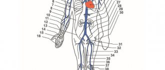
The circulatory system has a complex structure and consists of a network of vessels for different purposes: transport, shunting, exchange, distribution, capacitance. There are veins, arteries, venules, arterioles, capillaries. Together with the lymphatic ones, they maintain the constancy of the internal environment in the body (pressure, body temperature, etc.).
Arteries move blood from the heart to the tissues. As they move away from the center, they become thinner, forming arterioles and capillaries. The arterial bed of the circulatory system transports necessary substances to the organs and maintains constant pressure in the vessels.
The venous bed is more extensive than the arterial bed. Veins move blood from tissues to the heart. Veins are formed from venous capillaries, which, merging, first become venules, then veins. They form large trunks near the heart. There are superficial veins, located under the skin, and deep veins, located in the tissues next to the arteries. The main function of the venous section of the circulatory system is the outflow of blood saturated with metabolic products and carbon dioxide.
To assess the functional capabilities of the cardiovascular system and the permissibility of stress, special tests are carried out, which make it possible to assess the performance of the body and its compensatory capabilities. Functional tests of the cardiovascular system are included in the physical examination to determine the degree of fitness and general physical fitness. The assessment is based on such indicators of the heart and blood vessels as blood pressure, pulse pressure, blood flow speed, minute and stroke volumes of blood. Such tests include Letunov's tests, step tests, Martinet's test, Kotov's - Demin's test.
The meaning of arteries. The structure of the walls of large, medium and small arteries.
Arteries - blood flow centrifugally to the periphery.
The walls of the vessels have a three-layer structure:
1) Tunica intimate
- The inside is lined with endothelial cells - a constant laminar flow of blood (thanks to a continuous, smooth coating).
- Each endothelial cell produces biologically active substances (regulation of vascular tone, viscosity, blood clotting).
2) Tunica media
Parts:
- smooth muscle tissue (regulation of vascular lumen),
- connective tissue - elastic and collagen fibers (density - blood vessels do not dilate).
3) Tunica externa (adventitia)
- PBST is formed,
- Attachs PCT vessels movably,
- Pass through the nervi vasorum,
- Vasa vasorum passes through - outflow of lymph, nutrition of the middle and outer membranes.
In case of violation of vasa vasorum - obliterating endarteritis (replacement of sdtk)
According to the structure of the middle wall, there are three groups of arteries:
1) Elastic type
- Elastic fibers predominate
- The walls are thick about 15% of the outer diameter per wall,
- Common carotid artery, common iliac artery, aorta, subclavian artery, pulmonary trunk.
2) Muscular type
- GMT predominates,
- May narrow significantly.
Are divided into:
- Organ arteries (stomach, intestines). Regulation of blood flow to organs depending on functional activity.
- The arteries of the heart and cerebral artery are terminal arteries - with contraction of the tunica media, the lumen can completely close.
3) Mixed type
- Equal amount of muscle tissue and elastic fibers,
- Main arteries (axillary, femoral and their branches).
We recommend
Online English courses with strong teachers from Inglex. For visitors to our website we give 3 lessons using the promotional code WELCOME when paying for 5 lessons with Russian-speaking teachers
Start learning English
Interesting Facts
The heart begins to beat from the fourth week after conception and does not stop until the end of life. It does a gigantic job: per year it pumps about three million liters of blood and makes about 35 million heartbeats. At rest, the heart uses only 15% of its resource, and under load – up to 35%. Over an average lifespan, it pumps about 6 million liters of blood. Another interesting fact: the heart supplies blood to 75 trillion cells in the human body, except for the cornea of the eyes.