Causes
Various factors can contribute to the development of cardiac dysplasia in children and adults. The congenital form occurs due to mutation of the genes responsible for the formation of collagen. A failure may occur due to genetic predisposition or the impact of negative phenomena on a woman’s body during pregnancy.
The acquired form of cardiac dysplasia is associated with the influence of various unfavorable factors on a person, for example, junk food, alcoholic beverages, smoking, and poor environment.
Useful video
Watch the video about atrial septal defect:
If a cardiac aneurysm is detected, surgery may be the only chance for salvation, only with it the prognosis improves. It is generally possible to live without surgery, but only if the aneurysm, for example, of the left ventricle, is very small.
Cardiac MARS can be detected in children under three years of age, adolescents, and adults. Usually such anomalies go almost unnoticed. Ultrasound and other methods for diagnosing the structure of the myocardium are used for research.
In modern diagnostic centers it is possible to detect heart defects using ultrasound. In the fetus it is visible starting from 10-11 weeks. Congenital signs are also determined using additional examination methods. Errors in determining the structure cannot be excluded.
A cardiac aneurysm after a heart attack is considered a serious complication. The prognosis improves significantly after surgery. Sometimes treatment is carried out with medication. How long do people live with a post-infarction aneurysm?
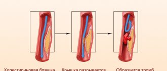
If a cardiac aneurysm has formed, symptoms may be similar to normal heart failure. Causes: heart attack, depletion of the walls, vascular changes. A dangerous consequence is a rupture. The earlier the diagnosis is made, the greater the chances.
If an aortic aneurysm is detected, the patient's life is in danger. It is important to know the causes and symptoms of its manifestation in order to begin treatment as early as possible. It's basically an operation. Rupture of the abdominal, thoracic and ascending aorta can be diagnosed.
The diagnosis of coarctation of the aorta can be detected in both newborns and adolescents. The signs of congenital heart disease are obvious. Diagnosis in children is carried out after birth and during examination. Treatment for coarctation of the aortic arch involves surgery. How to live after?
Damage to the heart with poor circulation is called cardiomyopathy in children. It can be dilated, hypertrophic, restrictive, primary and secondary. Symptoms include the standard set of signs of heart failure. Detected by Holter and ultrasound. Treatment may include surgery.
As a result of structural disturbances and stretching of the aortic wall, an aneurysm of the sinus of Valsalva may develop. If suspected, an examination should be carried out as early as possible, starting with echocardiography. Treatment consists of suturing the aortic wall.
Symptoms
It is not enough to know what it is - cardiac dysplasia; you also need to be able to recognize it. Symptoms do not appear in the early stages, so mild disease is detected in most cases completely by accident.
As the disease progresses, signs such as:
- Pain syndrome in the heart area.
- Heart rhythm disturbance
- Sleep disorders.
- Increased fatigue.
- General malaise.
- Cephalgia.
- Fainting.
The pathology may be accompanied by disturbances in the structure of the vascular components of the heart, which leads to deterioration of blood flow.
What is dysplasia?
The word dysplasia itself speaks of abnormal development of organs, body parts, and tissues. This is not necessarily a childhood disease; many ailments appear in adulthood, for example, cervical dysplasia. The following types of dysplasia are distinguished:
- Epiphyseal. The formation of bones in the embryo occurs in two stages: ossification of connective tissue with the formation of skull bones and ossification of cartilaginous tissue with the formation of the spine and limbs. Epiphyseal dysplasia - disorders in the development of the bones of the spine and limbs (spondylodysplasia of the knee and hip joint, cervical, thoracic, lumbar spine).
- Ectodermal. The disorders affect tissues developing from the ectoderm of the embryo: nervous system, tooth enamel, skin, sensory organs.
- Acromicric. It represents congenital anomalies in the development of bones: growth retardation, short hands, feet, facial abnormalities.
- Fibromuscular (fibromuscular). The disorders affect the muscular and internal elastic layer of the arteries.
- Fibrous. A relatively rare pathology characterized by the degeneration of normal bone into fibrous bone due to disturbances in the development of osteogenic mesenchyme in the embryo.
- Cervical intraepithelial neoplasia. Acquired disorders. The most common is cervical dysplasia.
- Focal cortical. Represents disorders of the cerebral cortex.
- DST (connective tissue dysplasia). Hereditary mutation of genes affecting the organization of collagen tissues.
- Cardiomyopathy (arrhythmogenic dysplasia of the right ventricle). Inherited heart disease.
- Bronchopulmonary. Occurs in premature infants after artificial ventilation.
The causes of dysplasia in a child are usually hereditary, prematurity, or associated with the course of pregnancy in a woman. This may include viral infections suffered during pregnancy, poor nutrition, the presence of bad habits such as smoking and alcohol in the expectant mother, as well as other features of pregnancy: oligohydramnios, toxicosis, abnormal position of the fetus.
Diagnosis and treatment
To detect dysplasia of the mitral valve of the heart or its other parts, doctors use instrumental examination methods. Good results are obtained by such diagnostic measures as electrocardiography and magnetic resonance imaging.
Treatment is prescribed taking into account the identified stage of the disease, the presence of concomitant pathologies, the general condition of the patient’s body, and his age. Conservative therapy is carried out, combining different methods.
Taking medications
Medicines are used to achieve the following goals:
- Restoration of normal metabolism in tissues.
- Strengthening the process of collagen formation.
- Elimination of symptoms caused by dysplasia.
- Normalization of the nervous system.
- Prevention of neurodystrophy of the heart muscle.
- Prevention or treatment of an infectious process in tissues.
Vitamin and mineral complexes are also prescribed to replenish the body with useful substances.
Therapeutic measures
Additionally, the doctor may recommend such measures as:
- Massage.
- Physiotherapeutic procedures.
- Physiotherapy.
- Diet.
Adult patients should forget about smoking and drinking alcohol.
Folk remedies
Alternative medicine methods can also be used as a supplement, but only with the permission of the attending physician. Under no circumstances should you try folk remedies on your own during therapy.
If the heart muscle and blood vessels are damaged, it is recommended to take herbal decoctions and infusions. Plants such as hawthorn, sage, motherwort, valerian, and wild rosemary have a positive effect on the functioning of this organ.
Treatment of children with pathology
If an asymptomatic cardiac aneurysm is detected, children should be under constant supervision of a pediatrician and cardiologist to prevent circulatory disorders. They are shown:
- food with sufficient protein content (chicken, turkey, fish), oatmeal and buckwheat porridge, freshly squeezed juices from fruits and berries;
- strict adherence to a daily routine with daytime rest;
- massage according to the classical method, acupressure, underwater;
- coniferous or mineral, pearl baths;
- magnesium electrophoresis, electrosleep.
The possibility of sports training is determined exclusively by a cardiologist, who takes into account the presence of palpitations, heart pain, fainting, arrhythmia on the ECG, and abnormalities detected by ultrasound. If there are significant deviations, professional sports are contraindicated, but mandatory dosed physical activity in the form of daily walks and therapeutic exercises is recommended.
Physiotherapy
Conservative therapy includes preventive courses of taking (3 - 4 times a year) the following drugs:
- magnesium salts (needed for collagen synthesis) – Magnerot, Magne B6, Magnicum;
- cardiotrophic agents - Riboxin, ATP, Carnitine, Cardonate, Kudesan, Cytochrome;
- multivitamin complexes containing group B - Pikovit syrup, Supradin kids gel;
- soothing - herbal mixtures with motherwort, lemon balm, sage and St. John's wort, wild rosemary.
Antiarrhythmic medications are prescribed only in the presence of rhythm disturbances; antibacterial therapy is required even for minor surgical interventions to prevent endocarditis.
Indications for surgical treatment arise in case of circulatory disorders, narrowing of the outflow tract of the right ventricle, valvular insufficiency due to enlargement of the right chambers of the heart, complex rhythm and conduction disorders. In such cases, a patch made of synthetic materials is applied to the place where the septum is thinned and fixed with separate sutures.
We recommend reading the article about carotid aneurysm. From it you will learn about the pathology, its types and causes of development, symptoms of aneurysm, diagnostic methods and surgery. And here is more information about coarctation of the aorta.
An aneurysm of the heart septum in a child occurs with insufficient development of connective tissue fibers and is classified as MARS syndrome. In most cases, there are no manifestations, but with a sharp increase in the load on the heart, a rupture of the septum can occur with the discharge of blood from the left half to the right. To detect an aneurysm, ultrasound of the heart with Doppler sonography is the most informative.
Mild and moderate course of the disease requires preventive courses of medication to strengthen the heart; in severe forms of the disease, surgery is indicated.
Prognosis and prevention
Heart damage has a favorable prognosis if the patient fully complies with all the recommendations of the attending doctor and is attentive to his health. It is quite possible to live a normal life with such dysplasia. If left untreated, the risk of complications is very high. For prevention, it is important to regularly check the body for pathological processes and promptly treat if disorders are detected. It is also necessary to lead a healthy lifestyle, especially for women during pregnancy.
Connective tissue dysplasia in the practice of a pediatrician
Translated from Greek as “deviation in formation”
“Medical Bulletin” has touched upon the topic of DST more than once.
But so far only isolated manifestations of this congenital pathology have been described. Thus, in “MV” No. 21 for 2010, Professor L.M. Makarov spoke about primary channelopathies, often leading to sudden cardiac death in children and caused by mutations of genes expressed in the myocardium. In “MV” No. 4 for 2011, Professor I.V. Viktorova described joint hypermobility syndrome, and in No. 17-18 for 2011, Professor E.A. Gallyamov spoke about the most important pathogenetic factor of gastroesophageal reflux disease - congenital hiatal hernias, formed due to the increased elasticity of the tissues limiting this hole.
These publications already show how widespread and significant diseases caused by CTD are. However, many years of research by Z.V. Nesterenko and the world science data that she analyzes indicate that DST affects not three, but much more body systems, and this problem is incomparably wider and more acute than it was thought about in 1989, when the Scottish doctor R. Beighton first proposed to designate the congenital pathology of TS, manifested by a decrease in its strength, by the term dysplasia, which translated from Greek means “deviation in formation.”
If proline changes to arginine
The frequency of detection of CTD (according to various authors) is quite high - from 26 to 80% for all ages and from 74 to 85% among children.
In the development of such dysplasias, mutations of genes encoding the synthesis and spatial structure of collagen and responsible for the formation of matrix components are of key importance. Accordingly, according to one of the first classifications of DST, they were divided into diseases caused by impaired synthesis or catabolism of the fibrous components of DST or its main substance.
But in the 1990s, a classification that is more common today was adopted. The first group of dysplasias includes fairly rare differentiated dysplasias (DDSDs), which have a monogenic and specific type of inheritance, more often autosomal dominant, and clearly defined clinical symptoms. These are Marfan syndrome, Ehlers-Danlos syndrome, osteogenesis imperfecta and several others.
To get acquainted with DDST, let's turn to the most common (frequency of occurrence among newborns is 5:100,000) of their representative - Marfan syndrome (MS). All proven cases of this syndrome are a consequence of a mutation in the fibrillin gene. It is localized in the long arm of chromosome 15, field 21 (15ql5-q21.3). The essence of the mutation is the replacement of the amino acid proline with arginine in the fibrillin protein. As a result, the synthesis of collagen type 3 increases and the content of collagen type 1 decreases. If normally the ratio of collagen-1:collagen-3 is 6:4, then with SMA it drops to 3:7.
The clinical picture in a typical case of SMA is manifested by a characteristic triad of signs related to the musculoskeletal and cardiovascular systems, as well as the visual organs.
Changes in the musculoskeletal system include: tall stature, asthenic physique (the length of the limbs is disproportionate to the length of the body), arachnodactyly (long thin fingers), chest deformity, high arched palate, kyphoscoliosis, ligamentous weakness. Lesions of the cardiovascular system include: dilatation of the aortic root, aortic regurgitation, dissecting aortic aneurysm, mitral valve prolapse, blood regurgitation due to insufficiency of this valve. A characteristic pathology of the visual organs is iridodenesis (tremor of the lens due to weakness of the ligament of Zinn), subluxation of the lens, a high risk of retinal detachment and high myopia.
Before the widespread use of surgical correction of cardiovascular pathology, almost all patients with SMA died before the age of 30-35 years. Moreover, the main group is still in childhood. However, in the 21st century, with adequate therapy, the life expectancy of most patients with SMA is only slightly inferior to this indicator in the general population.
Tall, freckled, stooped
The second group of DST, which makes up the disorders most often encountered by practical pediatricians, includes disorders united by the term undifferentiated dysplasia (UDTD). Unlike differentiated dysplasias, UCTD are genetically heterogeneous pathologies. The main characteristic of such dysplasias is a wide range of clinical manifestations without clear symptoms. That is, UCTD is not a nosological entity, and there is no place for it in ICD-10 yet. In turn, UCTDs are divided into 2 groups: diseases with an established and unidentified (and this is the vast majority of UCTDs) gene defect.
UCTD in a child can be diagnosed already at the stage of physical examination with a comprehensive assessment of the so-called phenotypic markers. External and visceral markers are distinguished, the predominance of which depends on what type of TS lesion occurs - dense or loose. The critical number of external markers that allows one to make a conclusion about the presence of UCTD is now considered to be 3-6. Based on the diagnostic significance of individual signs of UCTD, diagnostic tables are proposed with a score of external and visceral markers, as well as biochemical parameters.
For example, external changes in the skin in children with UCTD usually indicate damage to loose connective tissue and are characterized by the presence of hyperelasticity, increased extensibility, stretch marks, keloid scars, a pronounced subcutaneous venous network, characteristic pigment spots of the “café au lait” color, or depigmentation, a large number nevi.
But lesions of dense TS are manifested by changes in the skeleton: poor posture in the form of kyphosis and scoliosis, stooping, chest deformities, and flat feet. Such children, as a rule, are tall and asthenic in build.
All children with UCTD exhibit so-called minor developmental anomalies, or dysmorphia. The most common dysmorphias include: fair skin, fused eyebrows, wide bridge of the nose, hyper- and hypotelorism, blue sclera, epicanthus, abnormal tooth growth, deformed auricle, fused lobe, curved little fingers, incomplete syndactyly of the fingers, light or red hair color. Diagnostically significant for identifying UCTD is the presence of 6 or more dysmorphias in a child.
Markers external and internal
Let us now turn to the internal markers of UCTD, and we emphasize that there is a direct correlation between the number of external and internal markers.
If we talk about the integral indicators of internal UCTD markers detected by biochemical methods, then the most informative is determining the level of molecules formed during the breakdown of collagen. These are hydroxyproline and glycosaminoglycans in daily urine, and in blood serum - lysine and proline. A change in the ratio of collagens of different types in UCTD allows the use of the indirect immunofluorescence method according to Sternberg LA (1982) in diagnosis.
On the membranes of leukocytes, an increased representation of HLA histocompatibility antigens is usually detected - A28, B35, Cw5, Cw52, and on the other hand, a reduced number of antigens such as A2, B12, Cw3. The most promising, of course, are molecular genetic diagnostic methods that identify specific gene mutations. However, in cases of UCTD, these assays are still at an early stage of development.
One of the most often involved in the pathological process in UCTD in children is the cardiorespiratory system.
This leads to serious pediatric errors
With UCTD in children, the formation of the elastic framework of the lungs is disrupted at the beginning of life, which causes the valve mechanism of bronchial obstruction and the formation of emphysematous bullae due to rupture of morphologically incompetent interalveolar septa. The consequence of subpleurally located bullae can be spontaneous pneumothorax. A congenital defect in the cartilaginous and connective tissue framework of the trachea and bronchi leads to their increased mobility, the occurrence of bronchiectasis, and pneumosclerosis. Dyskinesia of the trachea and bronchi leads to the development of broncho-obstructive syndrome (BOS).
There is a high correlation between the severity of bronchial asthma in children and the manifestations of dysplasia. And the more common and severe the latter are, the earlier the child develops pulmonary hypertension and pulmonary fibrosis in addition to bronchial asthma.
Anomalies in the structure of the bronchopulmonary system in UCTD lead to a deterioration in the elimination of pathogenic agents in conditions of altered immune reactivity and contribute to the long-term persistence of bacteria and the formation of a recurrent course of pneumonia. It is also noteworthy that the level of atypical childhood pneumonia is growing, caused by intracellular pathogens - chlamydia and mycoplasma, with a predominant lesion of the interstitium of the lungs, with a recurrent course of which progresses, as in cases of asthma, pneumofibrosis, pulmonary hypertension.
With UCTD, congenital defects are often diagnosed: tracheobronchomegaly, tracheobronchomalacia, cystic hypoplasia of the lungs. Against the background of dysplasia, the listed diseases are especially often accompanied by the development of severe complications in the form of pneumofibrosis, pulmonary hypertension, bronchiectasis, and spontaneous pneumothorax.
Unfortunately, in contrast to the above-mentioned pronounced manifestations of UCTD in children, subclinical variants are usually not diagnosed. This often leads to incorrect interpretation of the pathological process and serious pediatric errors.
Truly small heart
Since 1987, the classification of diseases of the cardiovascular system of the New York Heart Association has identified the syndrome of connective tissue dysplasia of the heart (CDS), which accompanies both differentiated and undifferentiated dysplasia. In turn, cardiac dysplasia includes several syndromes.
Valvular syndrome combines isolated and combined heart valve prolapses and myxomatous valve degeneration. In approximately 70% of cases, the syndrome is represented by prolapse of the mitral valve, less often - tricuspid or aortic, enlargement of the aortic root and pulmonary trunk; ectopically located chordae, aneurysms of the sinuses of Valsalva. In some cases, such changes are accompanied by regurgitation phenomena, which is reflected in the indicators of myocardial contractility and volumetric parameters of the heart.
With thoradiaphragmatic syndrome, we are talking about the asthenic shape of the chest or its deformation, spinal deformities (scoliosis, kyphoscoliosis, hyperkyphosis, hyperlordosis), changes in standing and excursion of the diaphragm. Among children with CTD, the most common deformity of the chest is pectus excavatum, and the second most common is keeled. Deformations of the sternum, ribs, spine and the associated high position of the diaphragm reduce the chest cavity, increase intrathoracic pressure, disrupt the inflow and outflow of blood, and contribute to the occurrence of arrhythmias.
Vascular syndrome involves damage to elastic arteries, in which their walls expand and saccular aneurysms appear. Arteries of both muscular and mixed types are affected. As a result, bifurcation-hemodynamic aneurysms appear, as well as pathological tortuosity of vessels up to looping.
Thoradiaphragmatic heart syndrome (THDS) is formed in parallel with the progression of deformation of the chest and spine, against the background of valvular and vascular syndromes. In children with a typical asthenic constitution, the asthenic variant of STDS is more often manifested. In this case, the size of the heart chambers decreases, but while maintaining their normal thickness. In a word, a “true small heart” is being formed, functioning without serious deviations.
Unfortunately, in some children with chest deformation due to displacement of the heart, when it “moves away” from the mechanical influences of the chest bone, rotating and accompanied by “torsion” of the main vascular trunks, the so-called false stenotic variant of TDS is formed, which is especially severe.
Metabolic cardiomyopathy syndrome combines cardialgia, arrhythmias, and disorders of repolarization processes. The development of metabolic cardiomyopathy is determined by the influence of cardiac factors (valvular syndrome, variants of CVD) and extracardiac conditions (autonomic dysfunction, deficiency of micro- and macroelements, etc.). Cardiomyopathies with DST usually do not have specific symptoms.
Arrhythmic syndrome includes ventricular and atrial extrasystoles, paroxysmal tachyarrhythmias, pacemaker migrations, atrioventricular and intraventricular blocks, anomalies of impulse conduction along additional pathways, ventricular preexcitation syndrome, and QT interval prolongation syndrome.
A practical pediatrician should remember that in children suffering from CTD, cardiomyopathies and arrhythmic syndrome are very common (in 60-64% of patients). And it is they who determine the increased risk of sudden cardiac death in such children.
A universal remedy has not yet been found
A universal remedy that restores connective tissue for all forms of dysplasia has not yet been found. An individual treatment program is selected for each child with CTD. Its three main tasks are: improving the metabolic processes of connective tissue, eliminating existing complications and preventing new ones.
In any case, we are talking about the actually coordinated work of a team of pediatricians of several specialties. And about a very complex, complex treatment that affects the entire childhood period of life (in the Russian Federation - up to 18 years). We will have time to give only one example of such therapy, related to Marfan syndrome.
Non-drug therapy includes strict adherence to the child’s diet, including nutrition, where the emphasis is on the consumption of complete proteins and foods containing polyunsaturated fatty acids; and physical activity, certain types of which are prohibited for such patients. There is a list of professions for which schoolchildren suffering from CTD cannot be trained.
Drug treatment includes symptomatic administration of b-blockers. In the case of aortic dilatation, and especially in the presence of regurgitation, they reduce the ejection into the aorta and, accordingly, the load on its walls, correcting concomitant hypertension. It is believed that these cardiotropic drugs reduce the risk of sudden death in children with cardiac damage due to SMA and any other CTD.
Since it has been proven that the progression of skeletal pathologies in Marfan syndrome slows down when the deficiency of microelements (calcium, magnesium, zinc, copper) necessary for the formation of TS is eliminated, nutritional supplements containing the above substances, as well as hyaluronic acid, synthetic analogues of vitamins K and D3. In the blood of patients with SMA, there is often an increased level of somatotropic hormone. Therefore, to reduce its secretion, high-fat Omega-3 enzymes are prescribed in the diet from early childhood.
Let us now turn to therapy affecting connective tissue. It includes ascorbic acid in the form of special children's milkshakes, as well as correctors for impaired synthesis and catabolism of glycosaminoglycans - chondroitin sulfate and succinic acid. Another drug recommended as a corrector of bioenergetic processes is carnitine chloride.
What surgeons can do
For aortic aneurysm, dissecting aneurysm, aortic valve defect with symptoms of heart failure, and children with Marfan syndrome, only surgical treatment can help. Clear indications for prosthetics have been developed. For example, an aneurysmally dilated aorta must be replaced with an endo- or exoprosthesis. In case of mitral valve prolapse accompanied by “stable” regurgitation, valve replacement is not performed. However, with the rapid progression of regurgitation, up to the addition of left ventricular failure, valve replacement becomes necessary.
Surgical treatment of deformities of the chest and spine is an extremely traumatic procedure, carried out in several stages, often complicated by pleurisy, pericarditis, and pneumonia. The question of its feasibility was discussed many times at symposiums dedicated to DST. Specialists from various countries adopted a common position denying the advisability of such operations for any DST.
Dysplastic cardiopathy
From Greek “pathos” means illness, suffering. This Greek root is also found in the medical term “cardiopathy”. It is not difficult to guess that cardiopathy is some kind of change in the heart that is different from the norm. But what does such an entry in an outpatient card or medical history mean: “dysplastic cardiopathy”?
Agree, an incomprehensible, complex diagnosis can scare anyone. And given the fact that this cardiopathy in children and newborns is not at all uncommon, this diagnosis can completely shock young parents who are not sufficiently aware of what kind of disease this is and “what they eat it with.” Let's figure it out.
Features of the disease in adults and children
The disease can manifest itself immediately after a person’s birth or manifest itself gradually throughout life. In children and newborns, dysplasia of the connective tissues of the heart is most often present as a factor causing rhythm disturbances. This is: supraventricular or ventricular extrasystole. There may be congenital heart defects and other disorders.
In adulthood, identifying the cause of disorders, if it is connective tissue dysplasia, is just as important as in early years. The diagnosis is difficult to clarify.