Types of stroke and stages
To understand the relevance of MRI in the diagnosis of cerebrovascular accidents, it is recommended to know the classification of pathology. Let us note the varieties of the disease and its distinctive features.
Pathology is divided into the following forms:
- Hemorrhagic is a rupture of an artery with blood escaping beyond the vascular bed. As a result of this damage, an internal hematoma forms, which compresses the neurons and provokes symptoms. This form is divided into hemorrhagic stroke of the membranes and directly into the brain tissue.
- Ischemic stroke is, on the contrary, a lack of blood in a certain segment of the brain. This occurs due to blockage of the artery, as a result of which the tissues located along its course are cut off from the cardiovascular system and are left without oxygen and nutrients. This form is divided into microstroke, which is characterized by a mild course and a quick recovery period.
Brain tissue is fragile, and hemorrhage easily damages neurons.
Even the slightest oxygen starvation causes the death of cells that are not restored. Therefore, timely diagnosis and prevention of local diseases is important.
There are four stages of stroke:
- The most acute - develops in the first three days after the attack, is characterized by vivid symptoms that disorient the patient. There is a severe headache, loss of coordination, nausea and vomiting, loss of sensitivity up to the failure of half the body.
- Acute stage - from 3 days to 3 weeks. Characterized by a decrease in symptoms and gradual stabilization of well-being. Some functions are still lost.
- Earlier recovery - from the third week to six months. Minimal clinical signs are detected, and some of the lost functions return.
- Late recovery - sometimes lasts up to 2 years. Symptoms disappear, the return of function depends on the degree of neuronal damage.
The patient is under close medical supervision. First, rest and symptomatic therapy are indicated, then rehabilitation is carried out.
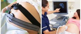
What forms are diagnostics prescribed for?
Magnetic waves are not suitable for all forms of stroke - they do not show fluid accumulations well and do not detect hemorrhage. Therefore, for the hemorrhagic form, computed tomography is a valuable diagnostic method.
MRI is done for ischemic stroke - in this case, the study is informative and will show:
- area of damage;
- level of artery blockage;
- degree of destruction of brain tissue.
To decide whether to prescribe a CT or MRI, the doctor identifies the type of disease based on clinical signs.
Ischemic is characterized by patient disorientation and mild headache.
MRI has several advantages in diagnosing stroke. The main thing is the absence of radiation, so the study is carried out with a minimum number of contraindications.
MRI for suspected stroke
Tomography is one of the most important methods for diagnosing stroke. When using the magnetic-nuclear effect, data is obtained by analyzing the electromagnetic vibrations of atomic nuclei that they emit in a magnetic field. Compared to computed tomography, the advantages of MRI are:
- determination of the area of necrosis if it is small or in the border zone with the bones of the skull;
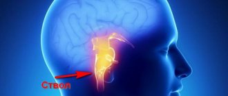
- obtaining clear images of the brainstem, basal and cortical regions;
- the ability to visualize deep brain structures,
- detection of arterial thrombosis;
- detection of cerebral edema (diffusion-weighted method).
The latter technology refers to a relatively new method of imaging the earliest stages of brain tissue damage. She is able to detect signs of a stroke a few minutes after its development.
Watch the video about stroke, its causes and symptoms, diagnosis and treatment:
Procedure
Unlike mid-field MRI, tomographs with a power of at least 1.5 Tesla are used to diagnose cerebrovascular accidents. Preference is given to high-field 3 T devices, which have the following advantages:
- pictures are taken with a large number of sections - this helps to thoroughly examine the lesion;
- the image is clear and informative - thanks to this, an accurate, detailed diagnosis is made;
- the examination time is reduced - this reduces discomfort, the necessary therapy is prescribed, and conclusions are drawn about the prognosis.
Since the study does not require preparation, the doctor makes a decision, and with the patient’s consent, a diagnosis is carried out.
The procedure for performing MRI for stroke includes the following steps:
- removing metal jewelry;
- the doctor makes sure that there are no metal crowns, a pacemaker or an insulin pump;
- the patient lies down on the platform, the head is carefully secured with a belt;
- The patient is placed in the capsule of the device, where images are taken.
The study will take 30-40 minutes. If the patient suddenly becomes ill, he uses the communication button and calls a doctor.
In an emergency situation, when minutes are counting and it is necessary to determine whether there is blood or a blood clot in the head, in this case a CT scan is prescribed (not informative in the first 6 hours).
How does the procedure work?
In case of a stroke, an MRI of the brain is performed and examination of its vessels using a special device - a magnetic resonance imaging scanner. After the patient is admitted to the hospital:
- Doctors decide whether an MRI is necessary.
- If the decision is positive, the patient is prepared for the procedure by removing metal objects from him. If possible, you should warn the MRI specialist about the presence of dental implants or structures made of materials that include ferromagnets (iron, steel).
- The patient is placed on the tomograph table, the head is fixed to prevent movement during the examination. The patient is pushed into the tunnel.
- Using a tomograph, a specialist takes layer-by-layer images of the brain and its vessels in several projections.
- The table with the patient is pulled out and the diagnostic results are prepared.
MRI of the brain is performed in high (1.5 Tesla) or ultra-high (3 Tesla) magnetic field modes. In case of stroke, ultra-high-field diagnostics are usually preferred. It does not harm the patient, but thanks to it:
- It is possible to obtain images of such sections that are impossible with high-field MRI. This mode allows you to carefully study the state of the brain even with a micro-stroke.
- The procedure time is reduced as scanning occurs faster.
- The results of the study are the most accurate, since the ultra-high-field scanning mode launches a program to eliminate distortions caused by the patient’s restless behavior.
- It is possible to accurately determine the volume of the affected area of the brain, which makes it possible to monitor the recovery of the organ during the rehabilitation period and promptly change treatment tactics in the absence of positive dynamics.
For angiography of cerebral vessels, intravenous injection of a dye can be used. MRI with contrast will take longer, but will accurately assess the condition of the arteries, veins and even capillaries of brain tissue.
Angiography with MRI
If it is necessary to assess the degree of cerebral circulatory impairment, angiography is performed with a contrast agent, which is administered a few minutes before the procedure.
The contrast stains the arteries and determines:
- the cause of ischemia (vasospasm, detachment of a blood clot, atherosclerotic plaque);
- level of blockage;
- area of the brain that is left without blood.
Allergy to contrast is rare; a minimal test is done to determine it. If there is no intolerance, a study is carried out.
Magnetic resonance imaging: pros and cons
Studies have shown that magnetic resonance imaging is more sensitive in detecting small, deep lesions and infarcts in the structures of the posterior cranial fossa.
It has been noted that signs of previous strokes persist on MRI images indefinitely in the vast majority of cases, so MRI can be used to determine the nature of a previous stroke at a later stage of the study.
Using MRI during the first hours after a stroke, it is difficult to distinguish a hemorrhage focus from an ischemic focus. Clear changes usually appear within 24 hours. However, in the future, typical changes on MRI persist for life, so the consequences of hemorrhage can be detected even years after its occurrence.
In addition, MRI is more accurate than CT in detecting even small areas of hemorrhage or ischemia in the brain stem and cerebellum, since this method does not interfere with bone structures.
MRI is often better than CT at identifying the underlying cause of intracerebral hemorrhage. For example, it identifies diseases of the venous bloodstream, multiple point metastases, characteristic signs of inflammatory vascular diseases, primary tumors and structural features of blood vessels, which, if ruptured, can lead to hemorrhage.
MRI can help answer the question of whether the lesion was ischemic or hemorrhagic when stroke patients present too late for CT to be used reliably.
Both CT and MRI use different examination modes.
For example, there is the so-called magnetic resonance angiography, with the help of which it is possible to obtain images of brain vessels in a magnetic field, safely for the patient.
In this case, it is possible to discern with a sufficient degree of certainty the structural features of the vessels, their narrowing, deformation or other changes. Sometimes the study is carried out with contrast enhancement, that is, an MRI is performed after intravenous administration of a special contrast agent, which allows you to more accurately see the condition of the brain vessels.
Deciphering the pictures
When there is a lack of blood circulation, the neurons feeding from the affected artery die.
In this case, the doctor looks at the intensity of the MRI signal, which should be attenuated.
An ischemic stroke looks like a white spot on pictures - a change in color indicates a softening of the signal from the tomograph.
When a pathological area is identified, the doctor studies the images in detail, determines the size and location, and writes a conclusion. Based on these data, a forecast is given.
Hemorrhages in the brain and its membranes are not always detected on MRI, so CT is performed first.
Ischemic stroke
Hemorrhagic stroke
If there are no changes on the x-ray, a magnetic tomography scan is prescribed.
Contraindications to MRI in neurology
Contraindications for MRI:
- Metal implants;
- Claustrophobia;
- Pacemakers, although new protocols allow imaging in selected cases;
- MR-incompatible prosthetic heart valves;
- Allergy to contrast agent;
- Severe obesity.
Patients with metal implants may experience many potential complications, such as pacemaker failure and related consequences. For patients with a metal prosthesis, it is recommended to check its compatibility with MRI.
Pacemaker
Cliustrophobic patients are unable to complete an MRI scan. In selected patients, gentle sedation or imaging in an open MRI system may be attempted. However, most open MRI scanners provide lower quality images.
In rare cases, patients may be allergic to a special substance (such as gadolinium) used in MRI. If any of these contraindications are present for stroke, a computed tomography (CT) scan of the brain is prescribed.
Features of examination after a stroke
After a stroke, MRI is an indispensable method that is superior to CT - changes in soft tissues are visible in the images for a long time:
- swelling - its reduction indicates the effectiveness of treatment;
- cyst - helps to give a prognosis;
- growth of neural networks - if available, the effectiveness of rehabilitation and restoration of lost functions is assessed.
MRI is recommended to be carried out annually; if your health worsens, the doctor will advise you to take pictures more often. Research in the late recovery period is valuable.
MRI is relevant for ischemic stroke; it is difficult to detect hemorrhages in the images. In the future, the tomograph will help determine the effectiveness of treatment and rehabilitation. Studies are carried out annually; on the advice of a doctor, it is sometimes prescribed more often.
Modern MRI
Advances in MRI include high magnetic field strengths (1.5-3.0 Tesla), which provide better image resolution, and the advent of open MRI for patients who are claustrophobic or overweight. Magnetic fields of 7.0 and 9.4 Tesla with increased signal-to-noise and contrast-to-noise ratios have recently been introduced into practice. However, some points: heterogeneous transmission fields and extensive contraindications for patient scanning limit their clinical use in acute stroke.
MRI
Hospitals can often monitor a patient's condition and provide treatment while an MRI is being performed, as MRI-compatible electrocardiographic monitors, intravenous infusion drips, and respirators have become available on the market.
Attention! Magnetic resonance imaging has some negative aspects: high price, scanning time and low sensitivity in diagnosing subarachnoid hemorrhages.