Blood flow speed is the speed of movement of blood elements along the bloodstream in a certain unit of time. In practice, experts distinguish between linear velocity and volumetric velocity of blood flow.
One of the main parameters characterizing the functionality of the body’s circulatory system. This indicator depends on the frequency of contractions of the heart muscle, the quantity and quality of blood, the size of blood vessels, blood pressure, age and genetic characteristics of the body.
Types of blood flow speed
Linear speed is the distance traveled by a blood particle through a vessel over a certain period of time. It directly depends on the sum of the cross-sectional areas of the vessels that make up a given section of the vascular bed.
Consequently, the aorta is the narrowest section of the circulatory system and has the highest blood flow speed, reaching 0.6 m/s. The “widest” place is the capillaries, since their total area is 500 times larger than the area of the aorta, the blood flow speed in them is 0.5 mm/s. , which ensures excellent exchange of substances between the capillary wall and tissues.
Volumetric blood flow velocity is the total amount of blood flowing through the cross-section of a vessel over a certain period of time.
This type of speed is determined:
- the difference in pressure at opposite ends of the vessel, which is formed by arterial and venous pressure;
- vascular resistance to blood flow, depending on the diameter of the vessel, its length, and blood viscosity.
What vessels look on the neck and why?
A doctor performs an ultrasound examination of the vessels of the neck
During an ultrasound examination of the vessels of the neck, the doctor must examine the following structures:
- brachiocephalic trunk;
- right and left subclavian arteries;
- right and left common carotid arteries;
- right and left internal carotid arteries;
- right and left external carotid arteries;
- vertebral arteries.
If necessary, the following can be additionally examined:
- jugular veins;
- veins of the vertebral plexus;
- supratrochlear arteries;
- ophthalmic arteries.
All of the above vessels are examined for the possible detection of the following pathologies:
- Atherosclerosis of extracranial arteries. It is possible to establish not only pronounced atherosclerotic changes, localization and size of plaques, degree of stenosis, complications, but also the initial manifestations of atherosclerotic lesions of the carotid arteries in the form of thickening of the intima-media complex. In the presence of significant stenoses and vascular occlusions, the functioning of cervical anastomoses, that is, bypass paths of blood flow to the brain, is assessed.
- Nonspecific aortoarteritis or Takayasu's disease. Using ultrasound, the doctor can distinguish aortoarteritis from atherosclerotic lesions and give a detailed description of blood flow disorders.
- Dissection. Ultrasound can reveal signs of arterial wall dissection during thrombosis of unknown cause or after injury.
- Arterial deformations. Ultrasound quite accurately shows the presence, shape and location of deformations of the examined arteries, as well as the effect of the identified deformations on blood flow.
- Steele syndrome or vertebral-subclavian steal syndrome. Ultrasound helps to establish the location of the lesion, the degree of narrowing of the artery, and the characteristics of hemodynamic disturbances in it.
- External compression of blood vessels by neighboring organs and tissues.
- Congenital anomalies of vascular development and their effect on the blood supply to the brain.
- Disturbances in the venous outflow of blood from the brain. Ultrasound helps to identify the signs and causes of this pathology.
But the main purpose of conducting an ultrasound examination of the extracranial arteries of the neck is to identify possible causes and further prevent the development of a dangerous disease - cerebral ischemic stroke.
Importance and severity of the problem
Determining such an important parameter as blood flow velocity is extremely important for studying the hemodynamics of a specific section of the vascular bed or a specific organ. If it changes, we can talk about the presence of pathological narrowing along the vessel, obstacles to blood flow (parietal blood clots, atherosclerotic plaques), and increased blood viscosity.
Currently, non-invasive, objective assessment of blood flow through vessels of different sizes is the most pressing task of modern angiology. The success of solving it depends on the success of early diagnosis of such vascular diseases as diabetic microangiopathy, Raynaud's syndrome, various occlusions and vascular stenoses.
Vascular resistance
Circulating blood encounters resistance along its path, which manifests itself as a result of friction of blood elements between themselves and the walls of blood vessels. The thicker the blood, the stronger the friction; this parameter is also influenced by the diameter of the vessel and the speed of blood flow.
Thanks to the heart, the blood quickly overcomes vascular resistance, as it pushes the fluid forward with pulsating movements. Resistance is stronger in those areas where smaller vessels depart from the arteries. The highest resistance is encountered by blood in the arterioles, since they have a minimal diameter and the blood moves quickly. Internal friction increases, and these vessels are prone to spasm. Resistance increases with distance from the aorta.
Why is blood velocity measured?
Measuring blood flow velocity is important for diagnostic medicine. Thanks to the analysis of data obtained as a result of measurements, it is possible to determine:
- vascular condition, blood viscosity indicator;
- level of blood supply to the brain and other organs;
- resistance to movement in both circles of blood circulation;
- microcirculation level;
- condition of the coronary vessels;
- degree of heart failure.
The speed of blood flow in vessels, arteries and capillaries is not constant and the same value: the highest speed is in the aorta, the smallest is inside the microcapillaries.
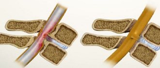
Anatomy of the vertebral arteries
The vertebral arteries are located on both sides of the neck
Two symmetrical a. Vertebralis are branches of the subclavian arteries on both sides. They pass through canals formed by openings in the transverse processes of the cervical vertebrae. Each of them gives five branches. The descending branches supply blood to the upper parts of the spinal cord and the medulla oblongata, the ascending branches enter the cranial cavity and participate in the formation of the arterial ring. The ring unites the branches of the carotid artery (carotid artery system) and the vertebral artery (subclavian artery system) on both sides. As a result, vital parts of the brain are supplied with uninterrupted blood supply, even if one of the four main arteries is damaged.
The risk of damage to the vertebral arteries occurs with various pathologies of the cervical spine. Symptoms of the pathology are usually associated with disruption of brain function.
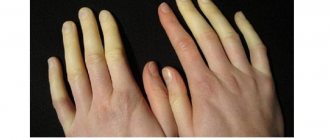
Why is blood flow velocity measured in the vessels of the nail bed?
The speed of blood flow in the vessels of the nail bed is one of the clear indicators of the quality of blood microcirculation in the human body. The vessels of the nail bed have a small cross-section and consist not only of capillaries, but also of microscopic arterioles.
When there are problems related to the circulatory system, these capillaries and arterioles are the first to suffer. Of course, it is impossible to judge the state of the entire system solely on the basis of a study of blood circulation in the nail bed area, but it is worth paying attention if the blood flow in this area is too low or high.
In medicine, to obtain the most reliable information, measurements of blood circulation parameters are carried out over large areas of the blood circulation.
Ultrasound signs of the main detected pathologies
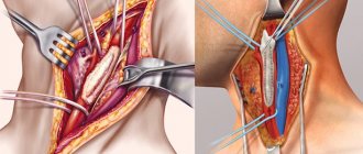
Atherosclerotic lesion of the vessels of the neck
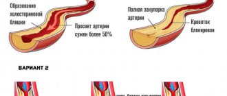
The main causes of vascular obstruction are most often atherosclerosis or thrombosis. They lead to stenosis or occlusion of the lumen of the vessel. Stenosis is an incomplete narrowing of the lumen. Occlusion is a complete blockage of the lumen of a vessel in any area, as a result of which blood cannot flow further. In the neck, atherosclerotic plaques most often form in the area of the bifurcation of the common carotid artery, the mouth of the vertebral artery, the siphon of the internal carotid artery, and the mouth of the subclavian artery. Doctors know these features, and therefore pay special attention to examining these particular places.
Carotid artery stenosis on ultrasound
The initial manifestations of atherosclerosis are characterized by an increase in the thickness of the intima-media complex from 1.0 to 1.5 mm. If the thickness of these layers is more than 1.5 mm, then they already talk about a plaque. During an ultrasound examination, the plaque may look completely different on the screen. They are homogeneous and heterogeneous, hyperechoic, hypoechoic and isoechoic. The most unfavorable are considered to be atherosclerotic plaques that are heterogeneous in structure and have an uneven surface. They are at high risk of complications.
In case of stenotic lesion of the artery, the doctor measures the degree of narrowing of the vessel on a longitudinal or transverse section of the vessel, and measures the extent of the lesion. Plaques up to 1.5 cm in length are considered local, and longer ones are considered prolonged. This parameter is extremely necessary for assessing the significance of lesions and planning treatment tactics.
Arterial thrombosis
Arterial thrombosis differs from atherosclerosis, as a rule, by the following ultrasound signs:
- occlusion predominates more than stenosis,
- the lesion is longer in length,
- more often the echogenicity of intraluminal formations is relatively homogeneous, the echogenicity varies depending on the stage of thrombosis,
- in the area of the beginning of occlusion - the surface is flat,
- with prolonged existence of thrombosis, hypoplasia of the artery develops.
Arterial deformities
Deformations are the second most common changes in the vessels of the cervical spine after atherosclerosis. They can be congenital or acquired. In children under 18 years of age, deformities are considered normal. Children are born with a short neck, and the vessels have the same length as those of adults, and in order for them to “fit” in the neck, they have different bends and deformations. As the neck itself grows, the vessels align and acquire a straight course. In older people, under the influence of changes in blood pressure, the vessels stretch and can become tortuous again. The following types of deformations are distinguished by shape:
- tortuosity is a deformation with an angle greater than 90 degrees, they are C- and S-shaped;
- bends - deformations with an angle of 90 degrees or less, they have the worst effect on blood flow, as they lead to a narrowing of the lumen at the bend;
- loops are circular configurations of the artery, often congenital.
During an ultrasound examination, as a rule, the course of the vessel is clearly visible, and it is not difficult for the doctor to determine the type of deformation, its location, and the size of the angle.
Nonspecific aortoartery disease (Takayasu disease)
Unlike atherosclerosis, which affects more men, Takayasu's disease is more common in young women. The main ultrasound sign of damage to the carotid arteries is uneven, diffuse, hyperechoic thickening of the wall of the common carotid artery. Moreover, unlike atherosclerosis, the thickening is circular in nature, that is, it affects all the walls of the vessel. It becomes difficult to distinguish the individual layers in the wall.
Metabolic angiopathy
Metabolic angiopathy is a complex of structural changes in the vascular wall of arteries caused by various metabolic disorders. Most often occurs in patients with diabetes. In this case, small pinpoint bright hyperechoic inclusions are visible in the vessel wall. Changes in the spectral characteristics of blood flow are characteristic: an increase in resistance indices is detected in the proximal part of the artery, a decrease in velocity in the distal part.
Arterial dissection
Dissection is called local separation of the wall as a result of its tear. Most often it occurs due to injury. At the site of dissection, detachment of the upper layer of the vascular wall occurs, blood begins to get under it and thrombose, forming a hematoma. During an ultrasound examination, the doctor sees a dissected wall with movable intima or the presence of a second lumen of a vessel with blood flow.
Cerebral venous circulation
There can be many reasons for disrupting the flow of blood from the brain. During an ultrasound examination, the transcript may contain the following criteria indicating stagnation of venous blood in the brain:
- an increase in the diameter of the internal jugular vein (more than three times the diameter of the common carotid artery) as a result of compression in the proximal parts or valve insufficiency,
- reduced diameter of the internal jugular vein as a result of congenital hypoplasia or compression,
- bidirectional flow (reflux) in the vein as a result of valve insufficiency,
- an increase in blood flow velocity in the internal jugular vein is more than 70 cm/s, in the vertebral vein – 30 cm/s,
- lack of blood flow in the internal jugular vein (thrombosis),
- an increase in the diameter of the lumen of the vertebral vein by more than 2.5 mm in the spinal canal,
- compression of the vertebral vein: its uneven diameter, arched course or acceleration of blood flow at the site of compression.
Spectral analysis
Electrocardiography is a method of graphically recording changes in the potential difference of the heart that arise during the processes of myocardial excitation.
The analysis of any ECG must begin with checking the correctness of its registration technique. Firstly, you need to pay attention to the presence of various interferences.
Video about Smetana Vysehrad Spa Hotel, Czech Republic
Attention! Only a doctor can diagnose and prescribe treatment during a face-to-face consultation with the patient. Medical news and articles about cancer treatment and disease prevention for adults and children. Foreign clinics and hospitals - treatment of tumors and rehabilitation abroad. When using materials from the site, the active reference is obligatory.
CONTENT
- Notations and abbreviations
- Main part
- Ultrasound examination of the main arteries of the extremities
- Duplex scanning of the arteries of the lower extremities
- Sample protocol for ultrasound examination of the main arteries of the lower extremities
- Duplex scanning of the arteries of the upper extremities
- Sample protocol for ultrasound examination of the main arteries of the upper extremities
- List of sources used