Neutrophils: general information
Not every patient knows about such a pathology as Pelger anomaly of neutrophils. What kind of disease is this? To answer this question, let's try to understand the structure and functions of neutrophils.
Neutrophils are the most numerous type of white blood cells. They are necessary for the body to fight pathogens of infectious diseases. These cells rush to the site of inflammation. and then absorb and digest dangerous bacteria and viruses. This process of neutralizing foreign agents is called phagocytosis.
The blood test usually indicates 4 types of neutrophils:
- Myelocytes. These are the youngest cells. Normally, they can only be present in the bone marrow. Their presence in peripheral blood always indicates pathology.
- Young cells. They may be present in a blood test in very small quantities (up to 1% of the total number of neutrophils);
- Band neutrophils. This is a small group of cells. They are 1 - 5% normally.
- Segmented cells. It is this type of neutrophils that predominates in the peripheral blood (40 - 70%).
Mature types of neutrophils include only segmented cells; all other varieties are considered young forms.
With Pelger's anomaly of neutrophils, changes in the structure of segmented cells are noted. They become similar to young forms. This often leads to errors in diagnosing various pathologies.
Pelger's anomaly of neutrophils - how does it manifest itself and for what reasons?
Pelger neutrophil anomaly is one of the rare blood pathologies that is inherited in an autosomal dominant manner. This condition is considered benign (it does not impair blood function and does not lead to deterioration of health) and consists of morphological changes in one of the types of white blood cells, neutrophils.
This pathology received its name in honor of its discoverer. It was not until 1928 that the Dutch physician Karl Pelger succeeded in fully characterizing atypical neutrophils.
After 4 years, the scientist Huet proved that this anomaly is hereditary. Abnormalities of neutrophil nuclei occur equally in both males and females.
The frequency of this pathology is recorded only once per 1000-1500 people.
Why might it appear?
The causes of the leukocyte abnormality have not been precisely established. Until now, scientists have only been able to find out the fact that pelgerization can be transmitted in an autosomal dominant manner. It can appear in both heterozygotes and homozygotes.
Hematologists have found that the main cause of pathogenesis is a violation of the segmentation process of already mature neutrophils. Since this pathology is inherited, it was found that genetic damage occurs due to changes in the structure of the gene responsible for the shape of the nucleus. The presence or absence of this pathology is easy to check if there is a blood test of both parents.
How does it manifest?
Before studying the manifestations of neutrophilic changes in the blood, you need to understand what function these cells perform. Neutrophils are leukocytes, blood cells that are responsible for ingesting pathogenic bacteria.
As soon as pathogenic bacteria or other pathogens enter the blood, neutrophils immediately begin to attack them.
Important! The normal content of segmented neutrophils in the blood is from 45 to 65% in the leukocyte formula.
If we talk about the absolute value, the number of neutrophils can vary from 2.0 to 5.5 Giga/liter.
Such an anomaly of leukocytes does not have specific clinical expressions, because their functionality is not impaired. A Pelger anomaly can be detected completely by accident when a person takes a test. Often in people who are carriers of this pathology, ESR and coagulability indicators remain normal. People with this abnormality have the same reactions to blood loss or infection.
Blood picture
Neutrophilic leukocytosis in the body develops due to various reasons:
- infectious diseases,
- gangrene,
- sepsis,
- peritonitis,
- allergies,
- myocardial infarction,
- skin diseases,
- neoplasms,
- long-term inflammatory processes,
- hematological diseases,
- helminthic infestations.
These diseases can be diagnosed in the early stages, but it is important that at this moment the Pelger anomaly is taken into account, which leads to a shift in the leukocyte formula to the left. This may lead to a misdiagnosis.
The whole point of this anomaly lies in only one thing - mature neutrophil cells can develop under the guise of young cells with a non-segmented nucleus. But at the same time, their granularity and chromatin density are similar to those of normal mature cells, which distinguishes them from myelocytes.
The nucleus of abnormal neutrophils may be:
- round,
- in the form of an oval or horseshoe,
- bean-shaped or dumbbell-shaped,
- bilobed or trilobed,
- in the form of an hourglass,
- with a constriction in the middle.
How is the diagnosis done?
To determine Pelger anomalies, diagnostics are carried out, which is based on the use of hematological analyzers. Blood that is submitted for analysis is checked for:
- core maturity,
- percentage of neutrophils,
- chromatin state.
In order for the analysis to be accurate, sometimes you have to take a blood test from your parents. This is especially important when the parents have a similar blood formula.
In addition to this congenital anomaly, hyposegmentation of neutrophils can occur in the following diseases:
- Hodgin's disease
- myxedema,
- flu,
- lupus erythematosus,
- tuberculosis,
- malaria,
- myeloid leukemia.
Hyposegmentation of neutrophils sometimes occurs due to medications or chemotherapy. Unlike a congenital anomaly, this condition goes away over time.
Is treatment appropriate?
The neutrophil abnormality does not require treatment because the functionality of the blood is completely preserved. Carriers of Pelger anomaly can be completely healthy people.
Important! In order to correctly diagnose various diseases in the future, it is appropriate to conduct an analysis for Pelger anomaly in children of those parents who already have this pathology.
Timely diagnosis will help make the correct diagnosis and prescribe the right treatment.
Pelger's anomaly is considered the primary signal that proves the presence of disorders of granulopoiesis. A few doctors around the world claim that this anomaly can affect a person’s growth and cause bone deformation. This fact has not been scientifically confirmed, so there is no point in talking about such changes.
Loading…
Blood picture
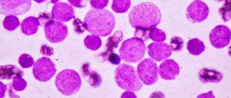
This congenital disorder can only be detected using a clinical blood test. If the nuclei of mature cells have few segments and change shape, this may indicate a Pelger anomaly of neutrophils. In the photo below you can see pathological changes in the nuclei.
The nucleus of Pelger neutrophils can have the following forms:
- oval;
- rod-shaped;
- horseshoe-shaped;
- bean-shaped;
- rounded.
In more rare cases, there are cores with a constriction in the center, similar to a gymnastic weight.
In this case, the core looks too short and consists of only one segment. It has a lumpy structure. Less common are nuclei of altered shape, having two or three segments.
If the nucleus of a segmented neutrophil has a round shape, then it can very easily be mistaken for a myelocyte. Only a detailed blood test can show that this is not a young, but a mature cell. The presence of neutrophils with altered nuclei often leads to errors in the interpretation of analysis results.
Why might it appear?
The causes of the leukocyte abnormality have not been precisely established. Until now, scientists have only been able to find out the fact that pelgerization can be transmitted in an autosomal dominant manner. It can appear in both heterozygotes and homozygotes.
Neutrophils
Hematologists have found that the main cause of pathogenesis is a violation of the segmentation process of already mature neutrophils. Since this pathology is inherited, it was found that genetic damage occurs due to changes in the structure of the gene responsible for the shape of the nucleus. The presence or absence of this pathology is easy to check if there is a blood test of both parents.
Causes
The anomaly occurs due to a violation of the formation of segments of neutrophil nuclei. The cause is a failure at the genetic level.
This disease is hereditary. It is transmitted in an autosomal dominant manner. This means that the pathology can be passed on to children if both parents are carriers of the defective gene.
The anomaly occurs in 1 child out of 1000 newborns. It is observed equally often in boys and girls. The pathology is benign. Many doctors consider Pelger's anomaly not a disease, but a constitutional feature.
Pelger's leukocyte anomaly
Pelger neutrophil anomaly is a benign hereditary pathology that is characterized by morphological changes in leukocytes. It was named after Karl Pelger, a Dutch doctor who first discovered atypical neutrophils in 1928.
Then in 1932, Huett proved the hereditary nature of the disease. Therefore, in some sources the disease is called Pelger-Huet anomaly. Occurs with a frequency of 1:1000-1500, equally common in men and women.
Previously, it was rare, but recently it has become more common, due to more extensive blood examinations.
Treatment
Pelger's anomaly does not require treatment, since blood functions are fully preserved. Its carriers are considered healthy people.
It is important to examine children to identify this blood anomaly, since the ratio of cellular composition does not undergo significant changes throughout life. Detection of an anomaly helps to avoid misdiagnosis of many diseases that give a similar blood picture.
Manifestations
There are no pathological symptoms with Pelger's anomaly of neutrophils. This congenital feature does not affect a person’s well-being in any way and does not pose a health hazard.
Changing the shape of neutrophil nuclei does not affect phagocytosis. Such cells retain the ability to fight foreign microorganisms and produce enzymes. This anomaly does not affect the state of immunity in any way.
A very small proportion of doctors believe that patients with Pelger's neutrophil anomaly are more likely to have short stature and a tendency to have bowed bones. However, these data have not been scientifically confirmed. Most people with this feature are of normal height and do not suffer from bone pathologies.
Clinical manifestations
Clinically, the disease is not expressed in any way, since the functions of leukocytes are not impaired. Leukocytes
are also capable of phagocytosing foreign cells and contain a set of enzymes identical to normal leukocytes. ESR is not increased, blood clotting is normal. Often this pathology is discovered by chance during a blood test. Due to the fact that there is a neutrophil shift to the left in the leukocyte formula, an infection can be mistakenly assumed. Reactions to blood loss, infection, etc. in people with this anomaly the reactions do not differ from healthy people.
The leukocyte formula is the ratio of several types of leukocytes in the human body
. According to some sources, there may be changes in the skeletal system: hyperkyphosis, short stature, skeletal deformities.
Consequences
What does Pelger's anomaly of neutrophils lead to? This violation does not cause any complications or negative consequences for human health.
Difficulties arise only when interpreting the results of a blood test. Very often, doctors mistake modified segmented neutrophils for band neutrophils. An increase in con can be a sign of the following diseases:
- oncological diseases of the hematopoietic system;
- bacterial and viral infections;
- injuries and burns;
- myocardial infarction;
- inflammatory processes in various organs;
- allergic reactions;
- sepsis;
- parasitic diseases;
- renal failure.
If the blood picture is altered, the doctor may suspect the above pathologies in the patient. In the history of medicine, there have been cases when patients with Pelger's neutrophil anomaly were mistakenly diagnosed with infectious diseases or leukemia. As a result, people underwent a course of treatment that was absolutely unnecessary for them.
This congenital feature often leads to erroneous interpretation of clinical blood test data. However, a qualified and experienced doctor never makes a diagnosis based on just one test.
Diagnostics
If the patient’s clinical analysis shows signs of Pelger’s anomaly, the doctor must indicate this in the transcript. To avoid errors in diagnosis, a person needs to additionally take a blood smear for the following indicators:
- kernel maturity;
- chromatin density;
- neutrophil ratio.
With Pelger's anomaly, the patient's chromatin density remains normal, the nucleus has an altered shape, but is old. This study helps distinguish altered mature cells from young forms.
If possible, a clinical test and blood smear should be taken from the patient's parents. Since the pathology is hereditary, the same anomaly is observed in the mother and father of the patient. This is an important diagnostic sign.
Is treatment necessary?
Clinical manifestations of Pelger's neutrophil anomaly are completely absent. A person with this feature is considered absolutely healthy and does not need treatment.
It is advisable to identify the anomaly in childhood. This will help avoid diagnostic errors in the future. If a patient with a Pelger neutrophil anomaly is scheduled to undergo a blood test, it is very important to tell the doctor about his congenital abnormality. Otherwise, the test results will be misinterpreted.
May-Hegglin anomalies. Treatment
Data in the literature are conflicting, but most patients with May–Hegglin anomalies do not appear to have clinically significant bleeding problems, and based on this, specific treatment for these patients is not required. Corticosteroids and splenectomy are ineffective. In rare cases, platelet transfusions may be required.
Patients with May–Hegglin anomalies who undergo vaginal or cesarean section do not have a significantly increased risk of bleeding.
In patients with May–Hegglin anomalies who are scheduled for surgery, it is important for physicians to obtain a personal and family history of bleeding tendencies. Intravenous desmopressin acetate may be a helpful step. Routine prophylactic platelet transfusions are not usually indicated unless reasonable to ensure that platelets are available if unexpected bleeding occurs.
Depending on the degree of thrombocytopenia and family history, people may be at increased risk of bleeding, in which case such people should avoid participating in contact sports entirely.
Similar anomalies
We examined the congenital type of Pelger anomaly. However, similar changes in neutrophils may also be observed in the following diseases:
- systemic lupus erythematosus;
- tuberculosis;
- malaria;
- flu;
- lymphogranulomatosis;
- decreased thyroid function;
- leukemia
In these cases, the changes are acquired. They are not observed constantly, but only with exacerbation of the underlying pathology.
There is also a very rare congenital disorder that geneticists call SOPH syndrome. It was first described in 2009. This pathology occurs among the peoples of the North, mainly among the Yakuts. Pelger's anomaly is only one of the manifestations of this syndrome. This genetic disease is accompanied by other pathological signs:
- short stature;
- sagging skin;
- atrophy of the optic nerves and retina.
If a child is diagnosed with a Pelger anomaly, then you should take a close look at his well-being and state of health. If no other pathological signs are observed, then there is no cause for concern. This constitutional feature does not affect either the duration or quality of life.
Alder Anomaly
Alder anomaly (Alder-Reilly anomaly)
– the cytoplasm contains numerous dark lilac granules consisting of acidic mucopolysaccharides. It is observed in genetic mucopolysaccharidosis of the Hurler, Gunter and Marote-Lamy type.
Sources
- https://LechiSerdce.ru/leykotsityi/5395-pelgerovskuyu-anomaliyu-neytrofilov.html
- https://www.clinlab.info/Hemocytology/Constitutional-neutrophil-anomalies-9
- https://redkie-bolezni.com/anomalii-meya-khegglina/
- https://fb.ru/article/450419/pelgerovskaya-anomaliya-neytrofilov-prichinyi-i-simptomyi
- https://krovotechenie.ru/issledovanie-krovi/pelgerovskaya-anomaliya-lejkotsitov.html
- https://serdechka.ru/krov/pelgerovskaya-anomaliya.html
- https://as-transfer.ru/pelgerovskaja-anomalija-krovi-chto-jeto/
- https://serdec.ru/krov/pelgerovskaya-anomaliya-leykocitov