Description of LVT
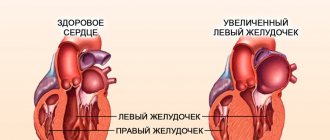
The heart has 4 chambers. If the organ is healthy, the blood in it flows from the atria to the ventricles. The movement of blood between the chambers is ensured by the heart valves, which open and close in response to the rhythm of the heartbeat. Due to trabeculae (chords), the elasticity of the valves is maintained.
LVDP of the heart in a child is a congenital anomaly that forms in the uterine period and is accompanied by the appearance of one or several chords at once. As a rule, this pathology does not require any treatment. But a child with an additional trabecula in the cavity of the left ventricle needs regular supervision by a cardiologist.
On a note! Trabeculae are partitions of bone or muscle tissue that form the frame of an organ. ICD 10 code – Q20.
The extra trabecula that forms in the heart is tendon tissue. Excess trabeculae are located differently to the vertical axis of the LV; they can be longitudinal, diagonal or transverse.
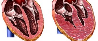
The most severe anomaly is transverse LVOT, as it can disrupt the heart rhythm. The diagonal and longitudinal trabecula of the left ventricle in a child are considered minor pathologies - they affect the movement of blood in the chambers.
1 chord or trabecula
Structure of the heart
Regarding the topic of minor anomalies in the development of the heart, which includes the additional chord of the cavity of the left ventricle, you can hear two concepts - chord and trabecula. These two words have different meanings and should not be confused. The heart is a hollow muscular organ, which is divided inside into four cavities - chambers.
The operation of the valves is carried out thanks to the existing chords and trabeculae. From the free edges of the valve flaps, tendon threads stretch like sails - in other words, chords. The notochord, attached to the trabecula, connects to the papillary muscle of the left ventricle. Here is a simplified diagram of the structure of the ventricular valve apparatus.
Is there any danger
Most cardiologists believe that extra left ventricular trabecula (LVAD) in a child is one of the normal variants. If doctors make such a diagnosis, parents need to explain the features of this anomaly so that they do not panic. The child does not require any intervention - neither surgical nor medicinal.
But there are isolated cases when LVPT is a factor contributing to the development of cardiovascular problems. A child with such a pathology may lose rhythm and experience the appearance of endocarditis, thrombophlebitis and other heart diseases. But it is impossible to predict in advance the development of such complications.
Forecast
The medical prognosis for additional left ventricular trabecula is favorable. A single longitudinal trabecula, which is common, does not cause virtually any inconvenience. People with this diagnosis live normal lives. But periodically - once a year - they should check the functioning of the heart to avoid various complications.
Serious complications occur extremely rarely. If a longitudinal chord (or several trabeculae) is detected, this can impede the functioning of the heart and interfere with the normal flow of blood into the ventricle.
Possible complications:
- Extrasystole is an arrhythmia in which premature contraction of the heart muscle is observed.
- Morgagni–Adams–Stokes syndrome, which causes fainting as a result of a sudden rhythm disturbance.
- Atrial fibrillation is a condition in which a person’s heart, for no reason, either beats quickly or stops.
- Even less commonly, the patient may experience pulmonary embolism, ischemic stroke, or cardiac arrest due to fatal ventricular conduction disturbances.
If the disease does not manifest itself in any way and is not complicated, then it is not a reason for refusing to vaccinate a child, from serving in the army, and it is not prohibited to become pregnant and give birth with it.
Clinical picture
One extra trabecula does not make itself felt in any way. Parents are often unaware of abnormal heart development. The pathology is detected when a planned ultrasound of the heart is performed - children should have it at 1 month and at one year of age.
If there are several transverse chordae at once, the child will most likely have problems with the functioning of the heart. Complications usually appear at 12-14 years of age. If there is an extra chord, connective tissue dysplasia may appear. Genetic pathology causes weakening of connective tissues in the heart, joints, ligaments and tissues. The disease requires immediate intervention.
Possible symptoms in children with LVAD:
- Tachycardia. Newborn babies and toddlers do not feel that there is any abnormality in their heart. During adolescence, children may feel chest pain and increased heart rate even with mild physical exertion.
- Decreased physical activity. Children with LVAD aged 13-14 years also often experience chronic fatigue.
- Problems in the gastrointestinal tract. Patients may suffer from heartburn, as they often have a bent gallbladder.
- If the extra chord in the heart is accompanied by connective tissue dysplasia, children may experience some deformities. For example, the back line sags, the gait becomes irregular, and the skeleton acquires abnormal flexibility.
If the child has not previously been diagnosed with LVAD, but the above symptoms appear, a doctor’s consultation is necessary. The heart will have to be carefully examined to assess its condition and functionality. The child will be prescribed timely treatment to prevent the development of complications.
Left ventricular trabecula: what is it, causes
Left ventricular trabecula is a minor cardiac anomaly that does not pose a threat to human life.
Recently, due to the development of technology and more thorough examination of newborns for congenital anomalies, the number of detected trabeculae in the left ventricle has increased significantly.
Many people do not distinguish between the concepts of “additional chord” and “additional trabecula,” but doctors say that these are different things. The notochord is a thin thread of connective tissue that extends from the mitral valve to the muscle tissue of the ventricle. By trabecula we mean septa, small outgrowths, cords, that is, the trabecula can sag into the cavity of the ventricle, rather than connecting the valve with muscle tissue. However, not all doctors recognize this difference and sometimes write “chord” instead of “trabecula” in the conclusion.
The left ventricular trabecula does not pose any danger to the child's life.
Most often it is found in children, since it is congenital. The heart, as you know, consists of four chambers: two atria and two ventricles. The ventricle and atrium are separated by a valve, which serves as a gate that allows blood into the ventricle and does not release it back into the atrium. This valve is connected to the muscle tissue of the ventricle by peculiar springs, chords, thin threads that stretch and open the valve, and then relax and close it. The valve opens and closes in accordance with heart contractions.
Useful video - Ultrasound of a child's heart.
The left ventricular trabecula is essentially another chord connecting the valve to the left ventricular muscle. This is not dangerous to the child’s health and may not manifest itself in any way throughout life.
Left ventricular trabecula is a minor cardiac anomaly; it is not a heart defect or a fatal disease. Often it does not even require special treatment, but it is recommended that a child with such an anomaly be observed and regularly shown to a doctor. The cause of left ventricular trabecula is heredity (in more than 90% of cases). It appears during embryonic development.
Signs of the disease
Left ventricular trabecula is a hereditary disease
The additional chord itself does not manifest itself in any way. It is quite rare to suspect left ventricular trabecula; most often, the disease is discovered by chance, during examination or echocardiography of the heart.
However, in some cases, left ventricular trabecula is accompanied by complications or occurs against the background of another, more extensive disease, such as connective tissue dysplasia. In these cases, the child will exhibit certain signs that require examination. Most of these symptoms are observed during the period of active growth, in primary school and adolescence.
If there is not one, but several additional chords, a connective tissue disease can be suspected, which will affect the entire body as a whole.
Signs of left ventricular trabecula:
- Pain in the heart area, tachycardia. Pain and rapid heartbeat can be observed during the period of active growth of the child, when there is an increase in the size of internal organs, growth of bones and muscle tissue. As a rule, the child complains of regular tachycardia and pain in the chest after or without physical activity.
- Fast fatiguability. If complications arise and increased stress on the heart, the child begins to get tired quickly, complains of chronic fatigue, causeless weakness, and quickly gets tired. For such complaints, pediatricians often prescribe an ECG and a blood test, which will help identify other disorders and detect trabecula.
- Limb deformity. With connective tissue disease, not only the heart suffers, but also the skeleton and muscle tissue of the whole body. A child with connective tissue dysplasia has abnormal flexibility, scoliosis, a wobbling gait, and a arched back line.
- Disruption of the gastrointestinal tract. Children with dysplasia often experience bending of the gallbladder, as well as gastroesophageal reflux, in which the contents of the stomach enter the esophagus along with gastric juice, causing irritation.
Causes of pathology
Many factors contribute to the development of additional trabeculae in the left ventricle of the heart in a child. The reasons for the development of LVAD are regularly discussed by professors of medicine. As a result of observations and studies, it was found that additional trabeculae in the LV can be inherited from the parents to the child.
But the genetic factor is far from the only one in congenital anomalies. Completely healthy women can also give birth to children with LVPT.
Factors influencing the development of an additional chord in the prenatal period:
- alcohol;
- nicotine addiction;
- exposure to radiation;
- poor environmental situation;
- depression;
- nervous overstrain.
On a note! If a woman has an extra chord in her heart, then there is a 50% chance that she will have children with the same pathology.
Is treatment required for additional trabecula?
If, during a routine ultrasound of the heart, an additional trabecula is found in the left ventricle, and the child does not have any cardiac abnormalities, then there is no need to treat such a disease. But regular examinations by a cardiologist should take place.
Intense physical and psychological stress is not recommended for children with this diagnosis. It is very important to organize the correct daily routine, provide a balanced diet, carry out therapeutic exercises with them, and protect them from stress.
In the case of connective tissue dysplasia and pathologies of the cardiovascular system, drug therapy is prescribed. If the case is especially severe, then surgical intervention is possible: removal of the trabecula.
Drug therapy regimen:
- Vitamin complex preparations. To maintain normal functioning of the heart and blood vessels, it is necessary to take vitamins B and PP. Possible injection or oral administration. Prescribed in courses lasting a month. Repeat throughout the year. Very important during the active growth of the child. These include “Multitabs”, “Vitrum Kids”, “Jungle” and many others.
- Medicines based on potassium and magnesium. They have a positive effect on the functioning of the heart. Prescribed in a course and in a dosage calculated by a doctor. An overdose is very dangerous for a child’s body. This group includes “Magneliz B6”, “Panangin”.
- Medicines to normalize metabolism. Improves the child's appetite and strengthens the body. Most often, children are prescribed L-carnitine and Elcar.
In addition to drug treatment, diet occupies a special place in complex therapy. Parents should minimize the amount of cholesterol in the child’s diet and increase the consumption of fresh vegetables, fruits, and berries. A healthy lifestyle is the key to a child’s well-being. Constant walks in the fresh air, proper nutrition, hardening, and adequate physical activity are welcome.
Diagnostic methods
Since trabecula is a congenital feature, it is usually detected during standard diagnostic examinations carried out in the first year of a child’s life. Typically, all newborns undergo echocardioscopy, and if dysplasia is suspected, an electrocardiogram (ECG) and other tests are prescribed.
If clinical symptoms appear that indicate complications of LVPT, the doctor may prescribe several cardiac studies at once. They do not require internal penetration.
Diagnostic procedure:
- Inspection. First, the doctor examines the patient to identify symptoms, their manifestation and intensity. Listens carefully to the heart.
- Echo-CG (ultrasound) of the heart. These are the most accurate and informative diagnostic methods. Echocardiography is an examination that includes both ultrasound and ECG, so it is considered more preferable. The technique is based on the reflection of ultrasonic waves by body tissues. An echographic examination is prescribed for patients with shortness of breath and interruptions in heart function. Echo-CG has no contraindications. But it can be difficult in patients with large breasts, as well as in patients with a deformed chest. The research results, along with a transcript, are given to the patient. It indicates various indicators - the size of the myocardium and ventricles, heart rate, volume of ejected blood, stroke volume of the heart.
- ECG. Electrocardiography is prescribed to evaluate cardiac activity. But the ECG cannot determine the presence of a jumper. The procedure only allows us to identify the complications that it caused. Heart monitoring – Holter – is often prescribed. It involves recording cardiac activity throughout the day.
Diagnosis of the disease
Additional trabecula are mainly found in newborns during a routine examination, or by chance in later life. If characteristic symptoms arise, then a cardiologist should be involved in identifying the pathology. He will examine and interview the patient. Having collected all the necessary information, the doctor will prescribe instrumental examination methods to determine the causative factor. Their list can be seen below:
- Ultrasound examination (ultrasound) of the heart muscle allows you to evaluate its rhythm and frequency of contractions. It is absolutely safe and helps to accurately determine the cause of the discomfort. Such an examination is prescribed mainly for people who have breathing problems, pain in the chest area or changes in its shape.
- An electrocardiogram (ECG) shows the functionality of the heart muscle. Based on the result obtained, the doctor will evaluate the correctness of the rhythm and the severity of its contractions. It will not be possible to detect trabecula using this method, but it will be possible to detect other cardiac problems caused by the anomaly.
Treatment
If there are no clinical symptoms with LVAD, no special treatment is required. Therapy is prescribed only for complications and the appearance of other trabeculae in the body.
If there are problems with the heart, the patient is prescribed medications. Operations involving the removal of trabeculae are used very rarely - only in the most complex and dangerous cases.
Therapy methods:
- Taking vitamins. To maintain cardiac activity, a course of vitamin therapy is prescribed. It is recommended to take it 2 times a year. Patients are prescribed vitamins B1, B2 and PP. They can be obtained in tablet form or as injections.
- Magnesium and potassium preparations. They are prescribed to normalize the condition and improve the conduction of impulses in the myocardial fibers. Medicines are taken in courses. The dosage is prescribed by the doctor. Recommended medications are Magnerot or Magne B6. Potassium- and magnesium-containing products should not be used uncontrollably; even with a slight overdose, problems may occur. This especially applies to children's bodies.
- Medicines to normalize metabolism. They are needed for general strengthening of the body, improving the patient’s condition, and increasing appetite. These drugs are also necessary to increase the cellular energy of the myocardium. Recommended drugs – Cytochrome C, Elkar, Ubiquinone.
- If patients experience symptoms of neurocirculatory disorders, Piracetam is prescribed.
On a note! Along with drug treatment, patients with complications of LVPT are prescribed a diet. Products with minimal cholesterol content are allowed.
In case of severe cardiac arrhythmia, patients are immediately hospitalized in the cardiology department. Inpatient treatment is prescribed if a person is diagnosed with atrial fibrillation. It usually develops in patients with multiple transverse chordae.
Course of therapy
Additional trabecula is not a deadly pathology. It is more likely an anatomical feature of the heart muscle than a developmental defect.
If the symptoms characteristic of the anomaly and concomitant pathologies are absent, then a treatment regimen is not required. It is enough for a person to come to visit the doctor at the time specified by him and be examined once a year.
Urgent care will be required if the patient, due to the additional trabecula, begins to develop other pathological processes associated with the cardiovascular system. The specialist will prescribe medication to relieve secondary pathologies. In situations where drugs are powerless, you need to consult a surgeon. It will remove the trabecula and eliminate the cause of the worsening condition. Surgery is used in extremely rare cases.
The treatment regimen for additional trabeculae involves a combination of medication and lifestyle adjustments. You can see it below:
- The use of vitamin complexes, especially group B, helps improve nerve conduction and the functioning of the cardiovascular system. You need to take pills or give injections for a month. It is advisable to conduct a course of vitamin therapy once a year. Children will benefit most from it during a period of rapid growth.
- Tablets based on potassium and magnesium improve the conductivity of impulses through the heart, stabilize blood pressure and eliminate arrhythmia. They are most relevant during puberty, that is, during puberty. The dosage should be selected by the attending physician, focusing on the results of the examination and the presence of other pathologies.
- Antioxidants are prescribed to improve metabolic processes and strengthen the immune system. The patient feels a surge of strength after taking them. It is not recommended to use such drugs in children under 18 years of age, with high blood clotting and in the acute period of myocardial infarction.
- Correction of diet is the basis of treatment. The patient needs to reduce the consumption of fatty foods, spices and confectionery in favor of steaming and saturating the menu with vegetables and fruits.
- Maintaining a healthy lifestyle will improve your general condition and normalize the functioning of your heart and nervous system. Experts advise doing exercises and walking more often. Gradually add running, swimming and other heart-healthy sports to your program. It is equally important to give up bad habits and sleep 8 hours a day.
The treatment regimen for adults and children is virtually identical. The only difference is the choice of medications, since many of them are contraindicated in childhood.
If various heart failures develop against the background of an anomaly, you will need to take antiarrhythmic, antihypertensive and diuretic drugs. They normalize blood pressure and heart rate, preventing the occurrence of severe complications (heart attack, stroke, heart failure). Such drugs should be prescribed by the attending physician. It is not recommended to use them on your own due to the abundance of contraindications and side effects.
Prevention measures
LVAD does not threaten human life. It does not prevent women from giving birth, or men from performing military duty.
Most often, LVDD develops due to heredity, and it is impossible to prevent its development. But since scientists believe that pathology can be triggered by unfavorable factors, pregnant women should avoid them in every possible way.
Prevention of LVAD concerns the behavior of pregnant women. They are recommended:
- follow the rules of healthy eating;
- regularly take a multivitamin complex;
- do not refuse feasible physical activity;
- do not smoke - you need to give up this bad habit even before pregnancy;
- avoid stress and heavy physical activity;
- take regular walks;
- do gymnastics, yoga.
To ensure that children diagnosed with an extra chord do not develop complications, parents must vigilantly monitor their health. Features of physical education of children with LVAD:
- Physical therapy classes are necessary.
- It is recommended to engage in dancing, yoga and other types of physical activity that strengthen the myocardium and prevent deterioration of the condition.
- Do not experience excessive loads. Professional sports are not recommended.
- Certain physical activities are prohibited - diving, snorkeling, parachute jumping.
Signs of the disease and diagnosis
Often, additional trabecula in the left ventricle do not manifest symptoms throughout life. Modern methods of examining children make it possible to detect the presence of anomalies in the first months of life. Only in certain cases the presence of trabeculae is accompanied by the development of a serious disease - connective tissue dysplasia. Then the child exhibits the main symptoms of the pathology:
- tachycardia and chest pain (in adolescence with intensive growth of muscles and bones, an increase in the size of internal organs);
- increased fatigue, especially after physical activity;
- disruption of the gastrointestinal tract (reflux, bending of the gallbladder);
- deformation of the limbs (connective tissue dysplasia is accompanied by problems with the spine: scoliosis, excessive skeletal flexibility, thinness).
If such symptoms are present, a thorough examination of the child is carried out, which allows us to identify serious abnormalities in the development of the heart.
An effective method for diagnosing additional trabeculae in the left ventricular cavity is ultrasound. Currently, every child aged 1 to 3 months is required to undergo an ultrasound examination to exclude heart defects and other anomalies that were not detected in the maternity hospital. In the absence of complications, the presence of an accessory chord is recognized by doctors as a variant of normal development, and the child is considered healthy. If symptoms of dysplasia are detected, additional studies are carried out:
- echocardiography;
- ECG of the heart without load and with load;
- bicycle ergometry (stress tests).
Symptoms
Although the disease can be diagnosed at any age, symptoms generally begin to appear in infancy. Some patients may have an undiagnosed form of LVHT, in such cases there are practically no symptoms.
The manifestations of LVH vary from one individual to another depending on the size and location of the non-consolidated myocardium. Symptoms are not strictly specific to LVHT, since they are often detected in other types of cardiomyopathy and diseases of the cardiovascular system, which often complicates diagnosis.
Clinical manifestations usually occur because:
- non-compact myocardium does not allow the heart to pump blood well throughout the body (which can cause symptoms of heart failure because the heart does not meet the body's needs);
- Myocardial trabecularity affects the normal electrical activity of the heart (which can cause arrhythmia, an abnormal heart rhythm).
Symptoms of LVH include:
- shortness of breath;
- fatigue (sometimes extremely pronounced);
- feeling dizzy;
- fainting;
- feeling of abnormal heartbeats (palpitations);
- swelling of the legs, ankles and feet (edema).
Causes of development, symptoms
- The main reason for the formation of an extra chord is heredity or predisposition. This anomaly is still developing at the time the embryo appears, so this pathology is difficult to determine at the initial stage of development.
- The next reason is the consequences of pathologies associated with connective tissue and dysplasia.
Symptoms that appear in childhood:
- chest pain
- fainting
- severe headaches
- fast fatiguability
- heart rhythm failure
- absent-mindedness
- there is no stable blood pressure, strong jumps are possible throughout the day
These same signs can also signal other heart diseases that are more life-threatening, so you should be examined by a cardiologist as soon as possible.
Additional symptoms include the following:
Eustachian valve
Eustachian valve
- valve of the inferior vena cava (valvula venae cavae inferior). This differently expressed fold of the endocardium, on average up to 1 cm wide, is located in the right atrium at the confluence of the inferior vena cava. During the period of intrauterine development, it directs a stream of blood from the vein to the oval opening, and when the oval opening is closed after birth, it loses its significance. In children it is more pronounced than in adults. In the absence of its involution, dilation of the inferior vena cava and partial obstruction of the flow from it occurs. Occurs in approximately 20% of subjects.
Additional chords in the left ventricle
(falshchords, or false chords) are tendon formations in the cavity of the left ventricle, which are located between the interventricular septum or the head of the papillary muscle and one or another wall of the heart and do not have a direct connection with the valve apparatus (Fig. 1.44). They are defined in the cavity as linear dense structures that do not thicken during systole. The detection rate of additional chordae is high—up to 90–98% of those examined.
Causes and prevalence
LVH is basically a genetic disorder caused by an altered or “mutated” gene. If the disease is genetic, it can be inherited (passed from parent to child) and is therefore often common in certain families. For this reason, it is recommended to screen first-degree family members (parents and siblings) if someone close to them has previously been diagnosed with LVHT.
The prevalence of LVH is not really known, although some have suggested that the disease affects men more often than women. Nowadays, more people are diagnosed with LVH because cardiography is now more advanced, so non-consolidated areas can be seen and diagnosed quite accurately.
LVHT is sometimes diagnosed in patients with dilated or hypertrophic cardiomyopathy.
Some statistics:
- LVHT is registered in 80% of cases in men.
- In children, LVH is most common, as it ranks third after hypertrophic and dilated forms of cardiomyopathy.
- Among diagnosed cardiomyopathies, LVHT is detected in 9.2% of cases.
- The total share of pathology accounts for 0.014% of cases (according to E. Oechslin).
Etiology of pathology - classification
Normally, the heart circulates blood from the atrium to the right and left ventricles. The outflow is carried out thanks to the heart valves, which work in the same rhythm with the contraction of the heart. Trabeculae are needed to support the valves and give them elasticity, they are located on each side of the ventricle. In medicine, trabeculae are also called chordae.
There are cases when, during the formation of the fetus in the womb, additional chords are formed in the baby’s heart; this phenomenon is very common in our time. If the extra trabecula is located on the left side, then doctors say that there is no threat to the functioning of the heart, but a location on the right can signal the development of serious cardiac pathologies.
During an ultrasound examination of a pregnant woman, the doctor must listen to the baby’s heartbeat, and if there are any abnormalities, the specialist will definitely notice them. Also, the heart rhythm can be disturbed during the growth of the child, during the period of sexual development of the body and during gestation. If there is a need for this, the doctor will prescribe an additional examination and establish a treatment method.
Features of the pathology
Additional left ventricular trabecula is often detected in infants. This phenomenon is associated with the improvement of medical equipment for diagnostics and more thorough examination of newborn children. The data obtained is usually enough for an accurate diagnosis, but there is no need to worry prematurely, since such a minor deviation cannot be dangerous to health.
From an anatomical point of view, the trabecula is an ectopic tendon formation. It is localized primarily in the left cardiac ventricle and has nothing to do with the valve. This anomaly is very often confused with a chord. You can distinguish them based on the following nuances:
- The chord is presented in the form of a thread connecting the mitral (bicuspid) valve and the ventricle.
- A trabecula is an abnormal growth, cord, or septum. It often simply hangs down in the cavity of the ventricle, without connecting it to the valve.
Some experts do not perceive such differences and write in their conclusion that the child has an additional notochord instead of a trabecula. This will not cause any particular harm, but it is still advisable to know what the difference is between them.
The main reason for the development of the anomaly is genetic predisposition. It is inherited in more than 90% of cases and occurs already in the embryonic period.