Why do an ECG test?
A referral for a cardiogram is issued in the following cases:
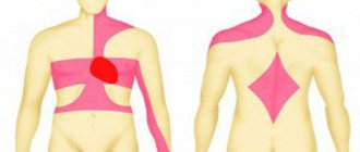
- Pain in the heart, shortness of breath while walking.
- If there are signs of arrhythmia, coronary artery disease, myocardial infarction.
- Before performing a series of operations, not only on the heart, but also on other important organs.
- In the presence of third-party diseases (ear, nose, throat) that cause complications on the heart.
- During the medical examination of pilots, athletes and drivers.
- To record heart activity.
- To diagnose diseases with symptoms of irregular heartbeat, dizziness, fainting.
- To regulate the operation of pacemakers and implants.
- A cardiogram is recommended annually for men and women after 45 years of age.
- During pregnancy.
Indications for use
Although cardiography is a routine research method, it also has indications.
To determine the cause of pain or discomfort in the chest area, the patient consults a therapist or cardiologist. The doctor initially collects anamnesis, examines, measures blood pressure and pulse, auscultates the heart, and then sends it for examination to find out what the cardiogram shows. Indications for ECG:
- chest pain (suspicion of angina or myocardial infarction);
- dyspnea;
- discomfort in the heart after viral or bacterial infections;
- pathological heartbeat, interruptions in the functioning of the heart muscle.
An ECG is mandatory in the following cases:
- when hospitalized in an inpatient department of any profile;
- before surgical interventions;
- during preventive examinations of adults;
- for schoolchildren when choosing a group of physical education classes.
An electrocardiogram of the heart is used both for the primary diagnosis of pathological conditions and for monitoring the dynamics of the disease. When prescribing drugs, the doctor relies on both the patient’s subjective sensations and ECG data, which reflect actual changes in the cardiovascular system.
Types of ECG tests
An electrocardiograph is used to perform the standard procedure. Such devices are widely used in cardiology hospitals, as well as in ambulances. Using suction cups, the electrodes are attached to the human body, and then electrical potentials flow through them.
The electrodes are usually called “leads”; a total of 6 of them are installed. The leads attached to the limbs are considered the main ones and are designated I, II, III and aVL, aVR, aVF. On the chest, the electrodes are marked V1-V6.
Each type of lead has a specific task, so individually the factors give different values. The doctor needs to combine all the information into one whole and decipher the cardiogram.
The graph is displayed on special graph paper. Each lead has its own schedule. In standard applications, the belt speed is set to 5 cm/s, which can be adjusted if necessary.
Holter monitoring
Unlike the standard procedure, which lasts several minutes, during Holter monitoring, information is recorded throughout the day. The duration of the procedure is explained by the need to obtain a complete picture of the processes occurring in the heart. This procedure can take readings not only when a person is calm, but also during physical activity.
Some diseases are difficult to detect during a regular cardiogram, since deviations can only appear during activity.
Other types of procedures
There are also specific procedures for obtaining a cardiogram:
- Monitoring with exercise. In this way, it is easier to establish pathologies in the functioning of the heart. A treadmill is used for such monitoring; it helps give the body the required load. The procedure is used in situations where pathologies appear only during accelerated heart function.
- Phonocardiography. Using this method, you can study not only electrical indicators, but also the noise that occurs in the cardiac region.
This method is used to diagnose heart defects.
Reading the ECG of the heart
Electrocardiography (ECG of the heart) is a method of graphically recording electrical processes occurring in the heart when it is excited. The method is based on the idea that the biocurrents of the heart have a regular distribution on the surface of the body, and can be diverted, amplified and recorded in the form of a characteristic curve - an electrocardiogram.
An electrocardiogram is a test that can give a person complete information about the functioning of his heart. Quite often, an electrocardiogram is done with physical activity. This is necessary in order to give a full assessment of the work of the heart during periods of active human activity.
The electrocardiogram consists of:
- ECG waves,
- segments (distance between two teeth)
- intervals (a combination of an ECG wave and a segment), reflecting the process of propagation of the excitation wave throughout the heart.
To make it clearer what are called waves, segments and intervals of an electrocardiogram, you need to study the diagram below.
The diagram shows an ideal view of the ECG of the heart. In reality, it can be very different from the ideal. For example, in the presence of atrial fibrillation (atrial fibrillation), there will be no P wave at all, and the distance between the R waves will vary greatly.
Waves, segments and intervals of the ECG of the heart
Prong R.
Atrial depolarization is recorded on the ECG in the form of a P wave. The ascending part of the P wave reflects the depolarization of the right atrium, the descending part - the left. In the diagram: pp - excitation of the right atrium; lp - excitation of the left atrium, which together give the P-wave.
Q wave
- associated with excitation of the interventricular septum. It has a small amplitude and is an optional tooth.
R wave
- caused by ventricular depolarization.
S wave
has a small amplitude and may often be absent.
T wave
Reflects the process of ventricular repolarization. The direction of repolarization waves is opposite to the direction of depolarization and is directed from the epicardium to the endocardium.
U wave
Variable, sometimes recorded after the T wave. The origin of the U wave is unknown, and ideas about its clinical significance are uncertain.
Segment P - Q.
This is the distance from the end point of the P wave to the beginning of the Q wave. The P - Q segment is recorded at the moment the impulse passes through the conduction system of the heart, when the potential difference is very small, therefore a horizontal line is recorded on the ECG.
Interval P - Q.
This is the distance from the beginning of the P wave to the beginning of the Q or R wave. It corresponds to the time of passage of the impulse through the atria, AV node, His bundle and its branches.
QRS complex.
It reflects the process of ventricular depolarization. The excitation process begins with depolarization of predominantly the left part of the interventricular septum in its middle third. Further excitation covers the apical region of the right and left ventricles. The base of the ventricles is the last to be excited.
Segment RS - T.
Corresponds to the period when both ventricles are completely covered by excitation. There is no potential difference and an isoelectric line is recorded on the ECG of the heart.
Q-T interval.
Characterizes the electrical systole of the ventricles.
Segment T - R.
Corresponds to the diastolic phase of the cardiac cycle.
In this case, the standard locations (leads) of electrodes from the limbs are: first (I) lead (right hand - PR, left hand - LR); second (II) lead (LR and left leg - LN) and third (III) lead (LR - LN).
Normal ECG of the heart
Waves of a normal human electrocardiogram (ECG).
| Teeth designations | Characteristics of teeth | Duration range, s | Amplitude range in leads I, II and III, mm |
| P | Reflects depolarization (excitation) of both atria, normally the wave is positive | 0,07—0,11 | 0,5—2,0 |
| Q | Reflects the beginning of ventricular depolarization, negative wave (directed downward) | 0,03 | 0,36—0,61 |
| R | Main wave of ventricular depolarization, positive (directed upward) | see QRS | 5,5—11,5 |
| S | Reflects the end of depolarization of both ventricles, negative wave | — | 1,5—1,7 |
| QRS | A set of waves (Q, R, S) reflecting ventricular depolarization | 0,06—0,10 | 0—3 |
| T | Reflects repolarization (fading) of both ventricles; the tooth is positive in I, II, III, aVL, aVF and negative in aVR | 0,12—0,28 | 1,2—3,0 |
When analyzing the ECG of the heart, the time intervals between certain waves are of great importance (see table. Electrocardiogram intervals). Deviation of the duration of these intervals beyond the normal range may indicate cardiac dysfunction.
Electrocardiogram intervals
| Interval designation | Characteristics of intervals | Duration, s |
| PQ | From the beginning of atrial excitation (P) to the beginning of ventricular excitation (Q) | 0,12—0,20 |
| PR | From the beginning of P to the beginning of R | 0,18—0,20 |
| QT (QRST) | From the beginning of Q to the end of T; corresponds to depolarization and repolarization of the ventricles (electrical systole) | 0,38—0,55 |
| ST | From the end of S to the beginning of T, reflects the phase of complete depolarization of the ventricles. Normally, its deviation (displacement) from the isoline should not exceed 1 mm | 0—0,15 |
| R.R. | Duration of the cardiac cycle (full cycle of the heart). Normally, these segments have almost the same duration | |
| TP | Reflects the state of rest of the myocardium (electrical diastole). This segment should be taken as the level of the isoelectric line in health and disease |
You can undergo a cardiological examination with an ECG of the heart during a cardio lesson or during cardiac testing at a cardio school.
The disadvantages of the electrocardiography method are:
- short duration of recording (which sometimes has to be supplemented by the Holter monitoring method - a method of long-term, 1-2 days, recording of ECG parameters)
- does not directly diagnose heart defects and tumors
- does not reflect hemodynamics
- does not reflect the presence of heart murmurs
- a test taken at rest may not reveal the existing disease (supplemented by a stress ECG)
Source https://www.fiziolive.ru/html/fiz/statii/valuation_cardiovascular_system.htm
(4,945 total visitors, 4 visits today)
Share with friends
Who orders the study and when?
A sheet with a referral for a cardiogram is issued by the attending physician or cardiologist. If you have complaints or heart problems, you should immediately go to the hospital for examination. This procedure allows you to check the condition of the heart, as well as determine the presence of abnormalities.
Using an ECG, a number of specific pathologies can be identified:
- formation of expansion in the area of the heart chamber;
- changes in the size of the heart muscle;
- development of necrosis in tissues during myocardial infarction;
- ischemic lesions of the myocardial wall.
Decoding the electrocardiogram
This procedure must be performed by a qualified medical professional - a clinical diagnostic doctor or a cardiologist, a therapist, who will subsequently prescribe effective treatment.
Some terms that are indicated in the cardiogram can be understood by patients themselves, including:
EOS - this indicator helps determine the location of the electrical axis, the heart muscle, and the functionality of its parts. The electrocardiogram may indicate a horizontal or vertical location with a shift to the right/left.
Heart rate is an indicator of the number of heartbeats. The norm is from 60 to 90 beats per minute. An increased heart rate is considered if it exceeds 91 beats per minute. An increased heart rate is called tachycardia, and a low heart rate (less than 59 beats/minute) is called bradycardia.
Non-sinus rhythm is an indicator of heart pathology in which non-essential electrical signals are produced outside the sinus node. Usually a sign of atrial fibrillation (old name: atrial fibrillation).
Regular sinus rhythm is an indicator of normal functioning of the heart muscle.
Atrial flutter is one of the types of arrhythmia and requires urgent medical intervention.
Ventricular hypertrophy – shows thickening of the walls of the ventricles or changes in their shape.
QT is an indicator of cardiac conductivity, and if abnormalities are visualized, frequent fainting can occur and can even lead to death.
Sinoatrial blockade - demonstrates disturbances in the conduction of impulses from the node to the atrium, often indicating the development of the following diseases: cardiosclerosis, cardiomyopathy, infarction, myocardium.
How to prepare for research
An ECG (interpretation in adults implies a precise procedure, the results of which determine the treatment) is carried out after the doctor explains the main nuances of the preparation so that the results of the study become as correct as possible:
- a few days before the ECG you should stop drinking alcohol;
- It is advisable not to smoke on the day of the procedure;
- It is recommended to do the procedure on an empty stomach;
- avoid physical activity the day before the ECG;
- avoid stress and overexertion;
- It is undesirable to use medications that affect the functioning of the heart before a cardiogram;
- Do not drink coffee or other caffeinated drinks before the examination;
- heavy foods and caffeine negatively affect the functioning of the heart, increasing heart rate, which affects ECG readings;
- Before an ECG, it is not advisable to apply fatty lotions and oil gels to the body. Components in cosmetics may interfere with the passage of impulses between the sensor and the body;
- when choosing clothes, preference should be given to spacious sweaters and pants that can be easily wrapped or removed;
- During the procedure, you must remove all metal jewelry.
Modern medicine makes it possible to easily and painlessly examine the work of the heart. To do this, in case of illness or for preventive purposes, a person turns to a cardiologist to receive a referral for analysis.
An ECG is performed in a specially equipped room in which an electrocardiograph is located. Modern devices are equipped with a thermal printing element that replaces a conventional ink unit. Using the thermal effect, a cardiogram curve appears on paper.
In the latest models of cardiographs, the result is not immediately printed on paper, but remains on the monitor screen. Using the program, the device itself decrypts the indicators and also saves the data on a disk or flash drive.
The device was first developed by Einthoven in 1903. Since then, the cardiograph has received many changes and improvements, but the principle of operation remains the same. Equipping the device with a multichannel device allows you to display results from several leads at once.
In 3-channel devices, the standard leads (I, II, III) are first deciphered, then the branches aVL, aVR, aVF coming from the limbs, and at the end the chest leads.
The ECG room is usually located away from electromagnetic fields and X-ray exposure. In the treatment room, the patient lies down on a flat couch. You should first remove your clothes down to your underwear, or open the places for attaching the electrodes.
The electrodes are made in a pear-shaped form with suction cups. Depending on the number of channels in the cardiograph, the color of the wires can be white or multi-colored.
In multi-channel devices, marking is performed as follows:
- V1-red wire;
- V2-yellow wire;
- V3-green wire;
- V4-brown wire;
- V5-wire black;
- V6 wire is blue.
Before starting the procedure, the doctor must check the quality of the electrodes adjacent to the body. The skin should be clean, free of sweat and greasy film. Some electrodes are placed on the lower legs and feet. For attachment to limbs, suction cups are made in the form of plates. Their purpose is to register standard leads.
Each mount has a specific color, which helps to avoid confusion during examination. The red wire is attached to the right wrist, the yellow wire to the left, in the area where the pulse is actively palpated, a green electrode is attached to the left limb below, and a black one to the right.
When studying the cardiogram, the right leg does not take part in the readings. Therefore, an electrode is attached to it for grounding.
The cardiogram displays a gear pattern with cycles, which is responsible for the state of the heart muscle during shock and during rest. This pattern is called the cardiac cycle; each lead usually contains up to 5 cycles. These readings are standard for a regular cardiogram, but in the case of symptoms of myocardial infarction or other heart disease, these cycles may be several times larger.
After printing the cardiogram, the person is freed from the suction cups. The resulting paper is signed and left for analysis. In specific cases, a cardiogram is prescribed after performing physical exercises. To obtain correct results, readings are taken before and after exercise.
Carrying out an ECG
Any doctor can give a referral for this procedure, however, most often this is the domain of cardiologists. But when you are referred for an ECG, which doctor performs this procedure, each hospital may answer you differently. This is mainly done by functional diagnostic doctors, but often this procedure is also performed by nurses.
So, the sequence of actions when performing an electrocardiogram:
1. The subject lies down on the couch. 2. The electrode attachment points are degreased with ethanol. 3. Then a conductive gel is applied to them (it is sometimes replaced with wet gauze wipes). 4. Electrodes are attached to his chest, hands and ankles, fixed with a suction cup. 5. Wires from the electrodes extend to the device itself, which receives and processes cardiac impulses. 6. Afterwards, the doctor turns on the device, which begins to record an ECG graph. 7. The output is a tape with graphs, after decoding which the specialist can prescribe and adjust further treatment.
If there are serious deviations in the diagram, the attending cardiologist should immediately get involved in assessing the results.
In order for the ECG procedure to be successful, it is important to follow some rules:
- During the procedure, the patient should breathe evenly and not worry. For this purpose, it is desirable that the subject lie on the couch for at least five minutes.
- The last meal before the procedure should be no later than two hours.
- The room where the electrocardiographic examination is performed must be sufficiently warm. Otherwise, physiological tremors caused by cold can distort the pattern of cardiac activity, which will be reflected in incorrect cardiogram data.
- Patients with severe shortness of breath during an ECG are recommended not to lie down, as usual, but to sit, since it is in this position that all cardiac arrhythmias are most clearly recorded.
In addition to how this procedure is done, many people have a question: how long does an ECG take? Let's answer: no more than a couple of minutes.
Despite the fact that this procedure does not bring any discomfort, it is still worth knowing one more thing about the ECG: how often can this examination be done?
People over 40 years old are recommended to have an ECG once a year.
How often to do an ECG for older people is decided by their attending physician, but it is recommended once a quarter.
Norms of ECG indicators
An ECG (interpretation for adults includes a number of indicators with an acceptable interval) is carried out according to values that indicate a healthy state of the cardiac system.
| Indicator designation | Range of acceptable values |
| P | 0.05-0.12 s |
| T | 0.14-0.27 s |
| Q | 0.04-0.06 s |
| QRS | 0.07-0.3 s |
| Heart rate | 63-85 beats/min |
| PQ | 0.11-0.19 s |
What can affect the ECG result?
The reliability of the data obtained is influenced by the following indicators:
- incorrect connection of the device to the human body; in some cases, the wiring may come off during the process of obtaining a cardiogram;
- if there are residues of lotion, soaps and other components on the skin that leave a protective film;
- experiencing severe stress on the eve of the procedure;
Stress and anxiety affect normal ECG interpretation in adults - taking antidepressants, nootropics and sedatives the day before;
- in case of anxiety or fear of medical procedures;
- if a person was rushing to the hospital, causing the heart rate to increase significantly.
Before lying on the treatment table, a person should spend 10-15 minutes in a calm environment. It is important that the person does not worry and his pulse is within the established limits.
How to do an ECG
To take an electrocardiogram, the patient is placed on a couch and a healthcare professional places electrodes on the legs, wrists and chest. If there is difficulty breathing in a horizontal position, the procedure is performed while sitting.
Rules for the procedure
To ensure good contact between the skin and the electrode, the attachment site is degreased with ethyl alcohol and a special conductive gel is applied.
After this, readings are taken using an ECG diagnostic device. The whole procedure takes about 10 - 15 minutes.
In order to get a reliable result, you need to be in a calm, relaxed state, and not hold your breath. Muscle tremors from excitement or cold can lead to data distortion.
The generally accepted leads are 3 standard, 3 reinforced and 6 chest. Each lead will record at least 4 cardiac cycles. After this, the device is turned off, the electrodes are removed, and the functional diagnostics doctor is given a signed tape, which he must decipher.
For information on the ECG recording method, watch this video:
Are there any special features during pregnancy?
In the body of a pregnant woman, the load on the heart muscle changes, as
it must provide blood supply to the fetus in the uterus. The electrocardiogram may show abnormalities that are not an indication of heart disease.
Therefore, starting from 3 - 4 months, when deciphering the testimony, an amendment is made for the presence of the gestation process.
When preparing and conducting the procedure itself, standard research techniques are used.
How to do an ECG for women
For women, the rules for installing electrodes are the same as for men. They should be located in the heart area, directly on the skin, so before performing an ECG, you must completely remove all clothing from the chest, including your bra. Please note that tights or stockings will interfere with attaching the sensors to the lower leg.
Decoding the results
An ECG (interpretation in adults takes into account three indicators: contraction interval, segmental factors and wave size) indicates the risk of developing arrhythmia.
Sinus rhythm of the heart
This factor is responsible for the systematic movement of both atria, which are active under the influence of sinus action. With its help, you can study how correctly the parts of the heart function, displaying the correct functioning of tension and relaxation of the heart muscle.
The highest teeth on the diagram are responsible for the state of the rhythm. Normally, the gap between the vertices should be standard or vary by no more than 10%. Otherwise, arrhythmia will occur.
Heart rate
Heart rate in a simple sense is referred to as pulse; it can be easily calculated during an ECG study. To do this, take the speed of the cardiogram, as well as the size of the segment between the high peaks.
Typically the process speed is 25, 50 and 100 mm/s. The frequency is determined by multiplying the recording duration by the length of the segment.
Conductivity
This factor shows the state of impulse transmission. In normal condition, pulses are transmitted in the same sequence.
The waves in the ECG sections are deciphered as follows:
- P-shows the process of the atrium. It can be used to determine the quality of the atrium's response to passing impulses. In the correct interpretation, the height of the tooth is no more than 2.4-2.7 mm, the apex has a rounded tip and the cycle duration is no more than 0.1-0.3 s. In a number of pathologies, the end of the tooth has a sharp end, which indicates the expansion of the myocardial component.
- Q,S-informs about the condition of the septum in the cardiac system. Normally, the Q value is negative, lasting up to 0.03 s. S indicates the end of ventricular excitation. This is a negative indicator, the depth of which should not exceed 2 mm.
- R-characteristic of the ventricle in an excited state.
- T-state of the ventricles at rest. Normally, the height of factor T should be the third part of the R wave. The apex has a smoothed shape, duration from 0.15-2.3 s. The higher the value, the more likely the presence of autonomic pathologies in the work of the heart.
- The RQ interval shows the duration of the pulse.
- QRST is the time during which the ventricular region contracts.
- ST is the time of transition of the ventricle to excitation.
- TP is the duration of diastole in the cardiac region.
ECG norm in adults (table)
Using a table of norms, you can conduct a sequential analysis of the height, intensity, shape and length of the teeth, intervals and segments to identify possible deviations. Due to the fact that the passing impulse spreads unevenly throughout the myocardium (due to the different thickness and size of the heart chambers), the main normal parameters of each element of the cardiogram are identified.
| Indicators | Norm |
| Prongs | |
| P | Always positive in leads I, II, aVF, negative in aVR, and biphasic in V1. Width - up to 0.12 sec, height - up to 0.25 mV (up to 2.5 mm), but in lead II the wave duration should be no more than 0.1 sec |
| Q | Q is always negative and is normally absent in leads III, aVF, V1 and V2. Duration up to 0.03 sec. Height Q: in leads I and II no more than 15% of the P wave, in III no more than 25% |
| R | Height from 1 to 24 mm |
| S | Negative. Deepest in lead V1, gradually decreases from V2 to V5, may be absent in V6 |
| T | Always positive in leads I, II, aVL, aVF, V3-V6. Always negative in aVR |
| U | Sometimes it is recorded on the cardiogram 0.04 seconds after T. The absence of U is not a pathology |
| Interval | |
| PQ | 0.12-0.20 sec |
| Complex | |
| QRS | 0.06 - 0.008 sec |
| Segment | |
| ST | In leads V1, V2, V3, it shifts upward by 2 mm |
Based on the information obtained from deciphering the ECG, conclusions can be drawn about the characteristics of the heart muscle:
- normal functioning of the sinus node;
- functioning of the conduction system;
- frequency and rhythm of heart contractions;
- the state of the myocardium – blood circulation, thickness in different areas.
What heart diseases can be detected using an ECG?
An ECG (interpretation in adults may indicate a number of pathologies) indicates dangerous diseases that require urgent intervention.
- Tachycardia. A disease that is characterized by an increase in heart rate, regardless of the person’s condition. The presence of pathology is characterized by a decrease in the distance between intervals, as well as a shift in RS-T.
- Angina pectoris. The ECG shows jumps in the amplitude of T waves, and ST factors also fluctuate.
- Arrhythmia is characterized by a disturbance in the activity of the heart rhythm and impulse formation. In this case, the distance between the RR interval changes and the PQ and QT interval fluctuates.
- Bradycardia. During this pathology, the patient's pulse slows down. Using an ECG, a reduced rhythm is established, and significant gaps between segments are visible. The amplitude of the teeth becomes uneven.
- Myocardial infarction. In this case, there is no R wave on the cardiogram. The ST segment is above the isoline, and T has a negative value.
- Extrasystole. It is characterized by a change in heart rate. The ECG shows a deformation in the image of the QRS waves, and there is also no P factor.
Electrocardiography: types of diagnostics
The first device capable of recording a high-quality ECG was a string galvanometer designed by V. Einthoven. Its basis was a very thin thread, which was in a magnetic field under a certain voltage. He created a new direction in the physiology of blood circulation - electrophysiology of the heart.
The first such equipment was very bulky and weighed 270 kg.
V. Einthoven identified the main waves, intervals and segments of the ECG, and also calculated their time intervals. He also proposed a system for placing electrodes on the surface of the patient’s body. These data are used by cardiologists to this day.
The jagged edges are the ups and downs of a graphic . In electrocardiography, a segment is a section of a straight line between two teeth. An electrocardiogram can show cardiac dysfunction in the early stages, as well as consider the possibilities for the development of serious pathologies.
However, an ECG does not always accurately determine the presence of a disease. For example, a disturbance in the rhythm of the heart (arrhythmia) during a study at rest may “lurk” and not manifest itself.
Therefore, the specialist chooses another examination method, there are several of them:
- At rest is the standard method used most often. The patient lies on the couch in a relaxed state.
- With a load - during this procedure, the doctor will first take electrocardiograph readings, then ask the patient to perform a simple physical exercise (bending, squats), and then examine him again using the device. In addition, it is possible to use other methods - bicycle ergometry and treadmill test. In the first case, a bicycle ergometer is used (a device similar to an exercise bike with varying resistance of the pedals), in the second, a treadmill (a moving track). For each type of examination, electrodes connected to a computer are applied to the patient's body. The doctor monitors and analyzes the readings during the procedure.
- Daily (Holter) monitoring is the longest time-consuming method. When using it, adhesive electrodes are attached to the body of the subject. They are connected to a device that is attached to the belt or worn over the shoulder on a belt. It weighs no more than half a kilogram, so it does not cause any particular inconvenience.
The patient should keep a diary in which he indicates information about changes in physical activity, emotional overload, time of taking medications, sleep and wakefulness. Here he describes pain in the heart area and a feeling of discomfort that can arise during certain activities.
There are two options for Holter monitoring: full-scale and fragmented.
The first continues continuously for 1-3 days, as a result providing accurate and complete information about abnormalities in the functioning of the heart.
Fragmented monitoring may extend over a longer period. It is resorted to only when cardiac dysfunction occurs infrequently. Electrocardiography in this case is carried out using a special device.
To record deviations, the subject turns on the ECG recording button when pain occurs. The apparatus for such research is very miniature: it can be a pocket version or a device in the form of a wristwatch.
Transesophageal ECG is recommended when the information content of standard methods is low.
A sterile electrode is inserted into the subject's esophagus. This is usually done through the nasopharynx, less often through the mouth. The patient must make swallowing movements. But do not be alarmed - the transesophageal electrophysiological examination of the heart (TECFE) probe is thin and its insertion usually does not cause difficulties. At the same time, electrodes are attached to the chest to record an electrocardiogram.
The electrode is inserted approximately 40 cm - where the heart is closest to the esophagus. After this, they begin to record a cardiogram, and the probe begins to send weak electrical signals to the heart, causing it to contract more often.
At the end of the study, the electrode is removed from the esophagus.
In electrocardiography, there are instrumental methods for studying the functioning of the heart muscle. These include, for example, phonocardiography. In this case, a special microphone records the sounds made when the heart muscle is excited and relaxed. As a rule, listening is carried out by an experienced specialist with good hearing, who is able to separate murmurs and heart sounds from pathological sounds.
The book by V.V. Murashko “Electrocardiography” contains other methods of conducting research. Its cost is low, but it will be very useful for those who want to master the basics of ECG.
What to do if deviations from the norm are found
The first cardiogram obtained does not always reflect the true picture of the patient’s heart condition. That is why, after receiving the results, it is recommended to undergo a second check. Some heart diseases are not detected during a routine examination and require more accurate tests.
After receiving bad results, some subtleties should be reconsidered:
- Time of day when the ECG was performed. According to the rules, the procedure is carried out in the morning, on an empty stomach.
- Emotional condition. If a person is stressed or worried, the doctor should know about this so that the ECG results do not change for the worse.
- It is worth remembering whether there was a meal before the first ECG. This harmless factor can greatly affect the readings, especially if the patient has consumed alcohol, fatty foods or coffee.
- In some cases, electrodes may come off during the procedure, which dramatically affects the interpretation.
Heart rhythm problems can be present in the lives of healthy people, which is normal. Therefore, if you receive negative results, you should not immediately despair, since the heart is a sensitive organ and its examination takes a lot of time.
Taking into account these factors, the procedure followed should be reconsidered. In any case, if there are complaints or symptoms, the patient is sent for a repeat ECG. Interpreting ECG readings in adults is a complex and painstaking task. The specialist is required to have a correct understanding of all angles and components. Please remember that readings and results may vary.
Article design: Mila Friedan
Where to go and how to prepare
The study of the blood vessels of the human heart is carried out in a public or private medical center. If the necessary technical base is available, the diagnostic procedure will be performed in a clinic at the place of residence. The doctor will give specific recommendations when making a referral. The physician will discuss the nuances related to preparation for cardiography:
- 4 hours before the procedure it is forbidden to eat;
- For minors, the procedure is performed during sleep;
- it is allowed to prescribe additional tests to clarify the information received;
- 12 hours before the procedure do not smoke or drink alcohol;
- 8 hours before the procedure, do not take medications that slow down blood clotting.
No other specific preparatory methods are required. An exception is cases when the cardiologist gives specific recommendations based on the results of the examination.