Hypotony of the eyeball or low intraocular pressure occurs due to injury or detachment of the retina. The pathological process leads to disruption of fluid production inside the organs of vision. The tone of the soft tissues gradually decreases, which gradually leads to the development of opacities and hemorrhages of the vitreous body. Due to hypotension, the eye slowly decreases in size and loses its spherical shape.
Share
Tweet
Share
Cool
Send
What is intraocular pressure?
It is not blood that circulates inside the eye, but intraocular fluid. Nutrients and a small amount of oxygen are dissolved in it. It remains completely transparent, so it does not interfere with the penetration of light through the transparent structures of the organ of vision to the retina. The latter is already intensively supplied with blood by the choriocapillaris.
Fluid flows from the eye through the pupil into the cavity of the anterior chamber and freely penetrates through the trabecular meshwork, moistening the surface of the eye. Excess flows into the external environment or through the nasolacrimal duct. Inside the drainage system, intraocular pressure, or IOP, is maintained to circulate fluid.
Indications for pneumotonometry
In order to prevent glaucoma and other pathologies of the visual apparatus, annual pneumotonometry is recommended for people over 40 years of age.
Measuring intraocular pressure is performed for various diseases and problems:
- dysfunction of endocrine organs;
- retinal detachment;
- abnormalities in the development of tissue structures of the eye;
- pathologies of the cardiovascular system;
- in the period after surgery or in the presence of complications;
- glaucoma.
Why is this indicator very significant for doctors?
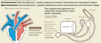
Intraocular pressure straightens the structures of the eye, maintains the tone of soft tissues and stabilizes the permeability of the choriocapillaris. Thanks to IOP, the retina remains in one position throughout life. Pressure indicators exist as a result of the synthesis and outflow of intraocular fluid. Normal IOP levels range from 11 to 21 mmHg. Art. As the pathological process develops, visual acuity and eye pressure change first.
Thanks to IOP fluctuations, ophthalmologists can determine the type of disease and prescribe a treatment regimen in a timely manner. If pressure readings increase, there is a risk of developing glaucoma. Hypotony of the eyeball indicates loss of soft tissue tone and impaired production of intraocular fluid.
What is IOP?
Intraocular pressure is the pressure of the fluid inside the eye. The constancy of this indicator ensures the normal functioning of the visual analyzer, due to which all structures of the eye are in stable and comfortable conditions. It is the IOP that determines whether the eye tissues will receive a sufficient amount of oxygen and nutrients, as well as the speed of metabolic processes.
How is it determined?
Intraocular pressure is measured in two ways:
- straight;
- indirect.
The direct method is to insert a pressure gauge needle into the anterior chamber of the eye through the cornea and take a direct measurement. In clinical practice, the manometric method is not used.
The indirect method is tonometric. The specialist uses special devices (Maklakov, Goldman, Pascal, ICare tonometer) that allow you to measure changes in the hydrodynamics of the eye as a result of pressure on the cornea. The doctor applies a certain force and looks at how much moisture is displaced from the chambers of the eye - the resulting volume is called tonometric pressure.
REFERENCE! There are contact and more modern non-contact tonometers, which do not require anesthesia to use.
The error in tonometric measurement is minimal and is less than 1 mm Hg.
What influences the value?
A change in IOP value—a deviation from the norm—can be physiological or pathological.
In the first case, we are talking about normal fluctuations in intraocular pressure during the day: due to playing sports, working at the computer for a long time, playing musical instruments, taking certain medications, drinking heavily and some other external factors.
The maximum permissible deviation is 3-4 mm Hg. during the day.
Pathological changes in IOP are associated with various diseases, and not only ophthalmological ones. Sometimes this symptom indicates the occurrence of:
- diabetes mellitus;
- diseases of the cardiovascular system (atherosclerosis, varicose veins, congenital heart disease);
- vegetative-vascular dystonia;
- kidney diseases (for example, glomerulonephritis);
- ophthalmological diseases (uveitis, astigmatism, glaucoma).
Glaucoma is the most common cause of increased intraocular pressure, and at the same time, increased IOP is a major risk factor for the development of glaucoma. This disease is very dangerous and, in the absence of adequate treatment, can lead to complete loss of vision.
IOP can have a persistently increased or decreased value due to genetic predisposition, hormonal imbalance, unfavorable environmental conditions, frequent stress, eye injuries (even in the distant past), improper work and rest patterns, and poor hygiene.
Causes of hypotension
A decrease in intraocular pressure is caused by mechanical damage to the tissues of the eyeball or the development of systemic pathologies. A decrease in ophthalmotonus most often develops against the background of diabetic coma, the final stage of acute renal failure. The disease occurs when there is low pressure inside the skull or vascular collapse.
Ophthalmologists identify the following causes of hypotony of the eyeball:
- Penetrating trauma. Damage to the soft tissues of the organ of vision is accompanied by a sharp drop in IOP due to the fact that fluid begins to intensively come out of the wound. The more extensive the injury, the faster hypotension develops. In critical situations, a gel-like substance of the vitreous body leaks out along with the intraocular fluid.
- Eye contusion. Severe damage to the organ of vision is accompanied by rupture of the sclera under the conjunctiva. Often lesions form under the external muscles of the organ of vision, because in such places the thickness of fibrous tissue is about 0.3-0.4 mm. The exit zone of Schlemm's canal is also considered a vulnerable area.
- Surgical intervention. Hypotony of the eyeball often develops as a complication of antiglaucoma operations. The risk of lowering IOP increases when using cytostatic drugs or expanding the burr hole for inserting surgical instruments. A decrease in ophthalmotonus may be due to a decrease in the volume of the gel-like substance of the vitreous body, which is typical for the intravitreal procedure and vitrectomy.
- Disturbance in the production of fluid inside the organ of vision. The production of biomaterial decreases with age-related changes affecting the ciliary body. Dysfunction of the intraocular structure also occurs when the ciliary muscle is damaged.
- Detachment of the retina and choroid. With retinal dissection, there is a risk of developing a sharp drop in intraocular fluid pressure, which is accompanied by severe hemorrhage into the vitreous cavity from the choriocapillaris.
- Formation of fistulas. Provoke the development of chronic hypotony of the eye. Neoplasms arise when the edges of a penetrating wound of the organ of vision are improperly sutured during surgery. In rare cases, in addition to fistulas, micro-tears occur.
Stages of the procedure
An optical ophthalmic device measures the fluid pressure inside the eyeball in 10 seconds.
To perform the procedure, the patient must remove glasses and contact lenses. The subject is seated in front of the device, fixing his head. The gaze is directed in front of you to the illuminated point of the device.
Next, a stream of air is supplied at a certain speed, which leads to a change in the shape of the cornea. At this stage, the tonometer processes the results obtained, recording them on a form. Based on the indicators, the diagnostician interprets the results.
Signs
A slight deviation from the norm in IOP indicators is accompanied by the appearance of floaters before the eyes and clouding of the visible image. The long course of the pathological process leads to acute headaches that radiate to the back of the head.
When performing precise work for a long time, requiring high concentration of vision, a person experiences rapid eye fatigue. As the pathological process progresses, patients note a decrease in visual acuity, complain of discomfort when moving the eyes and deterioration of lateral vision.
When you receive an injury or an infectious lesion of the organs of vision, pain and pain in the eyes occur. Invasion of pathogenic microorganisms is accompanied by redness of the conjunctiva and increased tearing.
When to contact an ophthalmologist for an IOP check?
It is mandatory to conduct a study of intraocular pressure indicators in the presence of diseases:
- Neurology;
- Diabetes mellitus;
- Vegetative-vascular dystonia;
- Glaucoma.
In addition, determination of ophthalmotonus indicators is necessary in the presence of factors:
- Dry eye condition;
- Stable visual impairment;
- Violation of the shape and structure of the pupil and eyeball;
- Pain in the head and eyes;
- Fatigue of the visual system in a short period of time;
- Clouding or redness of the eye.
| It should be remembered that the procedure is contraindicated when under the influence of drugs or alcohol, in the presence of psychiatric disorders, or infectious diseases of the visual organs. |
Diagnostics
The diagnosis of hypotony of the eyeball is made based on the collected medical history, physical examination and specific instrumental studies. At the first consultation, the ophthalmologist makes a preliminary diagnosis using palpation and makes a comparison with the opposite eyeball.
To accurately establish the type of pathological process and understand its etiology, resort to the following instrumental studies:
- Determination of IOP indicators. The main sign of hypotension is a drop in ophthalmotonus below 15 mmHg. Art. In this case, the true indicators do not exceed 8 mm Hg. Art. In case of penetrating injuries of the eyeball, there is a need for non-contact tonometry.
- Visometry. In severe situations with the rapid development of the pathological process, vision in the macula area disappears in more than 50% of patients. Visual acuity remains within normal limits only in 5% of cases. In 39% of people, the functional activity of the eyes decreases slightly, remaining within 0.1-0.2 diopters.
- Examination of the outer segment of the eyeball. When performing biomicroscopy, you can see foci of opacification and inflammation of the cornea. When injuries occur on the periphery of the eyeball, corneal defects occur. Due to the clouding of the transparent intraocular structures, it is difficult for light rays to penetrate through the pupil to the retina.
- Electronic tonography. During the procedure, the ophthalmologist determines IOP indicators, assesses the hydrodynamics of the intraocular fluid, its volume and production rate. It is strictly forbidden to use the technique in severe pathological processes.
- Ophthalmoscopy. A decrease in ophthalmotonus often leads to swelling of the retina in the zone of best vision. Long-term hypotonia of the organ of vision leads to degenerative-dystrophic changes in the retina. Ophthalmoscopy allows you to assess the extent of retinal damage and create a further treatment plan.
- Ultrasound of the organ of vision. The studies are carried out in parallel with measuring the size of the diseased eyeball, after which the data obtained are compared with healthy organs of vision. If the difference exceeds more than 3 mm, this indicates the development of severe hypotension.
Expert opinion
Danilova Elena Fedorovna
Ophthalmologist of the highest qualification category, Doctor of Medical Sciences. Has extensive experience in diagnosing and treating eye diseases in adults and children.
To assess the condition of the drainage system of the organ of vision, a Seidel filtration test is performed using a fluorescent sodium solution. It is administered for penetrating wounds of the cornea. The medicine is eroded due to circulating moisture in the anterior chamber and may show areas with microtraumas.
Is it possible to measure blood pressure at home?
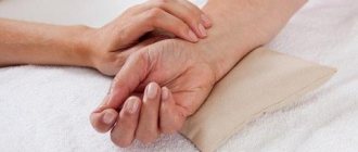
To determine hypotonia of the eyeball, it is necessary to palpate the organs of vision through closed eyelids. To carry out diagnostics, lightly press on the eyelid with the tip of your fingers. Normally, a person feels the elastic surface of the organ of vision, which gently springs. As IOP increases, the eye becomes hard to the touch, like the surface of a wall or stone.
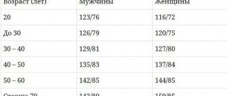
Features of IOP indicators depending on gender and age
Minor fluctuations in IOP can be considered a type of normal, but if the value is too far from generally accepted norms, certain diagnostics are prescribed. This takes into account the gender and age of the person.
In men
In men and women, normal IOP is approximately the same and depends on the method of measurement. According to Maklakov, this range is from 10 to 23 mm Hg. In men, daily changes in the indicator may be associated with sports, but they are less likely to experience hormonal disruptions, so there are no IOP surges due to this.
Glaucoma is less common in men, so until approximately 40-50 years of age, the rate remains at the normal level.
Among women
Normal IOP values in women are 10-23 mm Hg. In this case, all metabolic processes proceed normally, visual functions are performed in full. Just like in men, the indicator can vary during the day by 3-4 mm Hg: usually in the evening it becomes slightly lower, and in the morning it reaches a maximum.
But in women, various hormonal changes are more common; diabetes mellitus and hypertension are more often diagnosed, even at a young age. This is especially true when a passive lifestyle, poor nutrition and poor environment affect health, and such diseases become widespread among young people.
Changes in IOP occur during pregnancy, also in women who work more often at the computer and in certain diseases.
In young people
In healthy young people, until approximately 40 years of age, IOP remains at the same level - 10-23 mm Hg. All processes proceed in full, there are usually no difficulties with visual function: the eyes receive the required amount of microelements and oxygen.
If any diseases appear, IOP may be either less or more than normal.
After 40
For those who lead a healthy lifestyle, intraocular pressure can remain at the same level - 10-23 mm Hg.
How to increase IOP?
You can increase the pressure inside the eyes not only with the help of medications. An increase in indicators is observed with the introduction of foods high in antioxidants into the diet and daily eye exercises. Exercises will help increase the tone of the soft tissues inside the organs of vision.
Nutrition
During the period of drug therapy, it is necessary to balance the diet. Vitamins and mineral compounds will help normalize the production of intraocular fluid. To do this, exclude from the daily menu:
- animal fats;
- fast carbohydrates, sugar;
- confectionery;
- products with trans fats.
It is allowed to consume boiled chicken eggs, hot spices, salt, fruits and vegetables with beta-carotene on a daily basis. It is recommended to eat dark chocolate and hazelnuts between meals.
Advice! To increase the effect of diet therapy, you should drink multivitamin complexes and dietary supplements with a high content of retinol, vitamin C and alpha-tocopherol.
Gymnastics
To improve the circulation of intraocular fluid, pay attention to eye exercises for 15 minutes every day:
- Alternately close and open your eyes. Repeat the exercise 10 times.
- Blink frequently for 2 minutes. If you feel tired eyelids, you need to take a break. If this is difficult to do, it is recommended to blink quickly for 20 seconds. After this time, they switch to a medium pace, which also lasts 20 seconds, after which they return to a fast pace.
- Move the gaze to the right to the maximum extent, fix it on the extreme object, wait 5 seconds. Repeat the manipulations in the other direction. Then similar movements are made up and down.
- A person stands in front of a window and fixes his gaze on any object standing on the windowsill. After this, they turn their gaze to an object located on the street.
- The eyes are closed, after which they move the eyeball sideways, diagonally, counterclockwise and clockwise, down and up. You can draw numbers and geometric shapes.
- Hands stretch out in front of you and begin to move your fingers. At the same time, they try to carefully follow every movement with their eyes. During the exercise, the hands are gradually brought closer to the tip of the nose. While moving your hands, you cannot take your eyes off your fingers.
Methods for contact measurement of IOP indicators
The methods involve the impact of devices on the cornea of the eye to determine the state of IOP. Contact measurement methods are very unpleasant and often require instillation of painkillers. The disadvantage of such methods may be the likelihood of infection through the device.
Maklakov method
It is used in the presence of inflammatory eye diseases and after surgery. The procedure involves the use of anesthesia, as discomfort may occur.
The measuring device consists of several metal cylinders weighing 10 grams. The patient is placed on a horizontal surface. Weights are placed on the open eyelids, previously soaked in a special solution of pigment dye.
When the weight is pressed, the applied composition is imprinted on the apple. The weight is imprinted on a white sheet of paper. The final stage of the procedures is instillation of the eye with a disinfectant, which prevents the risk of infection.
Indicators are determined using a measuring ruler. The diameter of the print shows how much paint is left after placing the weight on the patient's eye. The larger the residue of the substance on the eyelid, the lower the IOP.
Currently, a portable device has been developed for conducting research using the Maklakov method. It is a ballpoint pen that applies pressure to the closed eyelid.
Goldmann tonometer
A slit lamp is used for research. Before starting the procedure, the patient needs to drip the eyes with anesthetic drugs, and also inject a special dye solution.
The device is brought to the cornea until it makes full contact. By squeezing the cornea, the device divides the presented image into two half-rings. Impact regulation occurs while the half-rings form a single whole. The scale determines the IOP indicator.
Schiotz and his method of measuring ophthalmotonus
The technique was developed for diagnosing the condition of IOP in the adult population. The procedure requires pre-treatment of the eyelid with anesthetic drops. A weight is applied to the apple of the eye, the pressure of which is prevented from being pressed. As a result, the needle of the measuring device moves to the side on the scale by which the IOP value is judged.
Dynamic measurement of IOP indicators
Dynamic contour tonometry is a contact technique for determining the state of ophthalmotonus, excluding the effect on the corneal membrane. The essence of the measurement involves applying the tip of the device to the apple of the eye. Thanks to the pressure sensor located inside the tip, measuring the indicators takes about 10 seconds. and is saved on the device’s memory card.
| The advantage of this technique is the simultaneous diagnosis of multiple indicators in one procedure, which makes it possible to determine the IOP status with high accuracy. |
Pneumotonometry
Contact method for diagnosing IOP indicators, determined by compressing air masses in the apparatus. The measuring device consists of a hollow tube and a slit lamp.
With the help of the device, an air flow is supplied, providing blood supply to the eye. An indicator of ophthalmotonus is the value of the ocular pulse.
| The measuring method is very painful and requires preliminary administration of an anesthetic drug. |
Tono-Pen
The technique involves diagnosing the condition of the apple of the eye using a portable device. The study is unpleasant and involves the administration of painkillers.
The measurement is taken by touching the tip of the instrument to the cornea of the eye. The study values are instantly displayed on the device display.
Rebound tonometry
The method is effective for diagnosing a number of ophthalmological diseases in the primary stages of development. The procedure is carried out without the use of painkillers. Involves disposable use of tips. The measuring device is located 3-10 mm from the center of the eye.
When the device is turned on, the probe moves at lightning speed to the cornea of the eye and then bounces off it. The speed of the device is directly dependent on IOP indicators.
Return to contents
Treatment
For hypotension, conservative therapy with medications is prescribed. The drugs should restore hydrodynamics in the drainage system of the eye, stop inflammation and reduce the risk of hemorrhages into the vitreous cavity.
Eye drops
To increase intraocular pressure, therapy is carried out with the following types of drugs in the form of eye drops:
- Cycloplegic mydriatics. Increasing the size of the pupil facilitates the circulation of intraocular fluid.
- Vasodilators. They improve blood supply to intraocular structures, increase vascular permeability, which causes plasma to escape. As a result, there is an increase in intraocular pressure.
- Antioxidants: Methylethylpyridinol. Remove free radicals and improve metabolism.
- Synthetic glucocorticosteroid drugs. The most commonly prescribed medications are dexamethasone phosphate. The drugs have an anti-inflammatory effect and prevent macular edema.
Folk remedies
Natural products based on medicinal plants are safe for the eyes and saturate the intraocular structures with vitamins and mineral components. Traditional methods of treatment are used only as additional therapy against the background of complex treatment with medications.
There are the following recipes for eye hypotension:
- A decoction of dill seeds. 20 g of the plant product is ground, after which 250 ml of boiling water is poured. Leave the solution for 30 minutes in a warm and dry place. Drink 4-5 times a day, 50 ml.
- Infusion of oak bark. 30 g of the crushed ingredient is placed in a glass jar, then 500 ml of hot water is poured in and left for 2 hours. The resulting infusion is taken once a day on an empty stomach before breakfast.
- Rosehip fruit drink, rich in antioxidants. A handful of fruits are placed in a glass jar, filled with hot water and left to infuse for 3 days in a cool place. Fruit juice is drunk throughout the day.
- A mixture of blueberries and black currants. Both berries are taken in equal proportions and then ground. Every 200 g of mass is mixed with 2 tsp. flower honey. Use 3 tbsp. l. 3-4 times a day.
- Herbal collection. Take the following ingredients in equal proportions: cinnamon powder, buckwheat, ground ginger root, motherwort, lemon balm sprigs, licorice. 2 tbsp. l. mixture of dried components is poured into 0.5 liters of boiling water. Take the medicine after 30 minutes. Drink up to 3 times a day, half an hour before meals.
The duration of therapy with folk remedies continues for 1-2 weeks, depending on the severity of the pathological process.
Useful video
How to treat low blood pressure:
Measuring IOP using non-contact methods
Non-contact diagnosis of ophthalmotonus is less accurate. The technique is used to study the intraocular globe in children and patients with diseases of the cornea.
| Diagnostic methods do not cause discomfort and do not have the risk of infection. |
Air flow
Measuring intraocular pressure using airflow devices is a popular way to diagnose IOP and examine the apple of the eye. The method involves the following steps:
- The patient concentrates his gaze on a point;
- The device delivers air flow to the center of the cornea;
- Depending on the degree of deformation, the IOP indicator is determined.
The device is able to easily detect elevated IOP values, whereas with low intraocular pressure, the measurements are not so accurate.
Optical coherence tomography
The method makes it possible to examine the condition of eye tissue and diagnose pathologies at an early stage. Measuring procedures using an infrared beam, which the doctor directs at the patient's fixed gaze.
Due to the projection of infrared radiation on the shell, a picture is formed, from which the doctor judges the condition of the IOP.
The measuring technique can detect glaucoma, visual nerve atrophy and other dangerous ophthalmological diseases in the early stages.
Portable devices
Portable tonometers are very effective when the patient needs to constantly monitor the pressure inside the eye. It is worth highlighting the ICare device, equipped with disposable sensors that are applied to the cornea for a moment and provide very accurate indicators of the IOP condition.
| The measuring method is absolutely painless and has no risk of infection. |
Reichert device for determining ophthalmotonus indicators
The Eye Response Analyzer measures the degree of flattening of the cornea. The device reflects two indicators of corneal hysteresis. This method of diagnosing IOP allows you to obtain information about the state of elasticity of the apple of the eye.
Transpalpebral tonometry
Non-contact method for studying the state of IOP through a lowered eye. Tonometry is performed using a Diaton apparatus. The device is designed to quickly determine IOP.
Advantages of the technique:
- Lack of contact with the cornea of the eye;
- Infection is excluded;
- No painkillers;
- Does not entail complications;
- It is performed in any position of the patient.
Electrotonograph
The device is used to diagnose glaucoma in the early stages of its development. The measurement takes 5 minutes by placing the device's sensor on the cornea of the eye. The device displays indicators of changes in IOP in a graphical form, and the final result is calculated by a computer.
Return to contents