What it is?
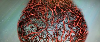
Atherosclerosis of the retina is a severe pathological situation, manifested in blockage of the peripheral vascular network in the area of the eyeball. The mechanism of action consists of several long stages that can take years:
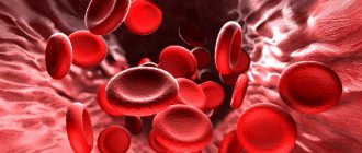
- When a carbohydrate enters the body, regardless of whether it is “simple” or “complex,” the carbohydrate is broken down into glucose. This makes it easier for the body to process and absorb it. When the lipid profile of the blood is disturbed, due to a malfunction of the liver or kidneys, glucose accumulates in clusters and clings to the deformed walls of the vessel. This is how the basis for the plaque is formed.
- Any free fractions traveling in the bloodstream may be attracted to the site of plaque formation, increasing its size.
- The capacity of the vessel decreases, and the specific area of passage of the blood composition decreases.
- When blood thickens, which goes hand in hand with atherosclerosis, blood cells stick together, forming a blood clot.
- The presence of an obstruction causes swelling and fluid accumulation at the site of the rhombus, loss of sensitivity, and tissue death due to insufficient blood supply.
- Under severe stress or anxiety, when the speed of blood circulation increases, the blood clot displaces and is carried away by the blood flow, and if it enters the ventricle of the heart, it can disrupt its functioning and lead to a heart attack.
Read the article on how to treat atherosclerosis or learn how to deal with this disease here.
Causes and risk factors
Atherosclerosis of the optic blood canals occurs in patients who are predisposed to increased levels of “bad” cholesterol in the blood .
For a long time, scientists believed that the disease only affected older people. However, modern research has revealed a pathological and long-term nature of development. Now it seems possible for doctors to identify the primary stages of the pathological process at a young age, in order to prevent the development of the disease at a more mature age.
The formation of lipid plaques on the walls of blood vessels is promoted by:
- Genetic and hereditary factors.
- Excess weight and metabolic disorders.
- Poor lifestyle, lack of physical activity.
- Unbalanced nutrition, when the daily diet is dominated by carbohydrates, refined foods, rich in sugar.
- Unhealthy habits such as using tobacco and alcohol products.
- Mechanical and thermal injuries.
- Lack of vitamins.
- Malfunctions of the central nervous system.
To combat the disease, it is important to know about the causes, risk factors and consequences of vascular atherosclerosis.
What to watch out for
Patients suffering from high cholesterol levels should monitor their levels, reduce them in time, and do this for life.
You should constantly take medications that contribute to the normal maintenance of the ocular circulatory system, anti-sclerotic agents and vitamins, since an advanced stage of the disease can lead to dire consequences, such as:
- Glaucoma;
- Eye infarction;
- Nerve atrophy;
- Thrombosis;
- Recurrent hemophthalmosis;
- Loss of vision;
- Irreversible blindness.
Adjust your diet, do as much physical activity as possible, take homeopathic remedies and vitamins, monitor your cholesterol levels, quit bad habits once and for all, and check your eyes periodically. With timely diagnosis and properly selected treatment, your eyes will remain the mirror of your soul for a long time!
Symptoms
Vascular atherosclerosis of the retina causes a narrowing of the lumen of the eye capillaries due to the accumulation of lipid-glucoid formations on their walls. There is a slowdown in blood circulation in the retinal tissue, dystrophy and atrophy, loss of vision (partial or complete), bleeding.
Symptoms of the initial stage:
- pain in the eye area;
- dizziness;
- headaches, migraine;
- chronic eye fatigue;
- blurred vision.
Familiarize yourself with the symptoms of vascular atherosclerosis and the pathogenesis of the disease.
Complications
As a result of birth trauma, pathology can also appear in newborns. The deviation manifests itself rarely, and, as a rule, does not require intervention and goes away within 2-3 days.
Ignoring the manifestations of atherosclerosis and starting treatment late often leads to the development of dangerous side effects. Due to improper treatment, thrombosis, occlusion of the inner membrane of the eye and nerve, hemorrhage in the vitreous body, atrophy of veins and capillaries can develop. Advanced atherosclerosis of the fundus vessels leads to complete blindness.
Diagnostics
with the diagnosis and treatment of atherosclerotic formations of the retina . During diagnostics, he uses modern clinical and instrumental techniques, which make it possible to identify disorders such as:
- changes in the size of the walls of the eye vessels;
- the measure of narrowing of the arteries and veins of the retina;
- structure and structure of blood vessels;
- the presence of blood clots and hemorrhages;
- the scale and nature of the gaps.
Note! Diagnosis begins with determining anamnestic information, studying the general picture of the disease and identifying accompanying ailments.
Additional Methods
- Ophthalmoscopy . Determines the number and basic characteristics of damaged vessels.
- Visometry . Makes it possible to assess the stage of vision loss and provides information about the condition of the retina.
- Computer perimetry , with the help of which local parts of the retina are studied.
- Ultrasound examination of the fundus and a conclusion about the condition of its veins and arteries.
- MRI of the eye , which will reveal the presence of disorders in the structural components of the organ, their location and features.
Timely diagnosis and proper treatment are the key to a full life.
How is lens sclerosis diagnosed?
To confirm the diagnosis, a specialist needs to analyze the condition of the lens under lateral illumination, using a 20 D lens. In addition, the lens is also examined in transmitted light, thanks to which it is possible to judge changes in its shape, if any.
An eye biomicroscopy procedure using a slit lamp is also mandatory. During the examination, it is possible to detect minimal changes in the color, shape and position of the lens. In addition, ultrasound A-scan, or echobiometry, is also performed. This diagnostic method allows you to specify the place where the lens compaction occurred, as well as determine the presence of opacification.
Treatment - specific tips and recommendations
The choice of treatment strategy for atherosclerotic lesions will depend on the results of the study, the presence of accompanying diseases and complications.
Therapeutic relief of the pathological condition is carried out by ophthalmologists, who, after a detailed study of each individual clinical case, decide on the choice of medications and treatment procedures or the urgency of surgical intervention.
Drugs used in the initial stages
Angioprotectors , protecting the weakened and lost elasticity of the vessel wall from ruptures.- Vasodilators that prevent spasms and improve blood circulation in damaged areas.
- Anti-sclerotic complexes for the direct elimination of fatty plaques.
- Antiplatelet agents , which help improve the rheological properties of blood and prevent the formation of blood clots.
To obtain the maximum effect from the drugs, patients are recommended dosage forms in the form of eye drops , as they go directly to the site of damage, providing an accelerated therapeutic effect. If there are diseases occurring simultaneously with this one, they are also treated to eliminate the chance of complications.
Important! Do not try to start treatment on your own. Incompatibility of drugs can lead not only to complete loss of vision, but also to the death of the patient.
Surgical treatment is used for patients in whom atherosclerosis has caused the development of a critical complication of retinal detachment. Among the most popular surgical interventions today are:
- laser coagulation of the retina;
- scleral ballooning;
- vitrectomy or removal of the vitreous.
Find out here whether treatment with folk remedies (herbs) is effective; what drugs are used for vascular atherosclerosis, read here.
Used in practice
- Mildronate , stimulant of central and local circulation. Price – 250 rub.
- Vazonit , Peripheral vasodilator. Cost 340 rub.
- Solcoseryl , ointment for disinfection. Price 350 rub.
- Arbiflex , anti-inflammatory drug. Cost 480 rub.
Laser therapy
A modern painless procedure that involves destroying deposits using a laser beam of variable wavelength.
The entire procedure takes no more than 20 minutes and has no contraindications. Because the procedure is expensive, it is optional and serves as an alternative to traditional surgery. After the procedure, the patient must also undergo consultations and be observed by an ophthalmologist for 2 months to monitor health and eliminate possible complications.
Consequences and complications
Increased blood pressure primarily damages the walls of blood vessels throughout the body. Examination of the fundus is one of the mandatory procedures when diagnosing hypertension and its consequences. With hypertensive angiopathy, arterial vasospasms, dilatation and inversion of the retinal veins are observed.
The thickness of the arteries, in relation to the size of the veins, decreases. The veins in the area of intersection with the arteries bend and move into the retina. Sometimes there is a loss of veins at the intersection with the arteries, swelling of the retina occurs, and hemorrhages may develop.
Hypertensive angiosclerosis
Since the causes are systemic in nature, angiosclerosis of both eyes is observed. In the early stages, there are no obvious external symptoms. As retinal damage progresses, double vision, blurred vision, or loss of vision may occur. Diagnosis of hypertensive angiosclerosis of the retina is carried out using fundus ophthalmoscopy and fluorescein angiography.
Reference! Treatment is based on therapeutic methods that involve preventing pathology.
Signs of the disease
Retinal atherosclerosis at the very beginning of the pathological process does not give specific symptoms. Manifestations of the disease are visible only during diagnosis; the doctor will determine the spastic conditions of the arteries and small blood vessels of the retina.
As the disease progresses and the amount of cholesterol deposits increases, the vascular walls become thicker. The patient notices a rapid decrease in vision, fog before the eyes, and rapid fatigue when working with visual strain.
Severe atherosclerotic changes are characterized by the formation of foci of hemorrhage, deposition of fat and protein over larger areas. The patient is diagnosed with a retinal infarction, in which the supply to the optic nerve stops.
The connecting cords provoke detachment of the retina, the discs of the optic nerves swell, and as a result, the diabetic faces partial or even complete blindness. The most dangerous complication of ocular retinopathy is acute blockage of the central retinal artery. The violation occurs instantly, literally in a few seconds. The patient will not feel a bit of discomfort.
Only in rare cases is acute blockage preceded by:
- flashes of light;
- temporary darkening of the eyes;
- sectoral (partial) vision loss.
The outcome is complete atrophy of the optic nerve and blindness. The ability to see can only be restored within the first hour from the moment of blockage; intensive therapy will be required. It is taken into account that damage to the blood vessels of the eyes may be the first symptom of an increasing acute vascular catastrophe - heart attack, stroke.
The disease is distinguished by the degree of damage. A diabetic can be diagnosed with a local degree of the disease if a quarter of the retina is involved in the pathological process. When atherosclerosis has occupied half of the retina, it is said to be widespread. If most of the problems are identified, a diagnosis of subtotal retinopathy is made; if the retina is completely detached, a diagnosis of total retinopathy is made.
Atherosclerosis of the vessels of the eyes can be mobile and rigid. The mobile form is observed when the patient spent the first two days in a horizontal position. The retina completely adheres to the lower layers.
Expert opinion
Guseva Yulia Alexandrova
Specialized endocrinologist
If this does not happen, a rigid form of the disease is identified.
Prevention and prevention of disease
Proper nutrition is the most important factor that will help cure an illness or alleviate its course.
Excessive consumption of sugars and carbohydrates leads to metabolic disorders. The body has no need to spend fat deposits, and it has plenty of prerequisites for fat accumulation. Consume more healthy fats Omega-3 and Omega-6, natural proteins of animal origin, exclude carbohydrates (porridge, bread, fruits, vegetables), starch, sugar from food. To normalize digestion, consume enough fiber, which is found in cabbage, green vegetables, parsley, and dill.
Eye gymnastics and exercises will also be useful.
In addition to preventing vascular disorders, this method allows you to avoid deterioration of vision. The doctor will recommend you a list of exercises that you will need to perform at least once a day. It is especially important to engage in eye exercises when working with monitors and other activities that cause strain on the visual organs. If your professional activity involves such stress, then eye gymnastics is simply necessary.- Limiting exposure to screens is very important in preventing illness.
Try not to spend a lot of time in front of monitors and displays unless you clearly need them. Read newspapers, books, preferably on paper, in natural light. However, prolonged reading can also negatively affect eye health, so you should limit your reading sessions by time.
Walking in the fresh air and exercising outside are also a good means of prevention.
It will keep the entire body in good shape, which naturally reduces the risk of developing vascular pathologies. Maintaining proper eye hygiene is also encouraged. Do not overuse makeup and neglect washing your face during the day.
Age-related changes in the lens of the eye
The lens is an important part of the eye, which from the birth of a person undergoes various physiological changes. For example, immediately after birth it has the shape of a ball, is soft and lacks color. However, as it ages, it becomes flat at the front of the lens. The color of the lens during this period is still transparent, but a yellow tint gradually appears, becoming more pronounced with age. Nowadays, surgery to replace the lens of the eye is very popular.
It must be said that not only the appearance of the lens changes, but also its elasticity; the lens becomes harder and harder over time. When a person reaches the age of forty, elasticity decreases to such limits that there is a threat of developing a pathology of a type such as age-related farsightedness, commonly known as “presbyopia.” After a person turns sixty years old, he develops sclerosis of the lens, which affects the decline in the functioning of this organ.