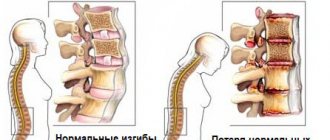
Etiology
The main factor is the lack of fusion of the sternal stripes during the period from the sixth to the ninth week of conception.
According to scientists, some factors may still affect the growth and progression of the disease:
- radiation exposure during pregnancy;
- drug use during pregnancy;
- exposure to toxins and hazardous metals.
To date, experts have not established the exact causes of the development of pathology.
Ectopia of the heart
What causes ectopic heart?
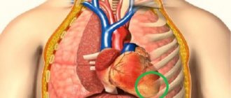
The sternum is a long, flat bone that connects to the ribs and forms the front of the rib cage, protecting the heart and lungs. If the fetus's chest does not develop properly, the heart may form outside of it.
In utero, the fetal heart begins to develop very early, while bones begin to develop later. The heart begins to beat during the first few weeks of fetal development, and the rib cage forms during the first trimester or the first 12 weeks of pregnancy.
Routine ultrasound is performed in the early stages of pregnancy and usually detects a misaligned heart. Sometimes an ultrasound will not detect a heart misalignment and the condition will be detected at birth. It is better if any problems with fetal development are discovered during pregnancy, then doctors can plan emergency care immediately after birth.
Classification
Ectopia of the heart is divided by scientists into three types:
- thoracic or abdominal ectopia of the heart;
- cervical ectopia of the heart;
- thoracoabdominal ectopia of the heart.
Thoracic ectopia - when the organ is not covered with tissue from the outside, the apex is usually directed up or down. Along with the thoracic form, the presence of concomitant diseases is noted, such as vascular transposition, pulmonary artery stenosis or ventricular septal defect. It is possible that the hernia of the umbilical cord is located adjacent to the organ itself, however, it is not covered with hernial membranes.
Cervical ectopia of the muscle has many similarities with thoracic ectopia, but localization of the organ in this case is possible, for example, between the mouth and the top of the muscle.
Thoracoabdominal ectopia of the heart is characterized by a cleft in the lower part of the chest, and the heart muscle itself is located in a membranous membrane. May be accompanied by ventricular septal defects and chest wall anomalies.
Causes of ectopia in a newborn
Normally, by the end of the second month, the sternum grows together and, together with the ribs, covers the heart. If this does not happen, then a cleft remains in the bone, and it extends to the front surface of the chest. The organ can enter the abdominal cavity through the diaphragmatic opening. And cervical ectopia occurs when development stops completely, when there is no movement from the original location of the cardiac rudiment in the pharynx.
There are also variants of dislocation under the stomach, at the site of the kidney.
Cervical ectopia
With complete thoracic ectopia, the heart is called naked. The chest is not fused, and there is no protection from the skin or pericardial sac on top. The incomplete form is characterized by a developed pericardium (there is partial coverage in front), or a thin membrane of the skin creates a translucent outer layer for the organ.
We recommend reading the article about congenital heart defects. From it you will learn about the causes, mechanism of development and classification of congenital heart defects, as well as the diagnosis and treatment of congenital heart defects. And here is more information about aortic heart disease.
Diagnostics
Of course, the abnormal location of the organ is noticeable visually, and in order to correctly carry out all surgical measures, the doctor will additionally need the results of radiography and ultrasound diagnostics, after which cardiac surgeons will be able to begin the operation.
One must be able to distinguish a complete ectopia of the heart from a partial one. With partial ectopia, the heart is covered with a membrane of a thin layer of skin, and the pericardial sac can be fully formed, but with complete ectopia, the protective layer of skin and the pericardial sac are not observed.
Can a child with ectopic heart disease live?
With a completely open version of ectopia, suppuration of unprotected tissue quickly occurs, which leads to death. When the heart is covered with skin, and especially located in the abdominal cavity, the prognosis is somewhat improved. Most babies are in an extremely critical condition at birth, but there are also cases where, after creating a protective layer over the myocardium, the child was able to save life until adolescence.
When the organ is located near intestinal loops or kidneys, there are patients who lived to adulthood. A thirty-six-year-old patient with ectopia cordis even had 4 pregnancies, and a man with such an anomaly managed to celebrate his 70th birthday.
Thoracic ectopia
Treatment
Treatment of ectopia of the heart is carried out only with the help of surgery. Creating a protective layer of skin for the heart muscle and preventing decreased venous return is the main goal of surgery. The operation should begin immediately after birth, since an organ that is not protected from the outside world cannot exist. The baby is ventilated because it is unlikely that his lungs are functioning properly and he is able to breathe on his own.
If there is free space in the middle of the sternum to place the heart muscle, the operation can be performed emergencyly, although often there is not enough such space in the chest of a newborn, and special protection is created from the outside.
It is possible that a baby diagnosed with ectopia will have to stay in the hospital for a long time, since the child needs constant monitoring and may need to undergo several additional operations.
Features of cervical and thoracic pathology
Location in the neck area refers to a severe developmental anomaly. The chest may have a normal shape or structural defects. There is localization between the left and right halves of the lower jaw, near the tongue, in the area of the palatine bone. The baby is usually stillborn or dies on the first day.
Thoracic dislocation in this disease is the most common variant. The front surface of the heart touches the chest, and the back is open or closed by the skin. There are often other anomalies in the structure of chambers, valves and great vessels, and internal organs. If part of the heart is in the chest cavity and the rest is in the abdominal cavity, then this disease is called Cantrell's pentad (five signs). It is characterized by:
- cleft sternum;
- diaphragm defect;
- umbilical cord hernia;
- thoracoabdominal (thoracoabdominal) localization;
- structural defect of the heart.
Forecast
The prognosis depends only on the type of pathology. If the presence of cervical ectopia is noticed in the fetus, many cases of stillbirth or death have been recorded during the first days of the baby’s life. In the case of the presence of other forms of ectopia, life expectancy is possible up to several months, however, one case of a person with abdominal ectopia of the heart living up to adulthood has been recorded.
The outcome of the pathology can be relatively favorable only if the anatomical location of the organ allows the operation to be performed effectively and allows time for this. If the location of the heart muscle is extremely unfavorable, for example, if it is not covered by skin, there is a high probability of death in the first hours of a newborn’s life.
Diagnostic methods
Ultrasound helps detect cardiac pathology. The diagnosis can be made starting from the 10th week of pregnancy. According to available data, several cases have been associated with chromosomal abnormalities - gene mosaicism, trisomy, Shereshevsky-Turner syndrome. This means that it is possible to calculate the risk of abnormalities even at the stage of medical genetic counseling.
Expert opinion
Alena Ariko
Expert in Cardiology
If cervical ectopia and an open heart are detected, termination of pregnancy is recommended; in the case of the abdominal form, it is possible to maintain the pregnancy if this malformation is isolated.
If ectopia is not diagnosed before birth, then its signs on the radiograph will be:
- absence of the entire mediastinum;
- increasing the level of the left part of the diaphragm (high dome);
- an increase in the size of the gas bubble in the stomach.
In Bolivia, a girl was born with a heart outside her chest (caution, photo)
In Bolivia, little Maria Gloria Almeida was born with an incredibly rare condition: her heart is located on the outside of her chest. The girl's weight was 1.8 kg, but despite all the difficulties, she survived.
The baby was rushed to Santa Cruz for emergency heart surgery.
Maria Gloria Almeida was born with a rare defect - ectopia cordis. This is a congenital malformation characterized by the location of the heart outside the chest through a defect in the sternum. The mortality rate for such a defect is at a fatal level.
Initially, doctors did not think the 1.8kg newborn would survive. But thanks to government funding, Maria's family was able to afford emergency surgery for their daughter. The girl was flown to a specialized center in Santa Cruz, located 800 km away. According to doctors, little Maria successfully underwent surgery.
Virmania Vargas, the mother of the girl and four other children, said her pregnancy was normal and her test results were also normal. Doctors were unable to determine the presence of a defect in advance. Experts estimate that ectopia cordis affects one in every 126,000 births, with girls being slightly more likely to be affected.
This condition is caused by the failure of the embryo to develop properly.
Those born with ectopic heart disease usually die in the first year of life, but in some cases successful surgery can be performed. Medicine knows of 50 cases where children with a similar defect lived to be 12 years old. Christopher Wall lived the longest with ectopia cordis, who underwent several operations and lived to be 34 years old.
We also wrote about how a girl born with her heart out in Russia survived.
Baby Bathsheba was born in Russia. The newborn girl's chances of life were reduced to almost zero, because the baby had a severe congenital pathology, due to which her heart formed right under the skin. When the mother of an unusual girl was pregnant, doctors were sure that the child would die immediately after birth. But a miracle happened! This year Virsavia Borun-Goncharova will turn 7 years old.
Rare disease
The girl was born with a cleft in the sternum, which caused her heart to fall outside of it.
This disease occurs in 1 in 1 million newborns. Bathsheba's intestines are also located incorrectly - they are located outside the abdomen.
Hope for recovery
The baby recently moved with her American mother to the USA.
Doctors do not intend to perform an operation on a girl with such a rare pathology for at least the next 2 years - the baby’s blood pressure exceeds normal levels.
Bathsheba is actively helped by caring people who donate money for treatment to charities created specifically for Bathsheba
Let us note that they have known about the girl for a long time, but she is not the only one. Another girl was born with her heart out. The first operation was performed by a team of 50 surgeons!
Spouses Naomi Findlay and Dean Wilkins from the UK refused to terminate their pregnancy at eight weeks, despite doctors warning that the child would be born with a pathology and a small chance of survival.
Baby Vanellope was born with a rare pathology: her heart was on top of her chest.
“Naomi and I held our breath, waiting for her to take her first breath. When she cried, we cried with her, praying that everything would be okay,” said the father of the newborn. 20 minutes passed, but Vanellope was still screaming.
At Glenfield Hospital, an hour after the girl was born, a team of 50 surgeons performed the first of three operations. Then two more operations were performed within three weeks.
Doctors expanded the baby's chest because there was no room for a heart. After this, within two weeks the heart naturally returned to the chest due to the effect of gravity. The skin on the chest was taken from the child's hands.
Vanellope still has several more surgeries ahead. Now surgeons have protected the girl’s heart with a special mesh, since the baby has no ribs.
In the future, doctors plan to 3D print a chest made of organic plastic that will grow along with Vanellope.
According to the doctors, they did not hope for a successful outcome of the operation. This pathology occurs only in 8 babies per million, and most often leads to the death of the child in the womb.
Even if the baby is born, the chances of survival are about 10%. Vanellope became the first child in the UK to be saved.
“I felt guilty for having bad thoughts because my daughter was fighting for her life and I was close to giving up. I’m glad I insisted on keeping the baby, I’m so glad!” - Naomi shares.
Previously, Sensum wrote that there were famous people who died from anorexia - “They warned girls not to repeat their fate”