External structure of the heart
The heart (cor) has the shape of a truncated cone, which is located in the anterior mediastinum with the apex to the left and down. The apex of this cone has the anatomical name "apex cordis", so you won't get confused. Look at the illustration and remember - the top of the heart is at the bottom, not at the top.
The top of the heart is called the base of the heart (basis cordis). You can show the base of the heart on the specimen if you simply circle the area into which all the major vessels of the heart enter and exit. This line is quite conventional - as a rule, it is drawn through the opening for the inferior vena cava.
The heart has four surfaces:
- Diaphragmatic surface (facies diaphragmatica). Located below, it is this surface of the heart that is directed towards the diaphragm;
- The sternocostal surface (facies sternocostalis). This is the anterior surface of the heart, it faces the sternum and ribs;
- Pulmonary surface (facies pulmonalis). The heart has two pulmonary surfaces - right and left.
In this picture we see the heart in combination with the lungs. Here is the sternocostal, that is, the anterior surface of the heart.
There are small projections at the base of the sternocostal surface. These are the right and left appendages (auricula dextra / auricula sinistra). I highlighted the right ear in green, and the left ear in blue.
What is special about the structure of a child’s heart?
Until the age of six, the heart is spherical due to the large atria. Its walls are easily stretched, they are much thinner than those of adults. A network of tendon threads is gradually formed, fixing the valve leaflets and papillary muscles. Full development of all heart structures ends by age 20.
The position of the newborn's heart in the chest is initially oblique, adjacent to the anterior surface. This is caused by an increase in the volume of lung tissue and a decrease in the mass of the thymus gland.
Until two years of age, the heart impulse forms the right ventricle, and then part of the left. The atria are the leaders in growth rate up to 2 years, and the ventricles after 10 years. Up to ten years, the LV is ahead of the right.
Chambers of the heart
The heart is a hollow (that is, empty from the inside) organ. It is a bag of dense muscle tissue, which has four cavities:
- Right atrium (atrium dexter);
- Right ventricle (ventriculus dexter);
- Left atrium (atrium sinister);
- Left ventricle (ventriculus sinister).
These cavities are also called the chambers of the heart. The human heart has four cavities, that is, four chambers. That is why they say that a person has a four-chambered heart .
On a heart that is cut in the frontal plane, I highlighted the boundaries of the right atrium in yellow, the left preseries in green, the right ventricle in blue, and the left ventricle in black.
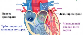
Right atrium
The right atrium collects “ dirty ” (that is, rich in carbon dioxide and poor in oxygen) blood from throughout the body. The upper (brown) and lower (yellow) complete veins flow into the right atrium, which collect blood with carbon dioxide from the entire body, as well as the great vein of the heart (green), which collects blood with carbon dioxide from the heart itself. Accordingly, three openings open into the right atrium.
Between the right and left atria there is an interventricular septum. It contains an oval depression - a small oval-shaped depression, an oval fossa (fossa ovalis). In the embryonic period, in place of this depression there was an oval hole (foramen ovale cordis). Normally, the foramen ovale begins to close immediately after birth. In this picture, the oval fossa is highlighted in blue:
The right atrium communicates with the right ventricle through the right atrioventricular orifice (ostium atrioventriculare dextrum). The flow of blood through this opening is controlled by the tricuspid valve.
Right ventricle
This cavity of the heart receives “dirty” blood from the left atrium and sends it to the lungs to be cleaned of carbon dioxide and enriched with oxygen. Accordingly, the right ventricle connects to the pulmonary trunk, through which blood will be sent to the lungs.
The tricuspid valve, which must be closed during the reflux of blood into the pulmonary trunk, is fixed by tendon threads to the papillary muscles. It is the contraction and relaxation of these muscles that controls the operation of the tricuspid valve.
The papillary muscles are highlighted in green, and the tendon filaments are highlighted in yellow:
Left atrium
This part of the heart collects the “ purest ” blood. It is into the left atrium that fresh blood flows, which is first purified in the small (pulmonary) circle from carbon dioxide and saturated with oxygen.
Therefore, four pulmonary veins flow into the left atrium - two from each lung. You can see these holes in the picture - I've highlighted them in green. We remember that arterial passes veins .
The left atrium communicates with the left ventricle through the left atrioventricular orifice (ostium atrioventriculare sinistrum). The flow of blood through this opening is regulated by the mitral valve.
Left ventricle
The left ventricle begins the systemic circulation . When the left ventricle pumps blood into the aorta, it is isolated from the left atrium by the mitral valve. Just like the tricuspid valve, the mitral valve is controlled by papillary muscles (highlighted in green), which are connected to it by tendon strands.
You may notice the very strong muscular wall of the left ventricle. This is explained by the fact that the left ventricle needs to pump a powerful flow of blood, which must be sent not only in the direction of gravity (to the stomach and legs), but also against gravity - that is, upward , to the neck and head.
Can you imagine how cunningly the circulatory system of giraffes is designed, in which the heart must pump blood all the way up the neck to the head?
The human heart: where it is located, structure
The heart is the main circulatory organ, thanks to which blood moves in a small and large circle, covering all organs and systems of the body. Its main function is to ensure continuous blood flow through the vascular bed. One part of it is responsible for venous blood, the other for arterial blood. The heart is capable of independently producing electrical impulses that allow it to contract with the required force and frequency.
- 1. Location of the heart
- 2. Structure 2.1. Right and left atrium
- 2.2. Right and left ventricle
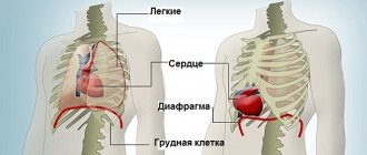
A person's heart is located in the chest. A more accurate localization is the mediastinum (the space between the thoracic spine at the back, the sternum at the front and the pleura at the sides). The pleura is the serous lining of the lungs. Below it borders with the tendon center of the diaphragm. The heart, with the vessels extending from it, occupies a middle location in the mediastinum. The rest of the space is allocated to the trachea, lymph nodes and bronchi of the first order. The heart is fixed in space using large-caliber blood vessels.
On the direct projection, one can note the predominantly left location, and on the right it protrudes from behind the sternum by 1-2 cm. The heart is a muscular organ, so it can enlarge due to overload, for example, in athletes. In women, the heart is smaller, its borders are slightly different - during percussion, the right border runs along the edge of the sternum.
Externally, the heart looks like a cone; in asthenics it is more elongated, in hypersthenics it has a rounded shape. The muscle walls are well developed and reach a width of up to 8 cm. The length is 12-16 cm, the transverse size is 8-10 cm. In the upper part there are 2 atria with vessels extending from them, and on the left there are 2 ventricles. On average, the mass of the organ ranges from 200 to 350 g, it depends on the gender and degree of training of the person. In professional athletes, the heart weight can reach 450 g.
The heart has 4 surfaces:
- diaphragmatic – flattened;
- costal – convex;
- right pulmonary – elongated and angular;
- the left pulmonary is rounded and shortened.
For their own nutrition, the vessels are located on the surface of the heart in special grooves that have the same name as arteries and veins. The coronary groove runs between the atria and ventricles. Anterior and posterior interventricular, respectively, between the ventricles in front and behind.
The internal structure has a cavity divided by partitions into 4 chambers: the right and left atrium, the right and left ventricle. The separation is carried out by the interventricular, interatrial and atrioventricular septum. In the latter there is an atrioventricular opening on the left and right, through which blood flows from the atrium to the ventricle.
The right atrium is shaped like an irregular cube. The muscle layer is 3 mm. It has 5 walls: superior, posterior, anterior, external and internal, common with the left atrium. From below, the absence of a wall is explained by the presence of an atrioventricular orifice. The upper part is somewhat expanded due to the venous sinus; the largest venous trunks are located there. Toward the anterior part, the atrium narrows, forming the right appendage, which with its upper part is adjacent to the aortic bulb.
The venous sinus is formed by the superior, inferior vena cava and the proper veins of the heart. Along the anterior lower edge of the vena cava there is an oval foramen. It is necessary to ensure intrauterine blood flow in the fetus; it often has several tendon strands. Subsequently, the hole closes and forms an oval fossa. The inner surface of the atrium has a relief structure due to the muscle ridges and pectineus muscles.
The left atrium has an irregular cube shape, with a muscle thickness of 2-3 mm. It has 6 walls: anterior, posterior, upper, left, the right is represented by the interatrial septum, and the lower is the base of the left ventricle. The anterior-superior part is angular and represents the left ear. which surrounds the trunk of the pulmonary artery. The posterior part of the upper wall has 4 openings for communication with the pulmonary arteries. The atrioventricular orifice is located on the lower wall. The inner surface is relatively smooth, with the exception of the left ear. Its ribbing is formed by the pectineus muscles and the remaining flap from the foramen ovale. If such an opening is not completely closed, has a slit-like lumen, the size of a pinhead, then it is visible from the left atrium much better.
Externally, the right ventricle is delimited from the left and atria by grooves. It has a cone-shaped shape, the base of which is adjacent to the right atrium, and the pointed part faces down and to the left. The thickness of the muscle layer is on average 5 mm. The anterior wall is convex, the posterior one is flat, and the middle one is represented by the interventricular septum. The cavity is visually divided into two sections. The posterior one is wide and communicates with the right atrium. The anterior section is narrow and oblong. A muscle shaft passes between them.
In the area of the atrioventricular orifice there is a valve formed by the endocardium, the inner muscle layer. Its structure contains muscle and collagen fibers connecting to the right atrium. The valve has three valves and chords extending from them, which connect to the papillary muscles. There are three such muscles in total: the anterior, major and septal.
The left ventricle has an oblong oval shape. The muscle layer can reach 14 cm. It has two sections: the posterior one communicates with the atrium, and the anterior one with the aorta. Along the perimeter of the atrioventricular opening there is a valve of the same name, which at the moment of heart contraction prevents the reverse flow of blood. The valve is represented by front and rear flaps. The inner posterior part of the left ventricle has a pronounced structure of muscle trabeculae, which intertwine with each other, providing greater contractility.
Contraction of the heart muscle is carried out using the conduction system of the heart, which is located mainly in the interventricular and interventricular septa and is represented by the sinus node, the His bundle and Purkinje fibers.
The outside of the heart is covered with a serous sac, the pericardium. It has two layers - the outer free one and the inner one, fused with the upper muscular layer of the heart.
Externally, the right ventricle is delimited from the left and atria by grooves. It has a cone-shaped shape, the base of which is adjacent to the right atrium, and the pointed part faces down and to the left. The thickness of the muscle layer is on average 5 mm. The anterior wall is convex, the posterior one is flat, and the middle one is represented by the interventricular septum. The cavity is visually divided into two sections. The posterior one is wide and communicates with the right atrium. The anterior section is narrow and oblong. A muscle shaft passes between them.
Table “Boundaries of the Heart”
| Borders | Location |
| Upper | Along the upper edge of the third rib |
| Right | Between the ribs (from the edge of the 3rd to the 5th rib). The border is shifted 1–2 cm from the right edge of the sternum |
| Left | From the 3rd rib to the top of the muscle sac |
| Lower | To the right from the 5th rib to the apex of the organ |
In the 5th intercostal space is the very top of the muscle sac. It is shifted more inward by 1.5–2 cm in the midclavicular line (left).
To determine the right, left, lower and upper boundaries, the percussion method is used (tapping the rib area with fingers from the edge of the sternum to the center). With such manipulation, a dull sound is formed in the area of the muscle sac (tapping the lungs provokes a clear sound), which makes it possible to fix the edges of the location of the vital organ.
Unconventional methods of treatment and prevention
Folk remedies help eliminate pain, spasms, and strengthen the heart muscle. But they can only be used after preliminary consultation with a cardiologist. The easiest way to eliminate heart pain is to mix tincture of valerian and hawthorn in equal proportions. Dissolve 25 drops of the mixture in 50 ml of water, hold it in your mouth a little, and swallow.
The main medications for the treatment of heart diseases are nitrates, which are divided into 3 groups depending on the main active ingredient:
Septa and grooves of the heart
The left and right ventricles are separated by a thick muscular wall. This wall is called the interventricular septum (septum interventriculare).
The interventricular septum is located inside the heart. But its location corresponds to the interventricular grooves that you can see from the outside. The anterior interventricular groove is located on the sternocostal surface of the heart . I have highlighted this furrow in green in the figure.
On the diaphragmatic surface of the heart is the posterior interventricular groove (sulcus interventricularis posterior). It is highlighted in green and indicated by the number 13.
The left and right atria are separated by the interatrial septum (septum interatriale), also highlighted in green.
From the outer part of the heart, the ventricles are separated from the atria by the coronary groove (sulcus coronarius). In the figure below you can see the coronary groove on the diaphragmatic, that is, the posterior surface of the heart. This groove is an important landmark for identifying the large vessels of the heart, which we will discuss later.
Circulation circles
Big
The powerful, large left ventricle launches arterial blood into the aorta - this is where the systemic circulation begins. It looks like this: blood is ejected from the left ventricle into the aorta, which branches into organ arteries. Then the caliber of the vessels becomes smaller and smaller, down to the smallest arterioles that approach the capillaries.
Gas exchange occurs in the capillaries, and the blood, already saturated with carbon dioxide and decay products, rushes back to the heart through the veins. After the capillaries, these are small venules, then larger organ veins that flow into the inferior vena cava (if we are talking about the torso and lower extremities) and into the superior vena cava (if we are talking about the head, neck and upper extremities).
In this figure, I have highlighted the anatomical formations that complete the systemic circulation. The superior vena cava (green, number 1) and the inferior vena cava (orange, number 3) drain into the right atrium (magenta, number 2). The place where the vena cava enters the right atrium is called the sinus vena cava (sinus venarum cavarum).
Thus, the great circle begins with the left ventricle and ends with the right atrium:
Left ventricle → Aorta → Large main arteries → Organ arteries → Small arterioles → Capillaries (gas exchange zone) → Small venules → Organ veins → Inferior vena cava/Superior vena cava → Right atrium.
When I was preparing this article, I found a diagram that I drew in my second year. It will probably show you the systemic circulation more clearly:
Small
The pulmonary (pulmonary) circulation begins with the right ventricle, which sends venous blood to the pulmonary trunk. Venous blood (be careful, this is venous blood !) is sent along the pulmonary trunk, which is divided into two pulmonary arteries. According to the lobes and segments of the lungs, the pulmonary arteries (remember that they carry venous blood) are divided into lobar, segmental and subsegmental pulmonary arteries. Ultimately, the branches of the subsegmental pulmonary arteries break up into capillaries that approach the alveoli.
Gas exchange occurs again in the capillaries . Venous blood, saturated with carbon dioxide, gets rid of this ballast and is saturated with life-giving oxygen. When the blood is saturated with oxygen, it becomes arterial . After this saturation, fresh arterial blood runs through the pulmonary venules, subsegmental and segmental veins, which drain into the large pulmonary veins. The pulmonary veins drain into the left atrium.
Here I have highlighted the beginning of the pulmonary circulation - the cavity of the right ventricle (yellow) and the pulmonary trunk (green), which leaves the heart and is divided into the right and left pulmonary arteries.
In this diagram you can see the pulmonary veins (green) flowing into the cavity of the left atrium (purple) - it is these anatomical formations that complete the pulmonary circulation.
Diagram of the pulmonary circulation:
Right ventricle → Pulmonary trunk → Pulmonary arteries (right and left) with venous blood → Lobar arteries of each lung → Segmental arteries of each lung → Subsegmental arteries of each lung → Pulmonary capillaries (entangle the alveoli, gas exchange zone) → Subsegmental/segmental/lobar veins (with arterial blood) → Pulmonary veins (with arterial blood) → Left atrium
Sequence of abbreviations
The chambers are filled with blood gradually, it passes from one to another under the influence of contractions. But at the same time, none of the cavities can be empty; they are constantly filled. Valves made of collagen, an elastic tissue, prevent physiological fluid from returning back. The time when each portion passes through a muscular organ is called a cycle.
Attention! The duration of the cardiac cycle is 6-7 seconds. During this time, all cameras have time to contract and relax.
The regulation system of a 4-chambered hollow organ consists of several valves:
- Tricuspid - located between the ventricle and atrium in the right section. When it is open, blood flow descends into the ventricle. After filling the cavity, its walls stretch and the valve closes.
- Pulmonary - opens as soon as the tricuspid closes, allowing physiological fluid to pass through for enrichment.
- Mitral - located in the left section, the properties are the same as the tricuspid, but there are only 2 leaflets.
- Aortic - opens when the right ventricle contracts. As soon as it contracts, it closes.
First, the atrium on the right contracts, pushing blood into the right ventricle. Then the left atrium contracts. Then they both relax, filled with venous blood again. At this time, the ventricles pump physiological fluid to the lungs for oxygen enrichment, and then into the systemic circulation. That is, the venous, right part, receives waste blood, sends it to the lungs, and from the lungs it goes to the right side and is pushed into the bloodstream.
Heart valves
The right atrium from the left, as well as the right ventricle from the left, are separated by septa. Normally, in an adult, the partitions should be solid, there should be no holes between them.
But there must be a hole between the ventricle and the atrium on each side. If we are talking about the left half of the heart, then this is the left atrioventricular opening (ostium atrioventriculare sinistrum). On the right, the ventricle and atrium are separated by the right atrioventricular orifice (ostium atrioventriculare dextrum).
Valves are located at the edges of the holes. These are clever devices that prevent the blood from flowing back. When the atrium needs to send blood to the ventricle, the valve is open. After the ejection of blood from the atrium into the ventricle has occurred, the valve must close tightly to prevent blood from flowing back into the atrium.
The valve is formed by leaflets, which are double layers of endothelium - the inner lining of the heart. Tendon threads extend from the valves, which are attached to the papillary muscles. It is these muscles that control the opening and closing of the valves.
Tricuspid valve (valva tricispidalis)
This valve is located between the right ventricle and the right atrium. It is formed by three plates to which tendon threads are attached. The tendon threads themselves connect to the papillary muscles located in the right ventricle.
On a section in the frontal plane, we cannot see three plasties, but we can clearly see the papillary muscles (outlined in black) and tendon threads attached to the valve plates. The cavities that the valve separates are also clearly visible - the right atrium and the right ventricle.
On a section in a horizontal plane, the three leaflets of the tricuspid valve appear before us in all their glory:
Mitral valve (valva atrioventricularis sinistra)
The mitral valve regulates blood flow between the left atrium and the left ventricle. The valve consists of two plates, which, as in the previous case, are controlled by the papillary muscles through tendon threads. Please note - the mitral valve is the only heart valve that consists of two leaflets.
The mitral valve is outlined in green, and the papillary muscles are outlined in black:
Let's look at the mitral valve in the horizontal plane. Let me note again that only this valve consists of two plates:
Pulmonary valve (valva trunci pulmonalis)
The pulmonary valve is also often called the pulmonary valve or pulmonary valve. These are synonyms. The valve is formed by three flaps that are attached to the pulmonary trunk at its origin from the right ventricle.
You can easily find the pulmonary valve if you know that the pulmonary trunk starts from the right ventricle:
On a horizontal section, you can also easily detect the pulmonary valve if you know that it is always located anterior to the aortic valve. The pulmonary valve generally occupies the most anterior position of all the heart valves. We can easily find the pulmonary valve itself and the three valves that form it:
Aortic valve (valva aortae)
We have already said that the powerful left ventricle sends a portion of fresh, oxygenated blood to the aorta and further in a large circle. The aortic valve separates the left ventricle and the aorta. It is formed by three plates that are attached to the annulus fibrosus. This ring is located at the junction of the aorta and the left ventricle.
When examining the heart in a slice in a horizontal plane, do not forget that the pulmonary valve is in front, and the aortic valve is behind it. The aortic valve is surrounded by all the other valves from this angle:
Internal anatomy and structural features of valves, atria, ventricles
Each part of the heart has its own function and anatomical features. In general, the LV is more powerful (compared to the right), as it forces blood into the arteries, overcoming the high resistance of the vascular walls. The PP is more developed than the left, it receives blood from the whole body, and the left one only from the lungs.
Which side of a person's heart is on?
In humans, the heart is located on the left side in the center of the chest. The main part is located in this area - 75% of the total volume. One third extends beyond the midline into the right half. In this case, the axis of the heart is inclined (oblique direction). This situation is considered classic, as it occurs in the vast majority of adults. But options are also possible:
- dextrocardia (right side);
- almost horizontal - with a wide, short chest;
- close to vertical - for thin people.
Where is a person's heart located?
The human heart is located in the chest between the lungs. It is adjacent to the sternum from the inside, and is limited below by the diaphragm. It is surrounded by the pericardium, the pericardium. Pain in the heart area appears on the left near the mammary gland. The top is projected there. But with angina, patients feel pain behind the sternum, and it spreads along the left side of the chest.
Where is the heart located in the human body?
The heart in the human body is located in the center of the chest, but its main part goes into the left half, and only one third is located on the right side. For most people it has an angle of inclination, but for overweight people its position is closer to horizontal, and for thin people it is closer to vertical.
Location of the heart in the human chest
In humans, the heart is located in the chest in such a way that its anterior and lateral surfaces are in contact with the lungs, and its posterior and inferior surfaces are in contact with the diaphragm. The base of the heart (from above) passes into large vessels - the aorta, pulmonary artery. The apex is the lowest part, it approximately corresponds to the 4-5 space between the ribs. It can be found in this area by lowering an imaginary perpendicular from the center of the left collarbone.
External structure of the heart
The external structure of the heart refers to its chambers; it contains two atria and two ventricles. They are separated by partitions. The pulmonary veins, the vena cava, enter the heart, and the arteries of the lungs, the aorta, carry the blood out. Between the large vessels, at the border of the atria and ventricles of the same name, there are valves:
- aortic;
- pulmonary artery;
- mitral (left);
- tricuspid (between the right parts).
The heart is surrounded by a cavity containing a small amount of fluid. It is formed by the pericardial layers.
What does a human heart look like?
If you clench your fist, you can imagine exactly the appearance of a heart. In this case, the part that is located at the wrist joint will be its base, and the acute angle between the first and thumb will be its apex. What is important is that its size is also very close to a clenched fist.
This is what a human heart looks like
Borders of the heart and their projection onto the surface of the chest
The boundaries of the heart are found by percussion, by tapping; radiography or echocardiography helps to determine them more accurately. The projections of the cardiac contour onto the surface of the chest are:
- right – 10 mm to the right of the sternum;
- left – 2 cm inward from the perpendicular from the center of the collarbone;
- apex – 5th intercostal space;
- base (upper) – 3rd rib.
What tissues make up the heart?
The heart consists of the following types of tissue:
- muscle - the main one, is called the myocardium, and the cells are cardiomyocytes;
- connective – valves, chords (threads that hold the valves), outer (epicardial) layer;
- epithelium – inner lining (endocardium).
Surfaces of the human heart
The human heart has the following surfaces:
- ribs, sternum – anterior;
- pulmonary – lateral;
- diaphragmatic – lower.
Apex and base of the heart
The apex of the heart is directed down and to the left, its localization is the 5th intercostal space. It represents the top of the cone. The wide part (base) is located on top, closer to the collarbones, and is projected at the level of the 3rd rib.
Human heart shape
The shape of a healthy person's heart is like a cone. Its tip is directed at an acute angle down and to the left of the center of the sternum. The base contains the mouths of large vessels and is located at the level of the 3rd rib.
Right atrium
Receives blood from the vena cava. Next to them is the foramen ovale, which connects the RA and LA in the fetal heart. In a newborn, it closes after the pulmonary blood flow opens, and then completely heals. During systole (contraction), venous blood passes into the pancreas through the tricuspid valve. The RA has a fairly powerful myocardium and a cubic shape.
Left atrium
Arterial blood from the lungs passes into the LA through 4 pulmonary veins and then flows through the opening into the LV. The walls of the LA are 2 times thinner than those of the right one. The shape of the LP is similar to a cylinder.
Right ventricle
It looks like an inverted pyramid. The capacity of the pancreas is about 210 ml. It can be divided into two parts - the arterial (pulmonary) cone and the ventricular cavity itself. In the upper part there are two valves: the tricuspid and the pulmonary trunk.
Left ventricle
It looks like an inverted cone, its lower part forms the top of the heart. The thickness of the myocardium is the largest - 12 mm. There are two openings at the top - for connection with the aorta and LA. Both of them are covered by valves - the aortic and mitral.
Why are the walls of the atria thinner than the walls of the ventricles?
The thickness of the walls of the atrium is less, they are thinner, since they only need to push blood into the ventricles. The right ventricle follows them in strength; it throws its contents into the neighboring lungs, and the left one is the largest in terms of the size of its walls. It pumps blood to the aorta, where there is high pressure.
Tricuspid valve
The right atrioventricular valve consists of a sealed ring that limits the opening and leaflets; there may be not 3, but from 2 to 6.
Half of the people have a tricuspid configuration.
The function of this valve is to prevent the reflux of blood into the RA during RV systole.
Pulmonary valve
It prevents blood from passing back into the pancreas after it contracts. The composition contains valves similar in shape to a crescent. In the middle of each there is a knot that seals the closure.
Mitral valve
It has two doors, one is in the front and the other is in the back. When the valve is open, blood flows from the LA to the LV. When the ventricle contracts, its parts close together to allow blood to pass into the aorta.
Aortic valve
Formed by three semilunar-shaped flaps. Like the pulmonary one, it does not contain threads that hold the valves in place. In the area where the valve is located, the aorta expands and has depressions called sinuses.
Adult heart weight
Depending on the physique and total body weight, the weight of the heart in an adult varies from 200 to 330 g. In men, it is on average 30-50 g heavier than in women.
Layers of the Heart
1. Pericardium . This is a dense connective tissue membrane that reliably covers the heart.
The pericardium is a two-layer membrane; it consists of fibrous (outer) and serous (inner) layers. The serous layer is also split into two plates - parietal and visceral. The visceral plate has a special name - epicardium .
In many authoritative sources you can see that the epicardium is the first lining of the heart.
2. Myocardium (myocardium). The actual muscle tissue of the heart. This is the most powerful layer of the heart. The most developed and thick myocardium forms the wall of the left ventricle, as we already discussed at the beginning of the article.
See how the thickness of the myocardium differs in the atria (using the example of the left atrium) and in the ventricles (using the example of the left ventricle).
3. Endocardium (endocardium). This is a thin plate that lines the entire internal space of the heart. The endocardium is formed by endothelium - a special tissue consisting of epithelial cells tightly adjacent to each other. The development of atherosclerosis, hypertension, myocardial infarction and other serious cardiovascular diseases is associated with endothelial pathology.
The structure of the human heart: diagram, blood circulation circles
The task of the circulatory system, the main center of which is the heart, is the delivery of oxygen, nutritional and bioactive components to the tissues of the body and the elimination of metabolic products. For this purpose, the system provides a special mechanism - blood moves through the circulation circles - small and large.
Small circle
From the right ventricle at the time of systole, venous blood is pushed into the pulmonary trunk and enters the lungs, where it is saturated with oxygen in the microvessels of the alveoli, becoming arterial. It flows into the cavity of the left atrium and enters the systemic circulatory system.
Big circle
From the left ventricle in systole, arterial blood travels through the aorta and then through vessels of different diameters to various organs, giving them oxygen, transferring nutritional and bioactive elements. In small tissue capillaries, blood turns into venous blood, as it is saturated with metabolic products and carbon dioxide. It flows through the vein system to the heart, filling its right sections.
Nature has worked hard to create such a perfect mechanism, giving it safety margins for many years. Therefore, you should treat it carefully so as not to create problems with blood circulation and your own health.
Share the article on social media. networks:
Topography of the heart
Remember in the last lesson on basic chest topography I said that without knowing topographic lines you will not be able to learn anything related to the chest cavity? Have you learned them? Great, arm yourself with your knowledge, now we will use it.
So, the boundaries of absolute cardiac dullness and relative cardiac dullness are distinguished.
This strange name comes from the fact that if you tap (in medicine this is called “percussion”) the chest, in the place where the heart is located, you will hear a dull sound. When percussed, the lungs are more sonorous than the heart, which is where this term comes from.
Relative dullness is the anatomical ( true ) boundaries of the heart. We can establish the limits of relative dullness during the autopsy. Normally, the heart is covered by the lungs, so the boundaries of relative cardiac dullness are visible only on the preparation.
Absolute cardiac dullness is the boundaries of the part of the heart that is not covered by the lungs. As you understand, the limits of absolute cardiac dullness will be less than the limits of relative cardiac dullness in the same patient.
Since we are now examining anatomy, I decided to talk only about the relative, that is, the true boundaries of the heart. After the article about the anatomy of the hematopoietic system, I generally try to keep track of the size of the articles.
Boundaries of relative cardiac dullness (true boundaries of the heart)
- Apex of the heart (1): 5th intercostal space, 1-1.5 centimeters medial to the left midclavicular line (highlighted in green);
- Left border of the heart (2): a line drawn from the intersection of the third rib with the parasternal line (yellow) to the apex of the heart. The left border of the heart is formed by the left ventricle. In general, I advise you to remember the third rib - you will constantly encounter it as a landmark for various anatomical formations;
- The upper bound (3) is the simplest. It runs along the upper edge of the third ribs (we see the third rib again) from the left to the right parasternal lines (both yellow);
- Right border of the heart (4): from the upper edge of the 3rd (again) to the upper edge of the 5th rib along the right parasternal line. This border of the heart is formed by the right ventricle;
- Inferior border of the heart (5): a horizontal line drawn from the cartilage of the fifth rib along the right parasternal line to the apex of the heart. As you can see, the number 5 is also very magical in terms of determining the boundaries of the heart.
Conduction system of the heart. Pacemakers.
The heart has amazing properties. This organ is capable of independently generating an electrical impulse and conducting it through the entire myocardium. Moreover, the heart can independently organize the correct rhythm of contraction, which is ideal for delivering blood throughout the body.
Once again, all skeletal muscles and all muscular organs are capable of contracting only after receiving an impulse from the central nervous system. The heart is capable of generating an impulse on its own .
The conduction system of the heart is responsible for this - a special type of cardiac tissue that can perform the functions of nervous tissue. The conduction system of the heart is represented by atypical cardiomyocytes (literally translated - “atypical cardiomuscular cells”), which are grouped into separate formations - nodes, bundles and fibers. Let's look at them.
1. Sinatrial node (nodus sinatrialis) . The author's name is the Kissa-Fleck knot. It is also often called the sinus node. The sinatrial node is located between the junction of the superior vena cava into the right ventricle (this place is called the sinus) and the appendage of the right atrium. "Sin" means "sine"; “atrium,” as you know, means “atrium.” We get - “sinatrial node”.
By the way, many who begin to study ECG often wonder what is sinus rhythm and why is it so important to be able to confirm its presence or absence? The answer is quite simple.
The sinus node is the first order pacemaker. This means that normally it is this node that generates excitation and transmits it further along the conduction system. As you know, in a healthy person at rest, the synatrial node generates from 60 to 90 impulses, which coincides with the pulse rate. Such ri, since it is generated exclusively by the synatrial node.
You can find it on any anatomical tablet - this node is located above all other elements of the conduction system of the heart.
2. Atrioventricular node (nodus atrioventricularis) . The author's name is the Ashshof-Tavara knot. It is located in the interprestrial septum just above the tricuspid valve. If you translate the name of this node from Latin, you will get the term “atrioventricular node,” which exactly corresponds to its location.
The atrioventricular node is a second- order pacemaker. If the atrioventricular node has to start the heart, then the sinatrial node is turned off. This is always a sign of serious pathology. The atrioventricular node is capable of generating excitation with a frequency of 40-50 impulses. Normally, it should not generate excitement; in a healthy person, it only works as a conductor.
The antrioventricular node is the second node from above after the sinatrial node. Identify the sinatrial node - it is the highest - and immediately below it you will see the atrioventricular node.
How are the sinus and atrioventricular nodes connected? There are studies that suggest the presence of three bundles of atypical cardiac tissue between these nodes. Officially, these three bundles are not recognized in all sources, so I did not separate them into a separate element. However, in the picture below I have drawn three green tufts - front, middle and back. This is approximately how these internodal bundles are described by the authors who acknowledge their existence.
3. Bundle of His, often called the atrioventricular bundle (fasciculus atrioventricularis) .
After the impulse has passed through the atrioventricular node, it diverges into two sides, that is, into two ventricles. The fibers of the conduction system of the heart, which are located between the atrioventricular node and the point of division into two parts, are called the bundle of His.
If, due to some serious illness, both the synatrial and atrioventricular nodes are switched off, then the His bundle has to generate excitation. third pacemaker . It is capable of generating 30 to 40 pulses per minute.
For some reason I depicted the His bundle in the previous step. But in this one I’ll highlight it and sign it so that you remember it better:
4. Legs of the bundle of His, right and left (crus dextrum et crus sinistrum) . As I already said, the bundle of His is divided into right and left branches, each of which goes to the corresponding ventricles. The ventricles are very powerful chambers, so they require separate branches of innervation.
5. Purkinje fibers . These are small fibers into which the bundle branches crumble. They entwine the entire ventricular myocardium in a fine network, ensuring complete conduction of excitation. If all other pacemakers are turned off, then Purkinje fibers will try to save the heart and the entire body - they are capable of generating a critically dangerous 20 impulses per minute. A patient with such a pulse requires emergency medical attention.
Let's consolidate our knowledge of the conduction system of the heart with another illustration:
Morphological features of the heart
If you examine sections of the heart under a microscope, you can see a special type of muscle that is not found in any other organ. This is a type of striated muscle, but has significant histological differences from ordinary skeletal muscles and from the muscles lining internal organs. The main function of the heart muscle, or myocardium, is to provide the most important ability of the heart, which forms the basis for the vital activity of the entire organism as a whole. This is the ability to contract, or contractility.
In order for the heart muscle fibers to contract synchronously, electrical signals must be supplied to them, which excite the fibers. This is another ability of the heart - conductivity.
Conduction and contractility are possible due to the fact that the heart autonomously generates electricity. These functions (automaticity and excitability) are provided by special fibers that are an integral part of the conduction system. The latter is represented by electrically active cells of the sinus node, atrioventricular node, the bundle of His (with two legs - right and left), as well as Purkinje fibers. In the case when a patient’s myocardial damage affects these fibers, heart rhythm disturbances, otherwise called arrhythmias, develop.
cardiac cycle
Normally, the electrical impulse originates in the cells of the sinus node, which is located in the area of the right atrium appendage. In a short period of time (about half a millisecond), the impulse spreads throughout the atrial myocardium and then enters the cells of the atrioventricular junction. Typically, signals are transmitted to the AV node through three main tracts - the Wenkenbach, Thorel and Bachmann bundles. In the cells of the AV node, the impulse transmission time is extended to 20-80 milliseconds, and then the impulses travel through the right and left branches (as well as the anterior and posterior branches of the left branch) of the His bundle to the Purkinje fibers, and ultimately to the working myocardium. The frequency of impulse transmission along all pathways is equal to the heart rate and is 55-80 impulses per minute.
So, the myocardium, or cardiac muscle, is the middle layer in the wall of the heart. The inner and outer membranes are connective tissue and are called endocardium and epicardium. The last layer is part of the pericardial sac, or cardiac “shirt”. Between the inner layer of the pericardium and the epicardium, a cavity is formed, filled with a very small amount of fluid, to ensure better sliding of the pericardial layers during heart contractions. Normally, the fluid volume is up to 50 ml; exceeding this volume may indicate pericarditis.
structure of the heart wall and membrane
Blood supply to the heart
From the very initial part of the aorta - the bulb - two large arteries depart, which lie in the coronary sulcus (see above). On the right is the right coronary artery, and on the left is the left coronary artery.
Here we look at the heart from the anterior (that is, from the sternocostal) surface. In green, I highlighted the right coronary artery from the aortic bulb to the area where it begins to give off branches.
The right coronary artery encircles the heart to the right and backward. On the posterior surface of the heart, the right coronary artery gives off a large branch called the posterior interventricular artery . This artery is located in the posterior interventricular groove. Let's look at the posterior (diaphragmatic) surface of the heart - here we see the posterior interventricular artery, highlighted in green.
The left coronary artery has a very short trunk. Almost immediately after leaving the aortic bulb, it gives off a large anterior interventricular branch , which lies in the anterior interventricular groove. After this, the left coronary artery gives off another branch - the circumflex . The circumflex branch goes around the heart to the left and back.
And now our favorite green color highlights the contour of the left coronary artery from the aortic bulb to the area where it splits into two branches:
One of these branches lies in the interventricular groove. Accordingly, we are talking about the anterior interventricular branch :
On the posterior surface of the heart, the left circumflex coronary artery forms an anastomosis ( direct connection ) with the right coronary artery. I highlighted the anastomosis site in green.
Another large anastomosis is formed at the apex of the heart. It is formed by the anterior and posterior interventricular arteries. To show it, you need to look at the heart from below - I could not find such an illustration.
In fact, among the arteries that supply blood to the heart, there are many anastomoses. The two large ones, which we talked about earlier, form two “rings” of cardiac blood flow.
But from the coronary arteries and their interventricular branches there are many small branches that intertwine with each other in a huge number of anastomoses.
The number of anastomoses and the volume of blood that passes through them are factors of great clinical importance . Imagine that a blood clot got into one of the large arteries of the heart and blocked the lumen of this artery. In a person with an abundant network of anastomoses, blood will immediately flow through bypass pathways and the myocardium will receive blood and oxygen through collaterals. If there are few anastomoses, then a large area of the heart will remain without blood supply, and myocardial infarction .
Venous drainage from the heart
The venous system of the heart begins with tiny venules that collect into larger veins. These veins in turn empty into the coronary sinus , which opens into the right atrium. As you remember, all venous blood of the entire body collects in the right atrium, and blood from the heart muscle is no exception.
Let's look at the heart from the diaphragmatic surface. The opening of the coronary sinus is clearly visible here - it is highlighted in green and is indicated by the number 5.
The great vein of the heart lies in the anterior interventricular groove . It begins on the anterior surface of the apex of the heart, then lies in the anterior interventricular groove, then in the coronary groove. In the coronary sulcus, the large vein bends around the heart backward and to the left, flowing on the posterior surface of the heart into the right atrium through the coronary sinus.
Please note - unlike the arteries, the great vein of the heart is located both in the anterior interventricular sulcus and in the coronary sulcus. This is still a large vein of the heart:
The middle cardiac vein passes from the apex of the heart along the posterior interventricular groove and flows into the right end of the coronary sinus.
The small vein of the heart (vena cordis parva) lies in the right coronary sulcus. In the direction to the right and back, it goes around the heart, flowing into the right atrium through the coronary sinus. In this picture, I highlighted the middle vein in green, and the small vein in yellow.
Heart fixation device
The heart is a critical organ. The heart should not move freely in the chest cavity, so it has its own fixation apparatus. Here's what it consists of:
- The large vessels of the heart are the aorta, pulmonary trunk and superior vena cava. In thin people with an asthenic body type, the heart is located almost vertically. It is literally suspended on these large vessels, in which case they are directly involved in fixing the heart;
- Uniform pressure from the lungs;
- The upper pericardial ligament (ligamentun sternopericardiaca superior) and the lower pericardial ligament (ligamentun sternopericardiaca inferior). These ligaments attach the pericardium to the posterior surface of the manubrium of the sternum (superior ligament) and the body of the sternum (inferior ligament);
- A powerful ligament that connects the pericardium to the diaphragm. I did not find a Latin name for this ligament, but I did find a drawing from my favorite atlas of topographic anatomy. Of course, this is the atlas of Yu.L. Zolotko. I have outlined the link in this illustration with a green dotted line:
Heart as an organ
So, the heart is nothing more than a hollow organ consisting of specific muscle tissue, which carries out the motor function. The heart is located in the chest behind the sternum, more to the left, and its longitudinal axis is directed anteriorly, to the left and down. In front, the heart borders on the lungs, almost completely covering them, leaving only a small part directly adjacent to the chest from the inside. The boundaries of this part are otherwise called absolute cardiac dullness, and they can be determined by tapping the chest wall (percussion).
In people with a normal constitution, the heart has a semi-horizontal position in the chest cavity, in people with an asthenic constitution (thin and tall) it is almost vertical, and in hypersthenics (dense, stocky, with large muscle mass) it is almost horizontal.
heart position
The posterior wall of the heart is adjacent to the esophagus and to the large main vessels (thoracic aorta, inferior vena cava). The lower part of the heart is located on the diaphragm.
external structure of the heart