Scientific editor: Strokina O.A., therapist, functional diagnostics doctor. February, 2020.
One of the most informative methods for studying the heart is scintigraphy (nuclear scanning). To carry out the procedure, a medication containing radioactive isotopes (radionuclides) is used. The drug is injected into the patient's body intravenously and, circulating in the bloodstream, is gradually absorbed by the heart muscle.
Based on the degree of saturation of myocardial tissue with radionuclides, experts evaluate its functionality: active absorption indicates normal heart function, and vice versa, “empty” areas may indicate ischemia (death) of cardiac tissue.
Perfusion scintigraphy is a specific method for diagnosing coronary heart disease (CHD) using radioactive thallium. The procedure is performed with functional tests, and unlike conventional electrocardiography (ECG) and even echocardiography (ultrasound of the heart) with physical activity, it allows you to most accurately determine the location of the ischemic zone.
Myocardial scintigraphy - characteristics
Myocardial scintigraphy
One of the most informative methods for studying the heart is scintigraphy (nuclear scanning). To carry out the procedure, a medication containing radioactive isotopes (radionuclides) is used. The drug is injected into the patient's body intravenously and, circulating in the bloodstream, is gradually absorbed by the heart muscle.
Based on the degree of saturation of myocardial tissue with radionuclides, experts evaluate its functionality: active absorption indicates normal heart function, and vice versa, “empty” areas may indicate ischemia (death) of cardiac tissue.
Perfusion scintigraphy is a specific method for diagnosing coronary heart disease (CHD) using radioactive thallium. The procedure is performed with functional tests, and unlike conventional electrocardiography with physical activity, it allows the most accurate determination of the location of the ischemic zone.
This examination technique consists of introducing into the patient’s body special radiotracers (radioactive isotopes), which are able to concentrate in undamaged myocardial cells. These drugs are radioactive and are capable of emitting gamma rays.
Next, the study is carried out in two stages: examination at rest and with a load on the heart. The received “signals” are recorded by a gamma camera and converted into statistical, dynamic and ECG-synchronized images. Myocardial scintigrams can be performed using:
- planar radionuclide research;
- SPECT (single photon emission computed tomography);
- PET (positron emission tomography);
- combinations of SPECT/PET, SPECT/CT or PET/CT.
They allow you to identify and determine:
- areas of myocardial ischemia, which is caused by damage to the coronary vessels;
- size and location of areas of myocardial infarction;
- degree of disturbance of the blood supply to the heart;
- possible risks of complications.
Myocardial scintigraphy also makes it possible to evaluate the effectiveness of drug therapy, rehabilitation measures, or various endovascular and surgical techniques for treating heart disease (balloon angioplasty, stenting or coronary artery bypass grafting).
There are two main ways to conduct research:
- Myocardial perfusion scintigraphy is a method for diagnosing coronary heart disease. Radioactive thallium is used for research. The method is considered more informative compared to electrocardiography, which is carried out under the influence of physical activity.
- With the use of vasodilating drugs. The injected agent allows you to expand the lumens of blood vessels and increase their permeability. In places where there is less accumulation of isotopes, the volume of blood throughput will be weak, which will indicate pathology in these areas.
Introduction to Terms
Myocardial scintigraphy with stress
If we speak correctly and accurately, the diagnostic method sounds like “myocardial perfusion scintigraphy.” The word "perfusion" means the blood supply to organs in natural conditions. From this it becomes clear that this method allows you to assess the blood supply to the heart muscle. The myocardium of the left ventricle is studied using radiopharmaceuticals that, when introduced into the body, are distributed in healthy, undamaged heart cells. Myocardial scintigraphy can be performed at rest, as well as under conditions of a specially created load (bicycle ergometry). In the latter case, the method is called stress myocardial scintigraphy.
Why is scintigraphy performed?
Myocardial scintigraphy helps detect coronary heart disease. Ischemic zones are often difficult to determine when performing an ECG, especially with concomitant pathologies. Ultrasound of the heart also does not always help in diagnosing this disease. Nuclear isotope scanning helps identify all areas that remain without power. This allows the doctor to determine the patient’s management tactics.
Myocardial scintigraphy is indispensable for identifying complications after acute conditions. A heart attack leaves behind non-functional areas, which are often difficult to identify on a cardiogram. In this case, myocardial scintigraphy will accurately show the “dead” zones of the heart muscle. It will also help identify areas that are subject to ischemia. In these places, further destruction of heart tissue is possible.
This type of diagnosis has become widespread in Europe and the USA. It is used for preventive examinations in patients who have a high risk of developing myocardial pathology (athletes, smokers, people with high cholesterol).
Also, annual scintigraphy allows you to evaluate the effectiveness of the therapy and determine the need for surgical treatment. This examination method is prescribed in the following cases:
- first diagnosis of a patient with cardiovascular pathologies;
- for preventive purposes for people at risk;
- the need to obtain a more accurate diagnosis if the symptoms are similar to other diseases;
- choosing effective tactics for treating the patient and his recovery.
- if there is a need to assess the functioning of the left ventricle with altered ECG indicators;
- to confirm or exclude the presence of angina pectoris;
- to determine the feasibility of using certain methods of therapy, such as coronary bypass surgery, angioplasty, stenting;
- in order to find out the reasons that contributed to the development of cardialgia.
- establishing the level of blood supply to tissues;
- general assessment of blood flow;
- detecting locations of scar areas after a heart attack or areas of ischemia that do not receive enough oxygen and the necessary nutrition during exercise.
In addition, the procedure is prescribed:
Scintigraphy is necessary because the choice of treatment methods and surgical intervention will depend on its results. The procedure can only be performed on the recommendation of a cardiologist when there is a need for:
Indications for prescribing scintigraphy
To undergo examination in the form of scintigraphy, appropriate indications are provided in the patient’s medical history. The cardiologist writes a referral for scintigraphy for the following indications:
- Examination of a patient if there are signs of ischemic heart disease. When ischemia is established, scintigraphy makes it possible to identify which coronary arteries are affected by the disease, the localization of damage is revealed, and the extent of damage is assessed. In addition, coronary angiography may be performed.
- Obtaining a diagnostic conclusion about the likelihood of myocardial infarction.
- An examination with radioisotopes is prescribed if it is necessary to decide on the appointment of an operation to restore the level of blood supply to the heart. The viability of the myocardial area is assessed. When pathological processes are identified that confirm the need for surgery (revascularization), the blood vessels for which surgery is indicated in the first place are determined.
- Checking the quality of revascularization in the postoperative period.
- As a preventive measure, the study can be prescribed to individuals who have an increased risk of problems with the cardiovascular system and individual vessels. These include:
- People suffering from diabetes.
- Patients suffering from excess weight.
- Patients with a history of hypertension of the second and third types.
- Carriers of unfavorable genetics (parents and grandparents had problems with heart health in their family).
- Persons who use alcohol, tobacco, drugs.
- People who regularly experience stress.
- Scintigraphy checks the ability of the heart to contract when excited.
- The operation of the pacemaker is checked.
- Using the procedure as an alternative to magnetic resonance imaging in cases where there are contraindications to the use of MRI.
- Examination of the heart for the presence of other pathological processes.
- For angina pectoris.
Indications for the study
It is known that the most accessible and informative method for diagnosing heart diseases is an electrocardiogram, which is performed in government institutions free of charge and takes only a few minutes of time. Myocardial scintigraphy, the price of which is quite high, is prescribed only in extreme cases, for the following indications:
- Determination of angina pectoris with questionable ECG indicators;
- Assessment of ventricular function and myocardial condition in the diagnosis of coronary artery disease;
- Determining the likelihood of complications;
- Evaluation of the effectiveness of the treatment.
Considering the high cost of the equipment used for the examination, such diagnostics may not be carried out in public medical institutions. In this case, the patient will most likely be referred to a specialized cardiology center that has the ability to provide the necessary services or to a private clinic.
It must be remembered that if myocardial scintigraphy is planned in Moscow, prices can be quite high, reaching 7-9 thousand rubles per examination. Therefore, if possible, you should call clinics located in the suburbs; perhaps they will be able to offer more favorable examination conditions.
If you are interested in the cost of diagnostics, be sure to clarify what type of diagnostics we are talking about. If stress scintigraphy is planned, the price will be slightly higher. In this case, it is worth asking the administrator in detail about all the details of the examination.
Carrying out radionuclide diagnostics
Myocardial scintigraphy is performed using special equipment under the supervision of a radiologist. The procedure itself can be divided into several stages:
- Taking cardiac parameters. The patient undergoes an ECG examination, which takes no more than a quarter of an hour.
- Administration of a substance to a patient. A medical professional administers an intravenous injection of a radiopharmaceutical.
- Waiting period. It is necessary to wait for complete penetration and distribution of the chemical compound in the body, in particular in the myocardial tissues (time range is from 20 to 30 minutes).
- Research at rest. It is the patient's responsibility to maintain a static horizontal position during the procedure. Any body movements can negatively affect the quality of the images, and, accordingly, the reliability of the results.
- Load scintigraphy. The patient performs a mini-training session on the simulator. As an alternative to sports exercises (if the patient is physically weak), a stress test is performed using injections of special cardiac stimulants. For provocative tachycardia, cardiotonic drugs are most often used: Dobutamine, Curantil, Dipyridamole. During the period of action of drugs or physical activity, the patient undergoes an ECG and blood pressure measurement.
At the moment of maximum increase in heart rate, the radiopharmaceutical is reintroduced. After the substance is dispersed in the cardiac tissues, approximately half an hour later, another procedure is performed on a gamma tomograph. In total, radionuclide diagnostics takes about three hours, depending on the physical condition of the person being examined.
Contraindications for the study
Despite the fact that the dose of radioactive substance introduced into the human body is very small and does not pose any health hazard, there are some contraindications that should be taken into account when signing up for myocardial scintigraphy in Moscow:
- pregnancy and breastfeeding;
- serious diseases that exclude severe stress on the cardiovascular system;
- infectious disease accompanied by fever;
- acute MI or acute heart failure;
- myocarditis;
- uncontrolled blood pressure;
- heart valve diseases.
In any case, before deciding to conduct an examination, the doctor must assess all the risks and compare them with the level of importance of this diagnosis in a particular case.
Only with full confidence in the appropriateness of the decision made can the patient be recommended to undergo scintigraphy.
OSG Security
After osteoscintigraphy, it is recommended to take a warm shower using soap or gel, wash your hair with shampoo, and wash the clothes you wore during the procedure.
Cotton balls after radiopharmaceutical injection, bandages or adhesive plasters (if used) must be disposed of in radioactive waste bins located in the clinic. Hazardous medical waste should not be brought home.
Also, to speed up the process of removing radiopharmaceuticals from the body, you need to drink more fluid. For the day after the examination, it is advisable to exclude or limit as much as possible contact with pregnant women or small children.
Osteoscintigraphy is the only highly informative method for diagnosing malignant diseases of bone tissue, absolutely painless and safe for the patient. Minimal radiation exposure allows for monthly examinations. Compared to traditional radiography, the radiation dose during scintigraphy of skeletal bones, performed with modern equipment, is tens of times less.
Myocardial scintigraphy for congenital heart defects
Myocardial scintigraphy in children is performed mainly to identify the origin of the left coronary artery from the pulmonary trunk. 201T1 is administered intravenously at rest, after which scintigraphy is performed in several projections or single-photon emission tomography. If there is a defect, a segmental perfusion disorder is detected.
This study allows us to differentiate between anomalous origin of the left coronary artery and other causes of impaired myocardial contractility in infants - myocarditis and dilated cardiomyopathy. In addition, scintigraphy is performed to detect myocardial ischemia and infarction in Kawasaki disease.
Myocardial scintigraphy is also performed after artery switching surgery during transposition of the great arteries. In the basin of coronary arteries damaged during surgery, a significant decrease in perfusion is observed.
The gallium isotope 67Ga selectively accumulates in foci of acute and chronic inflammation of both bacterial and non-bacterial origin. 67Ga uptake has been described in many inflammatory heart diseases, including infective endocarditis, myocardial abscesses, and pericarditis.
Animal experiments have shown that 67Ga accumulates in the myocardium during myocarditis, and clinical trials have shown a strong relationship between 67Ga accumulation and morphologically confirmed myocarditis.
Almost all reports on the use of 67Ga in myocarditis indicate its diffuse uptake by the myocardium, even in cases where ECG changes resemble myocardial infarction.
In case of morphologically confirmed myocarditis, scintigraphy with labeled antibodies to myosin has diagnostic value. In myocarditis, they accumulate in foci of necrosis, in contrast to 67Ga, which is tropic towards foci of inflammation. Scintigraphy with 201T1 in myocarditis has been studied very little. Focal perfusion abnormalities were identified, which, however, did not correspond to the distribution of blood vessels.
Indications
Myocardial scintigraphy is prescribed both to patients with already established diseases of the heart muscle, and to clarify the preliminary diagnosis. After carefully studying the results of the study, the doctor determines the further course of therapy and the need for surgical intervention.
Indications for the study are:
- Identifying the cause of chest pain.
- Prevention in a group of patients at risk of developing heart muscle diseases.
- Performing surgery on the heart or coronary vessels.
- Examination of athletes before competitions.
- Previously suffered a heart attack.
- Established angina.
Cardiac muscle diagnostics are also prescribed to patients to monitor ongoing treatment to determine its effectiveness. If angina is diagnosed, the study is carried out at the time of the attack. This allows us to assess the nature of the course and other features of the pathology.
Preparation for the procedure
After scintigraphy is prescribed, the patient is given some recommendations:
- Avoid eating a few hours before the procedure. On the last day before the examination, do not take caffeine-containing products: coffee, tea, chocolate, Coca-Cola, etc.
- Tell your doctor about all medications you are taking regularly, some of which he will suggest not using temporarily.
- Women of childbearing age should be sure that they are not pregnant.
Nursing mothers need to express all their milk before the procedure in order to feed it to the baby for 2 days after the procedure. The fact is that the radioactive marker will be present in breast milk all this time, so you cannot feed the baby with it for 2 days after the procedure.
- Men should notify their doctor in advance if they use Levitra, Viagra and similar drugs. The fact is that during the study, angina pectoris may appear, which will have to be treated with nitro drugs, and these, in turn, are incompatible with means of increasing erection.
- Patients with asthma should also notify a specialist about their illness.
- To prevent scintigraphy results from being distorted, the patient’s liver must be free of radionuclides. Therefore, studies such as computed tomography cannot be placed nearby in time, since after it the liver will still “glow” for at least an hour.
- It is also impossible to prevent radioactive substances from entering the gastrointestinal tract, which is facilitated by a ban on food and taking such medications 2-3 hours before the start of the procedure.
To check, it is better to take with you:
- Your insurance policy.
- A referral from your doctor with a statement of the task (why carry out the examination?).
- Previous findings of tests and procedures already performed and available medical reports.
- Current stress ECG tapes are perhaps only conclusions.
- Medications or drug plan.
- Comfortable clothes and shoes (for example, sweatpants).
- For ladies, a solid bra.
- Towel.
- Bring a change of clothes if necessary.
- Two high-calorie foods (such as butter, eggs, sausage or cheese sandwich, chocolate, cocoa).
Before planning or performing an examination, it is important for specialists to know what specific questions the attending physician has asked so that they can be answered during the procedure. The more information you can give the team about your symptoms and medical history, the more accurate the assessment will be.
Please prepare for the following questions:
- Have you had problems with your heart? What? When?
- Have you had a heart attack? When? What treatment? Is the doctor's report available?
- Is there a known heart valve defect? (Aortic stenosis)?
- Have special cardiac testing, such as cardiac catheterization, or invasive procedures (surgery, dilatation, stent) been performed previously?
- Do you smoke or have you smoked?
- Do you have abnormally elevated lipid levels and/or elevated cholesterol levels (please bring your medical record if you have one)?
- What medications do you take regularly (especially heart and blood pressure medications)?
- Are there previous cardiac findings (eg, stress ECG)? If yes, please bring them with you.
Contraindications and adverse reactions
Scintigraphy is contraindicated in patients with allergies to radioactive substances and pregnant women. If a woman is feeding her baby with breast milk, it is necessary to stop feeding for at least 24 hours to allow the medication to leave the body.
Side effects after the procedure include: allergies, increased or decreased blood pressure, frequent urination. At the first occurrence of symptoms, you should inform your doctor and undergo a course of symptomatic treatment.
More fresh and relevant information about health on our Telegram channel. Subscribe: https://t.me/foodandhealthru
We will be grateful if you use the buttons:
Preparations for SGM
As already mentioned, the radionuclide Tc-99m is used for cardiac scintigraphy. Until such a drug began to be used, the only substance was thallium-201. However, it had serious shortcomings. The drugs in which Tc-99m was used turned out to be devoid of such disadvantages.
Technetium is used as a radioisotope for two main reasons:
- Almost 90% of the gamma rays it emits have an energy of 140 keV, which is considered the ideal energy for a gamma camera.
- Technetium is very inexpensive compared to thallium.
In addition, the gamma rays emitted by Tc-99m are absorbed by soft tissue to such a high degree as thallium, so false positive accumulation defects resulting from the use of technetium in female and overweight patients are less likely to be observed.
Another important advantage is the short half-life (6 hours). When diagnosing heart disease, four groups of drugs are used in which technetium is used as a radioactive tracer. Myocardial scintigraphy is performed based on the following:
- Technetium pyrophosphate. Thanks to this drug, areas of necrosis that have formed due to myocardial infarction are visible.
- Teboroxime. This drug accumulates in the metabolically active tissue of the heart muscle.
- Methoxyisobutyl isonitrile. It accumulates in the same place as the previous drug. Its important advantage is its long half-life from the myocardium. There is also minimal redistribution after the initial entry of the drug into the heart muscle.
- Tetrofosmin. This is a new drug. Its properties are similar to the previous one, but it has its advantages. For example, after the first hit of the drug in the heart muscle, there is practically no redistribution. In addition, this drug is quickly eliminated from the liver.
When cells die, calcium slopes enter the cells in large quantities, and microcrystals of calcium phosphates are formed.
This technetium is deposited in them. However, this drug is used extremely rarely, since the area of necrosis becomes visible only a day or two after the occurrence of a heart attack, but after this time there is no longer any doubt about the diagnosis.
It is quickly captured from the blood and eliminated, which is why the drug is used only with gamma cameras, which have several detectors, making it possible to complete the examination even before the moment when most of the Tc-99m leaves the heart muscle.
Popularity of scintigraphy
Scintigraphic studies are widespread in America and European countries. In the United States, more than 17 million radionuclide studies were conducted in 2007. More than 15 million patients have successfully undergone scintigraphic diagnostics and been cured of serious diseases. In European countries over the same period (2007), specialists conducted more than 12 million studies.
Today in America more than 13 thousand gamma cameras are working properly. In the Russian Federation, scintigraphy is not as popular as in other countries. Only 200 gamma cameras were installed. In one year, about 1 million patients are sent for diagnostics.
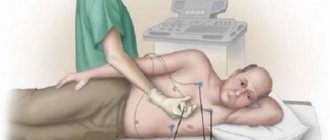
Carrying out myocardial scintigraphy
Initially, the procedure is carried out when the patient is in a calm state, and then he is offered normalized physical activity, against the background of which the work of the heart is monitored.
- Through a catheter, a small dose of a radioactive marker (thallium or technetium-labeled tetrofosmin) is injected into the patient's vein.
- This substance, along with the bloodstream, spreads throughout the body, entering the heart, where it is best adsorbed by intact myocardial cells.
- At this time, the specialist analyzes the distribution of the marker in the heart using a gamma camera that records the radiation of the radioactive substance, and the results are displayed in the form of dynamic and static images synchronized with the ECG.
- By determining the concentration of isotopes, it is possible to compare the blood supply in different parts of the heart. If the marker is actively absorbed, then there is a normal blood supply in this place, and where radionuclides are poorly absorbed, there are areas with ischemia or necrosis.
- After inserting the marker, about half an hour should pass, after which the gamma camera starts working, taking a series of pictures in which all zones are very clearly visible.
- The heart is then subjected to stress, and the procedure takes place against the background of partial decay of the radioactive element. The standard source of physical activity is a bicycle ergometer or treadmill.
- If physical stress is contraindicated for the patient, then they resort to a pharmacological option - they administer drugs that speed up and intensify heart contractions (dobutamine, dipyridamole, adenosine), while simultaneously monitoring blood pressure, pulse and taking an electrocardiogram.
- At maximum physical exertion, the marker is reinserted, and three axial scans of the heart are taken half an hour later.
If the patient feels chest pain, pressure, difficulty breathing, leg cramps, dizziness, he should tell the doctor about it right during the procedure.
The whole procedure takes quite a long time – from 2 to 4 hours. A big advantage is the non-invasiveness of scintigraphy (no tissue excision).
During physical activity, the heart needs more oxygen. Against the background of the load, blood flow begins to increase. If there are disturbances in the circulatory system, such stress will highlight the problem even more clearly.
To obtain natural stress, use a treadmill or exercise bike. When running, the intensity of the load gradually increases. All indicators will be displayed on the monitor. When maximum stress is reached, the patient is re-injected with a dose of the radioisotope substance.
If there are contraindications to physical exercise, medication is used. Drugs that imitate sports activities increase the pulse and contractions of the heart.
Based on the results of image comparison, the presence of ischemic foci in the heart is confirmed or refuted. Myocardial scintigraphy is a non-invasive method for assessing regional myocardial blood flow, myocardial function and viability.
The study consists of two parts: a heart scan at rest and a heart scan during exercise. For the purpose of visualization, an intravenous injection of a radioisotope is performed, which has the ability to accumulate in the heart muscle.
When making a comparison between images obtained during scanning at rest and during exercise, it is possible to identify the pathology of the coronary vessels, as well as determine the localization of the pathologically changed area.
This test allows risk classification, which helps assess whether a patient needs drug treatment or surgery, and also makes it possible to determine the location of myocardial infarction and its size.
For myocardial scintigraphy you need to make two recordings on different days! Please, if you are unable to attend your appointment on the appointed day, we ask that you notify us in advance (if possible the day before 17.00.)
Memo for patients undergoing examination. You should take with you to the examination:
- information about medications taken,
- preliminary test results.
The use of SPECT/CT significantly shortens the path to a correct diagnosis and selection of optimal treatment tactics with minimal radiation exposure and time costs. Procedure at rest:
- 4 hours before the scintigraphy procedure, do not eat, drink, smoke, or chew chewing gum!
- You can take your medications as usual.
The process of the procedure. The approximate time of the study is 2 – 3 hours:
- Consultation with a doctor.
- When examining the muscle at rest, isotopes are injected intravenously.
- At this time you lie down for about 10 minutes. on your back on the scanner table.
Procedure with physical activity. Before the procedure:
- If you are taking beta blocker medications, you may need to stop taking them for a while before your scintigraphy procedure. Be sure to consult your doctor.
- 4 hours before the scintigraphy procedure, do not eat, drink, smoke, or chew chewing gum!
- 12 hours before the scintigraphy procedure, do not drink coffee or eat products containing caffeine!
The process of the procedure. The approximate time of the study is 2 – 3 hours:
- Consultation with a doctor.
- The radioactive drug is administered at the very peak of physical activity.
- At this time you lie down for about 10 minutes. on your back on the scanner table.
The passage of the substance is recorded by a gamma camera.
Methodology
The radiopharmaceutical is administered intravenously. After the injection, the patient is allowed to go home for 3 hours or is asked to wait on the territory of the medical center. During this time, it is advisable to drink 1-1.5 liters of water to speed up the removal of radionuclides that are not absorbed by bone tissue. Immediately before the procedure, you need to empty your bladder.
Bone scintigraphy is performed in a sitting or lying position. The patient must remain completely still and cannot talk during the entire procedure (from 30 minutes to 1 hour). The medical staff of the diagnostic room is located in an adjacent room, from where they monitor the process and condition of the patient through a special window.
The gamma camera used for scanning is a large (50 cm in diameter) crystal that captures and records radiation across the entire surface of the patient’s body. The obtained data is processed by a special program and displayed on the radiologist’s computer monitor.
Where is scintigraphy done?
It is better to undergo scintigraphy at a medical center that specializes in radiodiagnostics.
Decoding the results
Analysis begins from the top of the heart muscle with gradual advancement to the base. When assessing deviations, they are divided into defects of a transitory or permanent nature. Compare images at rest and under load.
Emotional stress is characterized by transient defects and is not detected in a calm state. There are no constant changes, which will indicate the presence of a heart attack.
Analysis of radiopharmaceutical accumulation is done using a quantitative scale from 0 to 3 points. Such a system makes it possible to accurately determine the complexity of the violations occurring. The radioactive agent tends to accumulate only in places of active metabolic processes, that is, in tissues not affected by ischemia.
The scheme used in the analysis of scintigraphy indicators:
- photographs are studied and images are analyzed along three axes;
- do quantitative analysis;
- determine the location of defects in relation to the heart;
- assess the viability and determine the extent of damaged areas of the myocardium.
Among the factors that can distort the results of the survey are:
- increased tendency of tissues to absorb radionuclides;
- large mammary glands;
- high location of the diaphragm of a congenital nature;
- a lot of subcutaneous fat deposits.
Preparation
In order for the results of the study to be accurate, it is necessary to properly prepare for the procedure.
Experts recommend avoiding consumption of cola, chocolate and drinks that contain caffeine the day before the test.
The patient must inform the doctor about taking medications. In some cases, they should be abandoned, as the diagnostic results may be incorrect. The specialist should also be notified if there are allergic reactions to medications or food.
On this topic
- Scintigraphy
All about lung scintigraphy
- Olga Vladimirovna Khazova
- April 10, 2020
When prescribing myocardial scintigraphy to nursing mothers, if it is impossible to carry out another diagnostic method, the woman should stop breastfeeding for at least 2 days.
If a man takes Viagra and similar drugs, it is imperative to tell a doctor about it.
12 hours before the test you need to completely stop eating. In cases where the patient is diagnosed with diabetes, it is allowed to take a small amount of low-fat food.
Risks and Side Effects of Exposure
The radioactive drug Tc-99m-MIBI causes a very low radiation dose, which is the same as in conventional radiography. Your doctor will carefully weigh the risks and which test method is the most beneficial and least stressful in this particular case.
Compared to a radiocontrast agent, side effects such as allergies are extremely rare when exposed to Tc-99m-MIBI radionuclide. Bicycling itself rarely causes any complications, even in patients with heart disease.
This is important for the accuracy and completeness of the results you can achieve at the highest possible load. In only one case out of 10,000 - 20,000 examinations, the problem of cardiac arrhythmia may require treatment or a heart attack may be expected (odds of attack 1: 40,000).
If a stress test on a bicycle ergometer fails, for example, in case of orthopedic diseases, then the load can be simulated artificially using a special drug that is injected into a vein in the arm.
These drugs are also controlled in their effects. Side effects such as dizziness, palpitations, chest pain, and breathing problems are rare. With an antidote (reversing the effect of the drug) given immediately after the stress test, these side effects usually disappear immediately.
During the stress test, your stress ECG is also monitored. Of course, there is emergency equipment.
Myocardial scintigraphy: prices, addresses and online registration
Multidisciplinary medical center operating according to international quality standards. Provides consultative and diagnostic appointments for adults and children. Located 10 min. walk from Belorusskaya metro station and Mayakovskaya metro station. The clinic employs more than 100 specialists in the areas of gastroenterology, urology, gynecology, etc. It is possible to call a doctor at home. New modern equipment for ultrasound, DS (duplex scanning), X-ray, MRI, CT, PET-CT, bronchoscopy, densitometry, colposcopy, spirometry, cardiotocography (CTG), rheovasography (RVG), rheoencephalography (REG), sigmoidoscopy, 24-hour monitoring Blood pressure, 24-hour ECG monitoring (Holter).
Advantages and disadvantages
The most important advantage of scintigraphy is that thanks to it you can see how blood spreads through the myocardium. This study can take place both during physical stress and in a state of complete rest. Scintigraphy is a painless, 100% safe procedure.
The patient is prohibited from drinking tea and coffee the day before the study.
The radiopharmaceutical used for examination has no side effects, is not a contrast medium and does not cause allergic reactions. The amount of radiopharmaceuticals administered is minimal, and patient exposure to ionizing radiation is consistent with other similar studies. The only discomfort is caused by the needle prick.
But still, a few words should be said about the risks that myocardial scintigraphy carries. Let's consider the main ones:
- Pain. First, stress testing can lead to chest pain, cardiac arrhythmia and, in rare cases, heart attack. This is due not so much to the intensity of physical activity as to the patient’s cardiopathy.
- Allergy. Radiopharmaceuticals and radioactive drugs that are used during the examination can sometimes cause an allergic reaction. This occurs because the patient is highly susceptible to certain compounds or substances.
The examination begins with the injection of a special radionuclide drug into the patient’s vein.
- Dizziness and respiratory problems. Ultimately, drugs that mimic the heart's efforts during physical activity can cause side effects such as dizziness, rapid heartbeat, chest pain, and difficulty breathing.
It is important to remember that these risks are quite rare: in general, myocardial scintigraphy is a safe procedure.
It is important to remember that these risks are quite rare: in general, myocardial scintigraphy is a safe procedure.
When is scintigraphy indicated?
purpose of SG - assessment of the degree of myocardial damage due to chronic ischemia, infarction or post-infarction sclerotic changes
Myocardial perfusion scintigraphy may be recommended to the patient if it is not possible to confirm or refute the diagnosis using conventional diagnostic methods, for example, using an ECG or ultrasound of the heart. In such controversial cases, multislice cardiac tomography (MSCT) or scintigraphy is performed.
Thus, scintigraphy is indicated for the patient when it is necessary to diagnose the following diseases:
- Acute myocardial infarction,
- Previous myocardial infarction with developing post-infarction cardiosclerosis,
- Stable angina.
In addition, cardiac scintigraphy is performed to assess the effectiveness of therapy for coronary artery disease or acute infarction, as well as to establish a diagnosis of new-onset angina using stress tests with physical activity.
Thus, scintigraphy is indicated for the patient when it is necessary to diagnose the following diseases:
Reasons for incorrect research results
Sometimes it happens that the result is positive, but in fact, the reasons are completely different. This may be due to various factors. But the main reasons that lead to a false-positive result when myocardial scintigraphy is performed are:
- the patient’s large body weight is caused by obesity, since due to the large amount of fat, a poor image is obtained;
- the large size of the mammary glands, which in turn also obscure the view of the equipment;
- the diaphragm is placed too high, which also creates a barrier for viewing the picture of isot movement;
- the presence of a tendency to accumulate, which leads to the fact that the substance is poorly excreted from the body and remains with each subsequent study, distorting the result.
Cardiac studies, such as myocardial scintigram, always require attention and a very responsible attitude. Therefore, it is important to know how to prepare for it in order to get accurate results.
Source: » doctor-cardiologist.ru; beregi-serdce.com; diagnostics.ru; iserdce.ru; med-slovar.ru; prososud.ru; cardio-life.ru; nuklearnedizin-freiburg.de; infocardio.ru; serdcet.ru; serdcezdorovo.ru"
Contraindications
Myocardial scintigraphy, despite its safety, has a number of contraindications. The procedure is not prescribed in the following cases:
- Pregnancy period .
- Infectious lesions characterized by a significant increase in body temperature.
- Severe myocardial infarction
- Hypertensive crisis.
- Heart failure in the acute period.
- Diseases characterized by damage valves .
- An attack of atrial fibrillation.
The study is also not carried out in cases where the patient is diagnosed with myocarditis. Before performing the procedure, the doctor carefully examines the patient's medical history to determine the presence of contraindications.
results
To decipher the resulting images, the specialist uses specially developed computer programs and computer cards. They help to obtain more information about defects in the heart muscle.
First of all, the specialist compares images taken before and after physical activity. When studying them, it is possible to:
- Examine the heart along its axes.
- Determine the location of the pathological process.
- Determine the viability of the affected areas of the heart muscle.
- Determine the degree of accumulation of radioactive isotopes in various areas of the myocardium.
Only a highly qualified specialist can correctly decipher research data.
Possible complications
Myocardial scintigraphy is considered a completely safe procedure, even though the study uses radioactive isotopes. But the level of radiation does not exceed the radiation of the x-ray specimen. Thanks to modern equipment, it can be reduced to minimum values.
The isotopes that are introduced into the body are not dangerous to the patient’s health and are eliminated naturally within two days.
But after myocardial scintigraphy, side effects may develop, which is due to the individual sensitivity of the patient’s body to the drugs. Possible side effects include:
- Allergic reactions. Manifests itself in the form of a skin rash.
- Increased urination.
- A sharp change in blood pressure.
In exceptional cases, the development of a heart attack or life-threatening myocardial dysfunction is noted. This complication is diagnosed in 1 patient out of 30 thousand.
When using medications that mimic physical activity, side effects such as dizziness, difficulty breathing and pain in the chest area may also occur. They completely disappear after the specialist introduces a special solution that neutralizes the effect of the drugs.