Many people are interested in what this is - an enlargement of the left atrium. This means that this condition is pathological in nature, and pathology is often diagnosed in people of different ages. An increase in the size of the atrium on the left is regarded as a harbinger of quite serious diseases that are associated with the work of the heart vessels and its muscles. It is very necessary to identify this deviation in the patient as quickly as possible, and also to effectively eliminate it.
Description of the organ
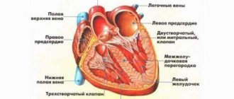
The left ventricle is a special chamber of the heart that begins the largest circle of blood circulation.
Due to the fact that this ventricle contracts, it pushes out the main amount of blood, which is supplied to various organs and, in fact, the brain, also feeding the entire heart. That is, this chamber plays an extremely important role in the full functioning of the heart, experiencing considerable stress. Therefore, it is the left atrium that begins to suffer the consequences first when any problems occur.
Possible complications
Hypertrophy triggers a whole chain of pathological reactions of the body that threaten human life:
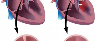
- Against the background of HLP, dilatation of the chamber cavity occurs with an increase in blood volume.
- This provokes stagnation in the pulmonary circulation, pulmonary hypertension.
- This, in turn, disrupts the functioning of the right side of the heart.
- Further insufficiency of the systemic circulation develops.
In the absence of timely treatment, all this ends in chronic heart failure with a fatal outcome.
Already at the stage of expansion of the left atrium, a person experiences a number of symptoms: shortness of breath both after exercise and without it, pain in the heart area, frequent pressure changes, angina pectoris.
Swelling of the extremities should also be a concern. In advanced cases, the patient’s breathing can be heard whistling even at a distance; it is difficult for him to perform daily work and take care of himself.
The essence of the disease
An enlargement of the cavity of the left atrium is also called hypertrophy, a disease caused by thickening of the left side of the ventricle, which causes loss of elasticity of the surface. With uneven enlargement of the septum, a person may also additionally experience impaired functioning of the mitral and aortic heart valves.
An increase in the left atrium is indicated by a thickening of the myocardium, which reaches at least 1.5 cm. This disease is called the fundamental cause of myocardial infarction and the preliminary death of young people who are very actively involved in various power loads.
Left atrial volume enlargement that is not treated in time may progress. Adapting to more difficult working conditions, cardiomyocytes begin the process of proliferation; the walls of the ventricle, located on the left side of the heart, become denser and may lose their elasticity.
The insides of the chamber do not change, and the septum, which is located between adjacent ventricles, can gradually expand, which sometimes causes disruption in the functioning of the aortic and mitral heart valves.
Due to the enlargement of the left atrium in a child and an adult, the vessels of the myocardium, which require an increased amount of oxygen, may contract.
It is noteworthy that the signs of this pathology are not always pronounced, which, of course, aggravates the general condition of the sick person. With timely treatment, even advanced disease can be successfully cured. After you have found out what it is - an enlargement of the left atrium, the signs of which are not always visible, it is worth talking about what causes them.
Description and mechanism of development
Left atrial hypertrophy (LAH) is a pathology that consists of excessive growth of the myocardium (the muscles of this chamber of the heart). This disease includes thickening of muscle tissue from 1.5 cm. HLP occurs in people of any age: it can be an adult or a small child.
The defect develops due to overload of the heart chamber with blood. At the initial stage, muscle tissue tries to adapt to new conditions and thickens, which allows the heart to function normally. If you continue to subject it to increasing intense loads, this will lead to the growth of connective tissue, dilatation (stretching of the atrium) and weakening of the chamber. The result is disruption of the left atrium and acute heart failure.
HLP occurs in two cases:
- during intense physical exercise;
- with a long course of third-party provoking diseases (for example, diseases of the cardiovascular or endocrine system).
Sometimes pathology is caused by a complex of factors. This type of hypertrophy is called mixed.
Classification
There are several types of left atrium enlargement. The disease is classified depending on the moment of its occurrence, stage of development, anatomical properties.
| Classification criterion | GLP type | Description |
| Moment of occurrence | Congenital | Associated with genetic mutations or disorders during intrauterine development of the fetus |
| Acquired | Triggered by bad habits (alcohol, smoking), third-party pathologies | |
| Stage of development | Increase in LP in compensation | It is asymptomatic. Deviations on the ECG are insignificant. No treatment is required, but specialist supervision is advisable |
| Subcompensation | Develops due to the long course of the primary disease. Characterized by nonspecific symptoms (chest pain, fatigue, tachycardia, shortness of breath). Requires therapy, possibly in a hospital | |
| Decompensation | Accompanied by pronounced symptoms from the cardiovascular and excretory systems. Requires urgent treatment in a hospital due to the high risk of death | |
| Terminal phase | Occurs 3-10 years after the onset of myocardial thickening. There is no treatment. The patient receives only palliative care. Life expectancy with this type of HLP is several weeks (less often, months) | |
| Anatomical properties | Symmetric | Uniform growth of muscle tissue. Asymmetry index – within 1.3 |
| Asymmetric | The thickening of muscle tissue is uneven. The asymmetry index is more than 1.3. Characteristic of congenital HLP |
Whatever type of left atrial hypertrophy is, its causes are different.
Causes
If a person has an enlarged left atrium, then the causes are sought in third-party diseases. The only factor that is not pathological is intense training. This does not mean that any sport is dangerous and entails this defect. We are talking specifically about exhausting activities at the limit of human capabilities. This applies, first of all, to professional athletes.
Interesting! If upon examination it turns out that the athlete’s hypertrophy is progressing, he is removed from professional sports.
Other causes of hypertrophy:
- arterial hypertension;
- atherosclerosis;
- hypertrophic cardiomyopathy;
- myocarditis;
- muscular dystrophy;
- tumors (myxomas);
- defects relating to the mitral valve;
- heart defects of various origins;
- diabetes;
- narrowing of the lumen of the aortic valve;
- advanced lung diseases;
- kidney diseases in advanced form;
- prolonged stress;
- sedentary lifestyle;
- excessive alcohol consumption;
- smoking.
Another reason for enlarged heart chambers is obesity. Often the atria become enlarged due to genetic defects. In this case, the pathology is inherited.
Symptoms
When the atrium is slightly enlarged, the patient usually does not experience any symptoms. They manifest themselves in later stages of the disease. Most often, hypertrophy is manifested by the following symptoms:
- pallor of the skin, mucous membranes;
- constant feeling of fatigue;
- pressing, burning or nagging pain in the left side of the sternum;
- labored breathing;
- change in voice timbre;
- atrial fibrillation (heart rhythm disturbance);
- disturbances in normal sleep patterns (insomnia or constant drowsiness);
- shortness of breath (first paroxysmal, then constant);
- blood pressure surges;
- frequent short fainting spells;
- cyanosis (blueness) of the nasolabial triangle.
Important! If you have at least one of the listed symptoms, you should consult a doctor.
Along with cardiac symptoms, manifestations of the underlying disease that caused the development of hypertrophy occur.
| Provoking pathology | Specific symptoms |
| Arterial hypertension | Decreased vision or flashing dots before the eyes, migraines, dizziness |
| Narrowing or blockage of the aorta | Pallor, wheezing and noises in the lungs, high pressure when measured in the upper extremities, low - in the lower extremities, migraines |
| Aortic valve insufficiency | Pale skin, dizziness or fainting, low diastolic blood pressure, pain with activity |
| Mitral valve insufficiency | Feeling of discomfort on the right side under the ribs, rapid heartbeat, cough, swelling |
| Narrowing of the lumen of the mitral valve | Swelling of the legs, cough (sometimes with hemoptysis), bright blush on the cheeks |
If hypertrophy is caused by a complex of reasons or is aggravated by a number of factors (for example, poor lifestyle, occupational diseases), the symptoms will be even more varied.
Diagnostics
The first stage of diagnosis is a general preventive examination of the patient. The doctor examines complaints and anamnestic data, measures blood pressure, feels the pulse and conducts auscultation (listens to the heart). With hypertrophy, a distinct noise will be heard, the nature of which varies depending on the primary disease.
To clarify the diagnosis, the following instrumental methods are used: chest x-ray, ECG, echocardiography. Optional: MRI, ventriculography, probing of cardiac cavities. Additionally, the attending physician prescribes blood tests (general, biochemical (detailed), for hormones), assesses the neurological status, etc. The research results are considered as a whole.
Signs on ECG
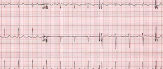
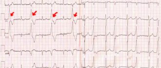
The simplest instrumental method for diagnosing HLP is electrocardiography. Based on the ECG results, the functional state of the heart structures is assessed. Left atrial hypertrophy manifests itself on the ECG with typical signs. The width, height and shape of the P wave changes. A characteristic “two-hump” appears. The QRS complex becomes wider.
Fact! An increased heart rate is recorded if an attack of tachycardia occurs at the time of the study.
Other signs of left atrial hypertrophy are determined by a cardiologist. In total, there are 10-15 deviations, which are quite difficult to decipher on your own. Only dynamic observation of indicators on the electrocardiogram will allow us to draw a clear line between GLP and overload.
Reasons for the increase
The risk of thickening of the atrium on the left increases significantly if relatives have previously been diagnosed with heart disease.
Among the main causes of slight enlargement of the left atrium, the following are very common:
- diabetes;
- overweight;
- ischemia and atherosclerosis;
- hypertension;
- heavy loads;
- peripheral vascular diseases;
- stressful situations;
- muscle dystrophy;
- aortic valve stenosis;
- Farby's disease;
- bad habits;
- mental instability;
- inactivity of a person;
- insufficient sleep.
The above factors provoke an increase in the pulsation of blood mass and an increase in the heart muscle, as well as excessive compaction of the left ventricular septum.
How to treat left atrial hypertrophy
Depending on the underlying disease, drug therapy, prevention of circulatory disorders using traditional medicine, or surgical intervention are prescribed.
Traditional therapy
The use of drugs is indicated for hypertension and secondary hypertension to unload the myocardium, slow down the frequency of contractions, and lengthen the period of relaxation (diastole) to facilitate the outflow of blood from the atrium. The following groups of drugs are used:
- beta-blockers (Concor, Betalok);
- calcium antagonists (Diacordin, Isoptin);
- ACE inhibitors (Tritace, Prestarium).
When heart failure develops, cardiac glycosides (Korglikon, Strophanthin, Digoxin) and diuretics (Lasix, Hypothiazide) are prescribed; in case of irregular contractions, antiarrhythmic drugs (Cordarone, Ritmonorm) and anticoagulants (Aspirin, Warfarin) are indicated to prevent complications. For myocarditis and rheumatic origin of heart disease, anti-inflammatory and antibacterial therapy is carried out.
Defects of valves and great vessels, abnormalities in the structure of the heart chambers, subaortic stenosis and the presence of a tumor are indications for reconstructive heart surgery.
Folk remedies
At the stage of full compensation, infusions and decoctions of plants can be recommended, which facilitate the work of the heart and strengthen the heart muscle, normalize the heart rhythm, and remove excess fluid from the body. Taking them helps stop the progression of the disease and the development of heart failure. For this purpose, herbal medicines are prepared from:
- marshweed grass;
- motherwort herbs;
- hawthorn fruit;
- mistletoe (berries, leaves and twigs);
- astragalus herbs;
- chokeberry berries.
A tablespoon of crushed plant material (one component or collection) is poured with a glass of boiling water and infused in a thermos for about 3 hours. After this, the warm infusion is drunk before meals, 1/3 cup three times a day for a month.
Diet
To prevent increased blood pressure and the appearance of edema, patients' diets are limited to 3-5 g of table salt per day and 1.5 liters of liquid (not counting first courses and drinks). The diet should be dominated by plant foods - vegetables, fruits, berries, cereals. The source of protein can be fish or chicken, cottage cheese and fermented milk drinks. Animal fats are limited, and fried and spicy foods, canned food and marinades, and alcohol are excluded from the diet.
Food intake should be small quantities 5 - 6 times a day, warm.
Allowed types of culinary processing are boiling in water or steaming, baking. Vegetable salads with herbs and vegetable oil, freshly prepared juices from berries, fruits, carrots and pumpkins are healthy.
Other reasons
The severity of this condition depends on the loads that force the myocardium to work much harder. Factors that provoke enlargement of the left ventricle include the following:
- atherosclerosis of the aortic valve;
- arterial hypertension (very common);
- glomerulonephritis.
The listed reasons can provoke the development of acquired enlargement of the left cardiac ventricle. Doctors also identify a number of hereditary anomalies, which causes the growth of the muscle layer of the left atrium:
- reduced aortic size;
- mutation of genes that control protein synthesis;
- defect of the septum between the ventricles;
- fusion of the pulmonary artery;
- aortic valve stenosis;
- mitral heart failure.
Causes of left atrial hypertrophy
Thickening of the muscular layer of the atrium occurs with a congenital anomaly of the structure of the heart, blood vessels, and can be a manifestation of acquired heart defects against the background of rheumatism. The most common causes of hypertrophy:
- narrowing of the aorta (coarctation);
- mitral, aortic valve disease (stenosis and insufficiency);
- underdevelopment (hypoplasia) of the left ventricle;
- open Botal's duct or oval window;
- change in position (transposition) of large vessels;
- persistent increase in blood pressure due to hypertension or symptomatic hypertension;
- tumors (myxomas);
- myocarditis;
- post-infarction changes - cardiosclerosis, left ventricular aneurysm;
- cardiomyopathy (especially with subaortic stenosis).
The development of myocardial hypertrophy occurs with increased load on the atrium. It is formed if an obstacle is encountered during the ejection of blood (mitral orifice stenosis), there is high pressure in the left ventricle, or there is a reverse flow of blood after contraction (with defects of the valves and vascular structure).
We recommend reading the article on myocardial hypertrophy. From it you will learn about the mechanism of development and types of myocardial hypertrophy, as well as the diagnosis and treatment of myocardial hypertrophy. And here is more information about heart dilatation.
Danger of disease
It is also worth noting what threatens an enlarged atrium (left or right). As a result, the patient experiences a malnutrition of the heart, and abnormal zones of hyperactivity are formed. According to doctors, a person may develop arrhythmia, and as a result of the increased volume of the heart muscles and impaired vascular blood flow, the risk of tissue necrosis and coronary heart disease increases. But if there is a lack of oxygen, the situation will worsen.
Left atrial hypertrophy without proper treatment can have dangerous consequences, resulting in a sharply increased load on the myocardium, even death. This especially applies to inactive and smoking people who also drink strong drinks. Myocardial infarction, ischemia, and even cardiac arrest may develop.
ECG changes
Hypertrophy is diagnosed using different methods, one of them is an electrocardiogram. Left atrial hypertrophy is not always visible on the ECG, so it is important to undergo a comprehensive examination. The method is cheap, 90% effective and reliable. The specialist uses a special device that uses electric fields to record the work of the heart.
The T, Q, P, R, S waves are distinguished. Together, the QRS segment shows how the heart muscles contract, ST and T indicate myocardial repolarization, P - what is the coverage. Overload of the left atrium on the ECG is determined by the P wave. If the left atrium, EMF increases, then the corresponding chamber of the heart is excited. If pathology is present, then the P wave shows this; on the right, excitation is not higher than normal. The P wave is elongated and bifurcated in leads I, II, aVL, V5, V6. The width of the tooth is more than 0.12 s.
Symptoms of enlargement
Over a fairly long period of time, hypertrophy of the left cardiac ventricle may not manifest itself due to compensation for the proliferation of cardiomyocytes during normal blood circulation. After you have found out what it is - an enlargement of the left atrium, it is worth talking about its obvious signs.
The first signs of thickening of the heart tissue, which should alert the patient, are not recommended to be ignored:
- increased fatigue due to increased physical activity;
- shortness of breath, breathing problems;
- periodic painful sensations (burning, pressing or squeezing) in the heart, especially after exercise;
- frequent dizziness and fainting;
- pressure surges;
- atrial fibrillation;
- feeling of a sinking heart;
- angina pectoris;
- increased swelling of the legs;
- bad dream.
Signs of left atrium enlargement depend on the level of septal compaction, thickening of the heart and myocardium. This disease may indicate interruptions and inadequate functioning of the cardiac system. Thus, the patient may not suspect a developing disease for many years. Heart failure may occur due to fainting, which occurs due to insufficient oxygen in the myocardium. Such an increase is usually a concomitant symptom of a certain disease.
Among such diseases are myocardial infarction, pulmonary edema, kidney disease, heart disease, etc. If any abnormalities are detected, it is very important to immediately contact a doctor. If treatment is not timely, there is a risk of complications resulting in death or disability.
Stages of hypertrophy
Hypertrophy of the organ in the left part can increase the mass of the right atrium. The task of both ventricles is to ensure every second, round-the-clock pumping of blood to the aorta. They must work uninterruptedly, only then will the heart fully perform its basic functions.
Increased contractions can cause increased physical activity and stressful situations. They are not dangerous and return to normal during a moment of rest or relaxation. If the increased contractions are caused by heart disease, they can lead to serious complications in the body, such as left atrial hypertrophy.
In medical practice, there are three stages of this condition:
- Formation stage. At this stage, there is a significant increase in the mass of cardiac cells, which contain a sufficient energy balance of glycogen, ATP molecules, and phosphocreatine.
- Compensation stage. Thickening of the walls of the ventricle occurs. At this stage, it can be observed that blood enters the myocardium from the deep network of the capillary system, as a result of which oxygen starvation often occurs.
- Stage of decompensation. It is characterized by the irreversibility of the processes of depletion of the heart muscle. At this stage, cell atrophy and replacement of these areas with adipose tissue are observed. This degree indicates the inability of the left ventricle to pump blood, which is the reason for the development of heart failure. This condition is especially dangerous for the patient, as it can lead to myocardial infarction.
In the compensation stage, the pumping function is performed by the myocardium, which makes it possible to consider this ability of the cardiac system to be unique. At this stage, the heart seems to stretch in length. This condition is called active dilatation.
In a child, atrial hypertrophy can compensate for various heart defects.
How to solve a problem
When detecting enlargement of the left atrium, it is necessary to accurately determine the cause of this disorder. The main thing in treatment is to normalize myocardial function and prevent possible complications as much as possible. This can be achieved through surgical or medical methods. Complex treatment consists of:
- normalization of the rest regime, improvement of lifestyle;
- weight control;
- maintaining a balanced diet;
- monitoring hormonal balance;
- moderate loads;
- minimizing stressful situations.
How is the treatment carried out?
If the underlying pathology is treated correctly, the prognosis for hypertrophy will be favorable. If the increase occurs as a result of a pathology associated with the respiratory tract, the doctor prescribes treatment taking into account the symptoms. If necessary, antiviral medications are prescribed. In particular, they are required if hypertrophy occurs against the background of a viral infection. Antibiotics are prescribed for bacterial infections.
Atrial hypertrophy is often associated with hypertension. Based on this, you need to take appropriate medications prescribed by your doctor. If hypertrophy occurs due to mitral valve insufficiency, the cardiologist will prescribe medications to support its function. In some cases, treatment is aimed at eliminating streptococcal infection: the patient can take Bicillin for 12 months. For advanced pathologies, doctors recommend surgery.
If atrial hypertrophy occurs against the background of mitral stenosis, surgical intervention cannot be avoided. Depending on the nature of the pathology, surgery is prescribed to restore the function of the valve or replace it. Treatment of hypertrophy is individual.
Treatment for enlargement
The goal of treating an enlarged left ventricle in the heart is to normalize the functioning of its muscles. First, the doctor must identify the cause of origin, as well as the existing features of the disease.
A variety of diagnostic procedures must be performed: the patient must have a complete blood test, blood pressure must be regularly monitored, an ultrasound scan, an echocardiogram and an electrocardiogram must be performed. Taking into account the data obtained, it is possible to accurately make a diagnosis, as well as determine the most optimal treatment regimen.
The basis of therapy are drugs with a negative effect, as well as those with an ionotropic effect - calcium blockers or antagonists (for example, Cardizem, Verapamil, Carvedilol, Procardia, Diltiazem). These drugs should be prescribed for life in optimal doses.
If disturbances in heart rhythm acquire serious consequences, antiarrhythmic drugs (Amiodarone, Ri, Disopyramide), as well as nitroglycein preparations, which allow dilation of the coronary heart vessels, can be prescribed.
When systolic dysfunction and dilatation of the cavities are detected, therapy for increasing the size of the left ventricle of the heart is performed according to generally accepted rules. For this, diuretics, ACE inhibitors, blockers, ongiotensin receptor antagonists, and spironolactones are used. Increased doses of ACE and saluretics usually increase obstruction.
Treatment of this condition using surgical methods is prescribed in case of ineffective drug treatment, especially in the presence of asymmetric hypertrophy of the septum between the cardiac ventricles, with clinical manifestations, severe obstruction and subaortic pressure.
Treatment methods
Depends on the root cause of the pathological condition. The point is to eliminate the factor of origin. Symptomatic measures are taken in parallel, but do not replace etiotropic effects.
- Arterial hypertension is treated with a group of pharmaceuticals. ACE inhibitors (Perindopril), calcium antagonists (Diltiazem, Verapamil), centrally acting drugs (Moxonidine), beta blockers (Carvedilol, Anaprilin), and diuretics (Veroshpiron).
They are used in combinations, but proper selection is required. Using the wrong regimen will lead to early kidney or heart failure.
- Defects of the aorta or heart valves. Surgery is indicated. Prosthetics, shunting and other methods are most widely used.
- Lung pathologies are eliminated by the use of short courses of corticosteroids, as well as bronchodilators. But with caution. Both of them affect the heart.
Symptomatic therapy:
- Antiarrhythmics to restore an adequate rhythm. Amiodarone. It is also possible to use Anapralin to relieve acute attacks of tachycardia.
- Diuretics for edema of the lower extremities and face.
- Cardioprotectors. Mildronate.
- Nitroglycerin to relieve pain.
Quitting smoking and drinking alcohol in any quantity is mandatory. Don't overexert yourself physically and emotionally.
Diet according to indications. It makes no sense to strictly limit your diet. If there are problems with lipid metabolism, statins are indicated. Sports in case of hypertrophy are contraindicated.
Performing an operation
What it is - an enlargement of the left atrium, and how to treat it with medication is clear. Surgical methods are also worth considering. Typically, transaortic septal myectomy according to Morrow is used to treat this disease. Also quite effective is the operation performed according to K. Borisov and L. Bockeria, as a result of which the surgeon eliminates the enlarged area of the left septum. An excellent alternative is septal transcatheter ablation.
These surgical techniques reduce obstruction by reducing the septum between the ventricles of the heart. Surgeons often use dual-chamber pacing, which has atrioventricular delay.
Diagnostics
Diagnostics are prescribed and monitored by a cardiologist. If the need arises, other specialists are involved in the research - a pulmonologist, a nephrologist and others. The examination is carried out on an outpatient basis, with the exception of acute conditions.
Diagnosis procedure:
- The cardiologist verbally interviews the patient and collects anamnestic data.
- The patient's blood pressure and heart rate are measured. In the presence of pathology, both indicators are above normal. Deviations are especially noticeable during acute periods.
- A doctor listens to the heart with a stethoscope. If there is HPL, noise is heard in the organ. This examination method is called auscultation.
- If listening does not allow an accurate diagnosis to be made, then the doctor refers the patient for examination, choosing the most appropriate and informative option. If there is a lack of information, additional studies are prescribed.
The task of diagnosis includes not only detecting the problem, but also determining its cause - why the left atrium is enlarged.
Diagnostic methods:
- Daily monitoring. An automatic tonometer records heart function 24 hours a day. The good thing about the method is that the fixation is carried out in natural conditions, when the patient leads a normal life, is not nervous, and performs everyday actions.
- Electrocardiography. According to the ECG schedule, the performance and functional state of all structures of the heart are assessed. The pronounced HPL on the graph is clearly visible to specialists.
- Echocardiography. This method is the main one for diagnosing LA hypertrophy. Thanks to visualization of the process, doctors determine the presence of a defect and the stage at which it is located. It is this diagnostic method that allows you to determine the cause of the pathology - Echo-CG diagnoses defects of the aorta and heart valves.
- Magnetic resonance imaging. MRI is prescribed only when necessary.
- Additional research. To have a more complete picture, the patient is prescribed a blood test - general and biochemical. You may also need an analysis of hormones, the patient’s neurological status and other studies.
As a rule, GLP is detected during preventive examinations. And if a person undergoes them regularly, then the pathology is detected in the early stages, when treatment can be quite effective.
Cardiomyopathy
This operation makes it possible to remove a more advanced hypertrophied area of the heart muscles. Often a complete or partial heart transplant is performed.
So, if the enlargement of the ventricles of the left atrium is caused by damage to the septum or heart valve, an organ transplant is performed first, which is simpler than whole heart surgery.
After performing cardiomyopathy, the patient should be monitored for life by a surgeon and cardiologist, and also take medications that will prevent thrombosis of the coronary vessels.
Symptoms
HPL is accompanied by a large number of symptoms. But their manifestation is observed only in the later stages, so the pathology goes undetected for a long time.
Symptoms:
- Heart rhythm is disturbed. Spontaneous attacks similar to tachycardia are observed. Their duration is determined by the shape and persistence of changes in the muscle layer. The attack can last up to half an hour or more. At the same time, other arrhythmic attacks can be observed - atrial fibrillation, ventricular fibrillation. With such symptoms, the risk of death is very high.
- Chest pain. It usually occurs at an advanced stage of the pathology. Appears after stress, physical exertion, suffocation due to lung diseases. The pain is pressing, pulling or burning.
- Weakness, fatigue and shortness of breath. It is difficult for the patient to walk, much less climb stairs. Every movement becomes difficult. The patient has a hard time withstanding not only physical, but also mental stress.
- Fainting. This dangerous symptom indicates complications in the brain. Ischemia of the nervous tissue begins.
- Paleness of the skin and mucous membranes. Cyanosis in the area of the nasolabial triangle. The symptom intensifies during attacks of tachycardia and acute chest pain.
A variety of symptoms creates a motley clinical picture, which is extremely difficult to understand. To identify the root causes of the pathology and make a diagnosis, it takes time, examinations and highly qualified doctors.
On a note! With advanced LA hypertrophy, shortness of breath is observed not only after physical exertion, but also at rest.