What is retinal angiosclerosis, how dangerous is it, and is it possible to get rid of it? These questions concern many hypertensive patients, since they often hear this diagnosis from an ophthalmologist. This problem is especially relevant for patients with an impressive history of the underlying disease. High blood pressure leads to pathological changes in blood vessels, and the eye area is no exception. The organ of vision is one of the first to be affected by hypertension, as a result of which the risk of impaired visual function, thrombosis and retinal dystrophy increases.
Description of the problem
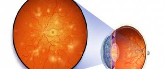
The retina is a tissue membrane located on the inside of the eye and is sensitive. Its function is to convert light signals into nerve signals, which in turn supply impulses to the brain.
The basis of the condition from nervous tissue provides a person with full vision. The structure includes ten layers: blood arteries, nerve cells, cellular tissue, etc. All this is necessary for the full functioning of the retina.
Numerous studies have shown that atherosclerosis of the eye vessels is a common problem that causes visual impairment. The disease can be caused by any part of the human body.
The most dangerous thing that can happen is retinal detachment. The process requires immediate intervention from medical personnel, otherwise the person will permanently lose his sight.
Reasons for the development of the disease
Sclerosis of the ocular vessels can occur for various reasons, among the most common are changes in the form of:
- development of arterial hypertension.
- generalized atherosclerosis.
High blood pressure primarily leads to damage to the walls of blood vessels in the human body. Ophthalmologists can detect the disease; this can be seen from changes in the blood vessels of the retina.
Another reason that can also trigger the development of the disease is high cholesterol over a long period of time. The reason for this may be deposits in the vessels; blood cannot circulate fully.
This can occur due to many factors:
| F actor | Explanation |
| Frequent overwork, lack of sleep, stress - all this negatively affects the condition of the body as a whole. |
| As a result of lack of physical activity, metabolic processes in the body are disrupted. |
| Excess body weight | Excess weight causes fat deposits and usually increases cholesterol levels. |
| Alcohol, smoking, and drugs are provocateurs for the development of vascular spasms (see Vascular spasm of the eye: symptoms and treatment - how to preserve vision?), as well as their lesions. |
| Poor nutrition and abuse of fast food are the cause of the appearance of vascular plaques and the appearance of bad cholesterol. |
With atherosclerosis, plaques form, they impair blood circulation, and oxygen does not flow in the required quantity. This is a direct path for blood clots to begin to form, blood clotting to be impaired, and pathologies of the vascular system to begin to develop. The fundus of the eye becomes vulnerable.
Interesting! Poor blood circulation leads to atrophy of retinal tissue, blood clots form, and small capillaries are affected.
Sclerosis of the fundus vessels may not be detected for many years, since the symptoms may be concomitant. Pathology can only be identified during a crisis, and it is often temporary.
Concomitant pathologies can provoke the development of the disease, among them the following can be distinguished:
- the patient has undergone surgery on the spinal cord or brain;
- there is a large vascular lesion of a sclerotic nature;
- heart disease;
- injuries, poor circulation;
- disorders in the endocrine system.
Only a doctor can understand what exactly caused the appearance of retinal sclerosis.
What is the disease and why is it dangerous?
Angiosclerosis is one of the pathologies of the vascular walls that cover the fundus of the eye. With the disease, the walls of blood vessels become dense and thickened, which leads to disruption of the blood supply to the membrane. The result is the formation of retinal edema.
Angiosclerosis is one of the stages in the progression of retinopathy in humans, characterized by damage to the retina of the eyeball. The disease is dangerous due to the fact that its further progression can cause complications - swelling of the retina, hemorrhage into it and the vitreous body. Such severe pathologies against the background of angiosclerosis are also possible, such as ischemia of the nerve endings of the retina and the formation of blood clots in the veins, as well as in the main artery.
The disease can cause a person to develop blindness. Angiosclerosis has a code according to ICD 10 under the designation H35 “Other diseases of the retina.”
Symptoms
Hemorrhage as a symptom of atherosclerosis
If we take into account that the cause of the development of the disease is systemic in nature, then two eyes are usually affected. It is almost impossible to detect symptoms at an early stage. If you can do this, you can quickly take appropriate measures.
Symptoms may include:
- periodic headaches, dizziness;
- there is acute pain in the orbital area;
- vision declines rapidly;
- general health worsens, fatigue in the eyes;
- at a late stage of the disease, double vision and blurred vision.
Due to the fact that the blood flow is slowed down, oxygen starvation occurs, the structure of the capillaries is disrupted, this is visible during diagnosis.
Severe forms of the disease are diagnosed more often. Severely impaired blood circulation behaves aggressively, the optic nerve may atrophy, hemorrhages or glaucoma may appear.
The course of development of the pathological condition
The disease does not develop suddenly. It goes through a number of successive stages, each of which is characterized by its own symptoms. It is important to understand that in the early stages the pathology can be successfully corrected, therefore, if even minor and weak signs appear, treatment must be started immediately. Angiosclerosis affects the retina of the eye, the organ responsible for adequate color perception and the formation of a three-dimensional image in the mind. Essentially, this is an inner shell that is penetrated by many tiny vessels. If their blood pressure rises sharply, characteristic problems appear, in particular, loss of visual acuity. In the most severe cases, the formation of a blood clot in the vessels of the retina is likely.
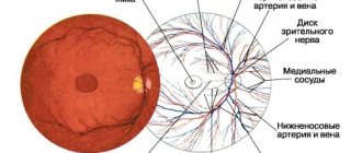
Retina, retinal veins
Ophthalmologists call two stages that pathology goes through in its development. The initial stage is easier. It is regarded as angiopathy. Sharp fluctuations in blood pressure provoke negative changes in the vessels, but their structure is not yet damaged. They may expand, which will be noticeable when examined by an ophthalmologist. The doctor will notice slight redness when examining the fundus. Such changes can affect not only the retina, but also the optic nerve. At this stage, the patient may not complain of pronounced symptoms, but the ophthalmologist will still prescribe treatment. It is at this time that the chances of normalizing the condition and preventing further development of the pathology are highest.
What does a doctor see for angiopathy?
The second stage of the disease is considered more severe. This is actually angiosclerosis. We talk about this condition if the blood vessels piercing the retina begin to intertwine. Their muscle walls undergo spasm, they narrow, and normal blood flow stops. Doctors conditionally divide the second stage of the disease into three stages.
- First stage . The lumen of the vein narrows. The nearby artery begins to put pressure on this vessel.
- Second phase . A large and deep bend of the vein is formed.
- Third stage . The bend of the vein becomes extremely pronounced. Part of this vessel becomes invisible and is visualized as a rupture.
Angiopathy
The later the pathology is detected, the worse it is for the patient. In later stages, changes become irreversible. This means that vision is lost forever and the person will never be able to see again.
Diagnostics
Example of a diagnostic event
In order to identify sclerosis, the patient must undergo a complete diagnosis. First of all, you need to study the overall picture, the symptoms.
A study is carried out, blood is given for tests in order to identify cholesterol deposits. The specialist collects information about previous diseases, heredity, etc.
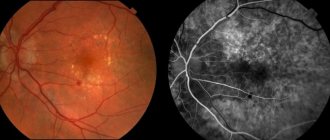
In order to determine at what stage of development the disease is, the fundus of the eye is examined and fluorescein angiography of the vascular system is performed. With the help of medications, namely tropicamine and atropine, the pupils dilate.
Thus, the research will be of high quality, but you need to prepare for the fact that your vision will deteriorate for a while and your eyes will become sensitive to light.
Fundoscopy is an examination in which the doctor looks into the back of the eye, so other pathologies can be detected. In addition, the following is carried out:
- Visometry - vision is determined in the affected area, a specialist examines the condition of the fundus.
- Computer perimetry – the periphery of the eye is examined.
- Electrophysiological diagnostics is the study of cells for their ability to “survive”.
- Tonometry - a special device measures the pressure in the eyes.
- Ultrasound diagnostics, MRI and CTM.
Thanks to numerous diagnostic methods, the presence of the following pathologies can be detected:
- the thickness of the walls of blood vessels, the presence of narrowing of the lumen in them;
- whether arteriovenous junctions are changed;
- the presence of blood clots, swelling in the optic nerve, hemorrhage;
- location of changes.
Detected atherosclerosis of the retinal vessels must be treated as soon as possible, otherwise complications cannot be avoided.
Angiosclerosis of the retina: causes, symptoms, treatment – CVZ
Angiosclerosis
- pathology of blood vessels, consisting of narrowing of their lumen and thickening of the walls. Such a picture can be found in any part of the body, including the fundus of the eye. In this case, doctors make a diagnosis that sounds like retinal angiosclerosis.
This condition is not an independent disease, but is one of the complications of systemic vascular anomalies. Against the background of sclerotic changes in blood vessels, irreversible retinal degeneration occurs. Particularly dangerous is a disorder affecting the central part of the photosensitive layer of the eye.
In this case, the patient is at risk of optic nerve dystrophy and complete blindness.
Complications
If sclerosis of the ophthalmic arteries is suspected, the patient is referred for examination to an ophthalmologist. The specialist examines the fundus of the eye, the optic nerve, and studies serious pathologies.
The disease is easily treatable at an early stage of development. It is enough to use medications; radical intervention is not required. With their help, you can stop the symptoms of pathology, prevent complications, and restore the previous functions of the eyes.
Interesting! Impaired vascular elasticity will cause optic nerve atrophy and glaucoma.
After complications appear, conservative therapy is required to stop pathological changes.
Stages of development of hypertensive retinopathy
Since retinopathy in most cases is a consequence of arterial hypertension, let us consider this disease in more detail. There are four stages of development of pathology:
- Hypertensive angiopathy . This is the initial stage of the development of retinopathy. All changes occurring during this period are reversible phenomena. At this stage, vasospasm of the retinal arteries is observed. Their thickness visually becomes smaller than that of the venous vessels. The veins expand, become tortuous, and their lumen narrows.
- Hypertensive angiosclerosis of the retina . At the second stage of retinopathy development, blood vessels lose their elasticity. As a result, the thickness of the arteries becomes heterogeneous: at some points they narrow, at others they widen. In some places, the vascular walls become denser and thicker.
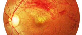
- Hypertensive angioretinopathy . Focal opacities are observed on the swollen retina. They cause a narrowing of the field and a deterioration in the quality of vision. In some places, hemorrhages occur in the retina, shaped like flames. Treatment may still be effective.
- Hypertensive neuroretinopathy . At this stage of the development of retinopathy, the optic nerve is affected and swollen. In the central part of the retina (on the macula, or macula), a pattern in the form of a half-star or star is observed.
Important! Retinopathy is dangerous because it can progress quickly and develop into neuroretinopathy within just one year. It is during this stage of development of the pathology that irreversible vision loss can occur.
Treatment methods
Atherosclerosis of the fundus vessels is treated in different ways, it all depends on the results of the examination, complications, the presence of concomitant diseases, etc. The therapy is carried out by an ophthalmologist; after studying the clinical picture, he decides whether medications need to be taken, and whether surgical intervention is required.
Atherosclerosis of eye vessels at the initial stage is treated with conservative methods. The following drugs are traditionally used:
| Drugs | Their role |
| The drugs are necessary to protect the vascular walls from ruptures. |
| Prevents spasms and improves blood circulation. |
| Normalize blood circulation, reduce the manifestation of symptoms of the disease. |
| Prevents blood clots from forming and improves blood properties. |
In order for medications to have the greatest effect, they are prescribed in the form of drops, which allows everything needed to be delivered directly to the eyes (see What to do if a blood vessel bursts in the eye: drops and other drugs that can eliminate the problem). In parallel with this, it is necessary to treat concomitant diseases.
The video in this article is an example of how the treatment is carried out.
To ensure that the drugs do not cause harm or cause side effects, you must carefully study the rules for their use; instructions are included in the package. The price of medicines varies. The dosage and course of treatment are prescribed by the doctor based on the individual characteristics of the person.
In relation to patients in whom the disease has provoked the development of serious complications, for example, retinal detachment, surgical intervention is used. Most often, laser coagulation of the retina and removal of the scorpoid body are performed.
What is retinal angiosclerosis and how to treat it
Angiosclerosis of the retina belongs to hypertensive diseases that cause organic changes in blood vessels. The pathology provokes enlargement and tortuosity of the arteries.
If the destructive process affects the retina (retina), its blood supply deteriorates and swelling develops.
The disease is also dangerous due to other complications, which include ischemia of the optic nerve, hemorrhages in the vitreous region, partial or complete loss of visual function.
Mechanism of development of angiosclerosis
The manifestation of angiosclerosis is preceded by periodic rises in blood pressure, causing narrowing of the vascular lumens and uneven thickening of the walls of blood vessels.
The pathological process worsens the condition of the retina, an important structure of the eye, the purpose of which is to conduct and transform light rays, provide color perception, and create a three-dimensional image.
The development of angiosclerosis in this part of the eyeball is dangerous due to thrombosis and defective visual perception.
Hypertensive angiosclerosis of the retina is characterized by gradual development. At the initial stage, the disease is characterized by good sensitivity to treatment and correction.
Reasons for development
The main causes of pathology include:
- arterial hypertension;
- generalized form of atherosclerosis.
There are also various factors, the presence of which significantly increases the likelihood of pathology in the retinal area. The risk of developing the disease increases if the patient has:
- injuries to the eyeballs, head;
- diabetes mellitus;
- cervical osteochondrosis;
- high intracranial pressure;
- impaired blood clotting;
- autoimmune diseases leading to inflammation of the inner choroid.
Angiosclerosis of the ocular retina tends to affect people with bad habits (alcohol, nicotine addictions) and the elderly.
Changes in retinal vessels in the presence of arterial hypertension
With a tendency to increase blood pressure, damage to the vascular walls in various body systems is observed. The fundus of the eye becomes one of the main areas experiencing the negative effects of hypertension.
Often, ophthalmologists diagnose hypertension after identifying angiosclerosis of the retinal vessels, which provokes thickening and hardening of the arterial walls.
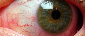
In the presence of pathology, the retinal vessels change their usual characteristics and are distinguished by the presence of a whitish reflection (the “silver wire” symptom).
The veins acquire atypical curves and move deeper into the retina. Sometimes they completely disappear from the area of intersection with the arteries.
In some cases, the walls of the arteries accumulate lipid deposits, due to which they acquire a peculiar (golden) hue.
In addition to angiosclerosis, hypertension leads to other changes affecting the vessels of the fundus - hypertensive forms of angiopathy, retinopathy, neuroretinopathy.
Retinal changes in generalized atherosclerosis
The generalized form of atherosclerosis is a vascular disease that occurs with the appearance of atherosclerotic plaques. The pathological process is associated with impaired lipid metabolism.
Generalized atherosclerosis affects various groups of blood vessels and is often chronic.
Affecting the retina, it provokes vascular angiosclerosis, hemorrhages in the vitreous body, and in the conjunctival area.
Symptoms and diagnosis
The onset of the disease often does not show any obvious signs. This feature is explained by the presence of minor changes in the blood vessels.
At first, the patient feels increased eye fatigue and sees spots, “floaters” or other inclusions in the field of vision. Such a condition is often not given due importance, and all negative phenomena are attributed to ordinary fatigue.
The presence of progressive retinal angiosclerosis is confirmed by the presence of the following symptoms:
- increased tension in the visual organs;
- nagging pain;
- blurry image;
- periodic dizziness, headaches;
- incomplete field of view;
- non-standard reaction of the pupils to lighting (their atypical dilation or narrowing).
Less common signs of pathology include memory impairment, tinnitus, problems falling asleep, and periodic nasal bleeding.
To diagnose retinal angiosclerosis, the patient is prescribed the following:
- Computed tomography.
- Ultrasound.
- Electrophysiological study (EPS).
The condition of the fundus must be examined. This procedure, called ophthalmoscopy in medicine, allows the specialist to get an idea of the type of vascular disorders and optic disc defects.
One of the most expensive diagnostic methods is retinal tomography. This method of examining the organs of vision allows you to most accurately assess the structure of all layers of the initial part of the visual analyzer, identify swelling and detect even minor pathological changes.
Treatment of retinal angiosclerosis
Pathology can be most successfully treated at an early stage of its development. The drug course prescribed for retinal angiosclerosis consists of the use of the following pharmacological products:
- eye drops that lower IOP (Fotil, Brimonidine);
- angiotensin II receptor antagonists (Candesartan, Losartan);
- beta blockers (Bisoprolol, Carvedilol);
- calcium antagonists (Amlodipine, Felodipine);
- diuretics (Hypothiazide, Indapamide).
For general toning of the visual system, special ophthalmic complexes or instillations with fortified preparations are prescribed.
To treat and prevent retina pathologies associated with generalized atherosclerosis, drugs that lower blood cholesterol are used. These include statins, represented by Atorvastatin, Mevastatin, Rosuvastatin.
In case of complications, they resort to laser therapy and injection of corticosteroids.
Hyperbaric oxygenation, a modern method based on the use of increased oxygen pressure, is considered an effective therapeutic agent.
A radical therapeutic measure is surgical intervention prescribed to patients with a complicated diagnosis.
Additionally, patients are given recommendations regarding the proper organization of their daily routine and a healthy diet.
In case of retinal angiosclerosis, physical inactivity should be avoided, salty foods that cause fluid retention in the body, swelling and an increase in blood pressure should be avoided, smoked foods, canned food, fatty meats, spicy, sweet, and baked goods.
Instead of junk food, a variety of vegetables, fruits, cereals, dietary meats and poultry should be present on the patient’s table.
Prevention of the disease
The main prevention is aimed at achieving normal blood pressure levels. For this purpose, it is recommended to adhere to a healthy lifestyle, avoid severe stress, nervous strain, and give up alcohol and cigarettes.
It is important to promptly identify and treat diseases that lead to the development of hypertension. Such pathologies include diseases affecting the nervous, cardiovascular, endocrine, and urinary systems.
To prevent sudden surges in pressure, an effective course of physiotherapy is indicated, including galvanization, electrosleep, hydrokinesis, and radon baths.
Patients who experience initial manifestations of angiosclerosis should immediately seek help from an ophthalmologist. Only through timely receipt of medical care can dangerous complications and irreversible deterioration of vision be avoided.
Source: https://oculistic.ru/bolezni/drugie-zabolevaniya/sovremennyj-podhod-k-lecheniyu-angioskleroza-setchatki-glaza
Treatment with folk remedies
In folk medicine, it is believed that vascular atherosclerosis is a condition that is easy to control and correct without the help of drugs. Doctors do not share this opinion and believe that such remedies can be a complement to the main treatment.
It is useful to include the following foods in your diet:
- parsley juice, fresh fruit or vegetables;
- dill seed tincture;
- teas made from currant leaves or rowan berries.
It is not recommended to eat fatty foods, because it is because of this that cholesterol deposits can be deposited.
It is no less useful to take herbal infusions; methods of preparing them can be as follows:
Option #1:
Take one hundred grams of St. John's wort, chamomile and immortelle, put in a container, pour half a liter of boiling water, leave for two hours. Strain the resulting product and drink it throughout the day. Complete the treatment in two weeks.
Recipe No. 2
Take twenty grams of lemon balm and valerian. Pour a glass of boiling water and leave for several hours. The course of treatment is twenty days, during this time drink a little tincture immediately before meals.
Before using alternative medicine, you should consult a specialist, as many of them may have contraindications.
The photo below shows the result of the treatment:
The result of the treatment with a favorable outcome
Prevention
There are few preventive actions against atherosclerosis of retinal vessels, but several points are important to observe:
- No bad habits. It is important to give up alcohol, smoking, and drugs.
- Proper nutrition - complete avoidance of fried and fatty foods. There should be enough fresh fruits and vegetables in the daily menu. Lean meat and seafood are healthy.
- Blood cholesterol levels should be controlled.
- For prevention purposes, it is recommended to take vitamins, homeopathic remedies, and anti-sclerotic drugs.
- Body weight control, as mentioned above, extra pounds can also negatively affect the condition of blood vessels.
- No stress, no lack of sleep.
- Blood pressure control.
Atherosclerotic damage to the ocular arteries can be stopped in time; the disease is not a death sentence. It can be corrected with medication. The prognosis depends on how early the pathology was detected and whether the patient complies with the doctor’s instructions.