What it is
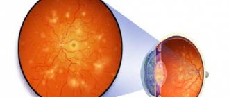
Thrombosis of the central vein is a violation of the patency of a blood vessel.
The disease progresses rapidly and often develops secondary against the background of existing complications of other diseases. Blockage in a certain area of the visual apparatus provokes backflow of blood into the capillaries and an increase in blood pressure in patients.
The degree to which the visible quality is reduced in full depends on which part of the vein is affected. If a lateral vein becomes blocked, doctors slowly but succeed in partially or completely restoring the functions of the visual apparatus.
In case of central blockage, the prognosis is the most unfavorable.
The organ atrophies. The retina undergoes degenerative changes.
More often, CVS develops in older people; it provokes a disruption of the circulatory process and a decrease in organ functions.
At the first appearance of unpleasant symptoms, you should consult a doctor, undergo diagnostics and the proposed treatment.
Classification
The disease is divided by type into:
- non-ischemic, in which the occlusion is partial, there is no hemorrhage and only a small part of the blood flow is affected;
- ischemic with complete occlusion, extensive hemorrhage and large lesions.
The disease occurs in stages:
- Prethrombosis state. The veins gradually expand, and slight stagnation appears.
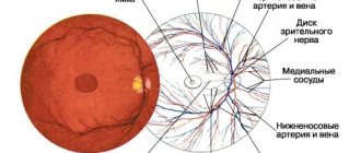
- Blood circulation is impaired, the vascular walls are tense, a yellow spot appears on the central vein and severe hemorrhage. The optic disc acquires unclear boundaries.
- The most difficult stage is when the ischemic type of disease begins to progress.
Why there are risk groups
Development may be preceded by:
- diabetes;
- hypertension;
- atherosclerosis;
- influenza, otitis with infection of the paranasal sinuses, oral cavity;
- blood pressure surges;
- high intraocular pressure or pressure exerted on the eyeball from the outside, for example, by a tumor body.
The risk group includes people:
- leading a passive lifestyle;
- suffering from obesity, cardiovascular diseases, failure of the function of the endocrine glands.
Occlusion is caused by arterial hypertension, glaucoma, cataracts with stable levels of high blood eye pressure.
Thrombosis of the eye is a blockage of the vein of the superior temporal or inferior branch of the vessel. This is the main reason for the violation.
Causes
Retinal vascular thrombosis is always a consequence of long-standing chronic eye pathology and/or systemic therapeutic diseases.
Risk factors and precursor diseases of retinal thrombosis:
- Atherosclerosis . The deposition of “harmful” lipids in the inner lining (intima) of blood vessels leads to damage to their walls. In response to this, inflammation occurs, which provokes the migration of coagulation factors to the site of damage and increased thrombus formation.
- Diabetes. This disease not only aggravates the course of atherosclerosis, but also contributes to the fragility and pathological tortuosity of blood vessels. There is even the term “diabetic retinopathy” - pathological changes in the vessels of the retina as a result of damage by structurally altered glycosylated (saturated sugars) proteins.
- Arterial hypertension . People with high blood pressure should be especially wary of retinal vascular thrombosis. Due to hypertension, the smallest vessels are damaged, blood supply is disrupted and the formation of blood clots is accelerated.
- Vasculitis - from Latin the term literally translates as “inflammation of blood vessels.” It occurs as an allergic reaction or as a result of connective tissue and blood diseases (hemorrhagic vasculitis, systemic lupus erythematosus, scleroderma, etc.).
- Protruding eyes due to long-term and persistent thyrotoxicosis . Excess thyroid hormones affect the periorbital tissue - it begins to grow. The eyeball literally “sticks out” outward. The vessels cannot keep up with it - they burst and thrombose.
- Tumors . They can grow both from eye tissue and metastasize from other organs. Sometimes a piece of tumor that gets into the vessel blocks its lumen. Read more about neoplasms of the eyelids and eyeball →
How it develops
Organ blockage is characterized by a transient course. If central occlusion is involved in the pathology, then 2/3 of the central nervous system is immediately affected.
Extensive hemorrhages begin to appear. Vision and color vision can deteriorate in a matter of hours.
If occlusion in the periphery is involved in the process, the sharpness of the image decreases slightly. However, patients begin to experience dark spots and fogs, and blurred objects.
With partial occlusion in the early stages, symptoms may be completely absent and appear only when the lumen is clearly narrowed by 80-90%.
Vision deteriorates slowly as the disease progresses. This is the only early symptom that is difficult to track.
Only as it progresses does the general well-being of patients deteriorate sharply.
Patients have no special complaints at the stage of prethrombosis, when the vision only periodically becomes blurred and acuity decreases.
At the peak of disease progression, the macular area begins to swell. The boundaries of the optic disc become unclear. The vitreous body is subject to hemorrhage.
Patients have partial loss of vision, and dark circles appear before the eyes.
Reference! Symptoms become noticeable at the progression stage, when the destructive process of CVS reaches a certain level. If the macula area is not involved in the pathology, signs may be completely absent for a long time. The disease is identified by a specialist ophthalmologist during a routine examination.
The dangerous thing is that people often ignore a slight decline in vision and do not consult a doctor. Meanwhile, with incomplete thrombosis, visual functions gradually fade away and are impaired.
Degenerative processes may become irreversible.
Symptoms
This disease passes without any special symptoms. The development of thrombosis can occur in just a few hours, manifest as deterioration or even cause complete loss of vision in one eye, and in some cases both.
With thrombosis of the branches, symptoms may manifest as:
- dark spots;
- fog in the eyes;
- distorted vision;
- sensation of pain when blinking, as if sand had gotten into the eyes.
However, more often, for example, if the center of the retina is not affected, vision is preserved and the disease can only be detected during examination. Therefore, it is so important to periodically visit an ophthalmologist, especially for patients at risk.
Why is it dangerous?
If treatment is not carried out in a timely manner, then the consequences of a blood clot in the eye are:
- optic nerve atrophy;
- glaucoma;
- macular fibrosis with the formation of a membrane against the background of collagen accumulation, which reduces the quality of the image.
The disease takes a relapsing course with severe swelling of the macula and pathological changes in the fundus of the eye.
At first glance, harmless swelling, puffiness and the periodic appearance of dark circles ultimately result in serious consequences. Degenerative processes begin to develop. Manifestations of iridocyclitis and uveitis are possible.
If the functioning of blood vessels is impaired, partial or complete loss of vision may occur.
The note! Thrombosis of the central retinal vein and its branches is in almost all cases considered an emergency condition when the decision on treatment must be made immediately. This will increase the chances of preserving the functions of the organ in case of venous narrowing, and will avoid complete blindness in case of damage to the central artery.
Signs of the disease
Symptoms completely depend on the degree of occlusion or the location of the thrombus. In thrombosis, almost the third part of the retina is involved in the pathology.
Common manifestations of the disease:
- blurry black spots, fog before the eye;
- distortion of the perception of colors, visibility and outline of objects;
- lacrimation;
- photophobia.
In most cases, patients do not complain of poor vision at an early stage.
The main symptoms of retinal vascular thrombosis are clearly expressed with complete occlusion, blocking the lumen by 96-98%.
Although this is a rare occurrence. Typically, partial occlusion is observed with a narrowing of the lumen by 60-70%.
Types of thrombosis
Depending on the degree of damage to the retinal veins, two types of thrombosis are distinguished:
- Ischemic. With this type, most of the vessels in the eyeball are damaged. A serious hemorrhage in the retina is diagnosed, and visual function is seriously reduced. Irreversible consequences may develop if treatment is not started on time.
- Not ischemic. A small area of blood vessels is affected, there is no hemorrhage, visual impairment is minimal and unnoticed by the patient.
In medicine, there is also the concept of complete and incomplete thrombosis of the central vein. The first group includes the ischemic form, the second – non-ischemic. The degree of ischemia directly affects the quality of a person’s vision.
When and who to contact
An ophthalmologist carries out diagnostic measures and prescribes treatment. If this is not available, you can first visit a therapist, who, if indicated, will refer you for consultation to an endocrinologist, neurologist, or cardiologist.
It is necessary to consult a doctor if you experience:
- partial loss of field of view;
- the appearance of black flies or circles at night and in the morning;
- frequent dizziness, headaches;
- squinting;
- impaired visibility of objects in front of you;
- extensive hemorrhages;
- decreased visual acuity.
As people age, blood vessels inevitably wear out.
Stress and mental disorders only aggravate the situation: they provoke narrowing and spasms, fragility and a decrease in their elasticity, and blood clots.
Diagnostics
An experienced doctor can quickly and easily make a diagnosis as soon as he conducts a visual examination of the fundus using a Goldmann lens.
If in doubt, he redirects to the following ophthalmological procedures:
- Visometry to identify the degree of deviation from the norm.
- Perimetry for the purpose of recognizing the boundaries of the field of view of that space, if you focus your gaze on one point.
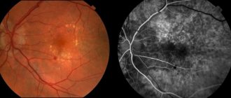
- Ophthalmoscopy to determine changes in blood vessels, the degree of hemorrhage and general condition.
- Biomicroscopy to visualize the vitreous body and determine the degree of its opacity.
- Fluorescein angiography, allowing for an accurate diagnosis.
Blood pressure indicators must be recorded, i.e. An ECG is performed.
Treatment methods
To understand how to treat eye thrombosis, the answer is step by step. Doctors take measures to:
- restoration of blood flow in the injured vascular area;
- reduction of edema;
- elimination of hemorrhages;
- normalization of blood microcirculation in the central nervous system area.
Atrophic processes develop quickly, therefore, after diagnosis, conservative therapy should be carried out immediately in order to:
- promote the resorption of hemorrhages on the eyelids;
- nourish and improve the trophism of the main components of the visual apparatus;
- relieve swelling.
Medication
Prescribed medications:
- drugs for dissolving blood clots;
- direct acting coagulants;
- hormonal drugs to relieve inflammation and swelling of the central nervous system;
- antiplatelet agents that improve blood circulation;
- vitamins, mineral supplements to improve immunity.
The disorder requires an integrated approach to therapy.
If hypoxia of the vascular wall occurs, drugs administered intravenously or glucocorticosteroids administered for edema are prescribed.
Injections of anti-inflammatory drugs are indicated to relieve inflammation and pain.
Traditional methods
Treatment with folk remedies for eye thrombosis will help significantly improve blood circulation and lower blood pressure. Infusions, decoctions, and teas are prepared from medicinal herbs and plants. Honey and beebread help prevent hemorrhage and strengthen the walls.
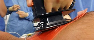
If macular edema continues to increase after treatment, then the only correct solution is laser coagulation with separation of the central zone from the affected one.
Features of symptoms
Symptoms of retinal vein occlusion develop over several hours or several days. Patients suddenly notice a dark veil before their eyes, others see distorted vision. The most important symptom is decreased visual acuity. However, severe symptoms are usually not associated with the vein occlusion itself, but are caused by macular edema.
Retinal thrombosis is painless in most cases. As the eye disease progresses, the patient becomes blind. Because symptoms usually occur in only one eye, the doctor must first determine whether the condition is an ocular thrombosis or a stroke.
The problem with the diagnosis is that in most cases the person does not experience any complaints. Therefore, in case of unclear symptoms, you should always undergo a comprehensive examination to rule out serious illnesses.
Forecasts
If you start treating the disease in a timely manner, the prognosis is positive. To consolidate the result, doctors perform laser coagulation of the retina 2 months after drug treatment. This makes it possible to reduce the likelihood of relapses.
It is especially difficult for people at risk. For hypertension and diabetes, it is important to monitor blood pressure and blood glucose levels. Periodically consult a doctor for examination.
The pathology develops at lightning speed. It is impossible to prevent its development, so preventive measures are not provided.
The violation causes irreversible changes: it reduces the quality of the visible, provokes atrophy of the optic nerve and complete blindness. If the retinal neurons begin to experience oxygen starvation, it is unlikely that it will be possible to correct the situation with medications or even surgery.
The outcome of the disease is fully influenced by time. Only timely diagnosis and proper treatment will help patients avoid the development of cataracts and partial or complete loss of vision.
Prevention measures
Retinal thrombosis can actually be prevented. You just need to undergo annual examinations and follow your doctor’s prescriptions. Methods for preventing retinal vein thrombosis depend on the presence of a specific risk factor and concomitant pathology.
- In case of hypertension, medications are needed to normalize blood pressure. There are many of them; an individual combination is selected for each patient. You should consult a cardiologist regarding the effects of specific medications.
- For all types of diabetes, the main task is to achieve a constant normal blood glucose level. This can be achieved through diet, adequate physical activity and carefully selected medications. For type 1 diabetes, you need to establish the dosage of insulin, for type 2 diabetes, the type and frequency of use of glucose-lowering drugs.
- Any eye diseases require increased attention. Under no circumstances should glaucoma be advanced. Not only does it threaten thrombosis of the blood vessels of the eye, it also leads to a complete absence of lateral vision. People with various types of retinopathy (diabetic or hypertensive) need to be checked by an ophthalmologist once every six months.
- Correction of hormone levels. If the thyroid gland is overactive, medications that reduce thyroxine levels are needed. Women are not recommended to get carried away with oral contraceptives - they increase the risk of blood clots.
- Prevention of increased aggregation (“sticking together”) of platelets - take Aspirin (ThromboASS or Plavix) daily, 1 tablet per day. This is especially necessary for those who suffer from cardiovascular diseases.
Vision is a special sense organ, without which a person loses the ability for self-care and normal social life. Patients with eye diseases should understand that thrombosis of the blood vessels of the eyes leads to irreversible changes. No operation will return or “resurrect” the retinal neurons that died as a result of oxygen starvation. It is better to start preventing retinal thrombosis right now.
Author: Maria Vasilyeva, doctor, especially for Okulist.pro