Anatomy of the heart and lungs
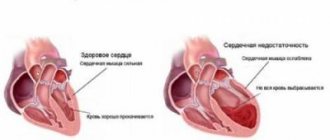
The heart is a hollow muscular organ into which blood enters from the venous vessels and is pumped into the arterial system. It is located in the chest, behind the sternum, mainly on the left. The heart wall consists of 3 layers:
- endocardium – internal lining, represented by endothelial cells;
- myocardium is the muscle layer itself, consisting of cardiomyocytes. These are special cells that ensure the non-stop functioning of the heart. There are 2 muscle layers in the atria, and three in the ventricles, due to which blood is pushed out of the cavities;
- epicardium - the outer layer, represented by the serous membrane.
The heart cavity is represented by four chambers, of which 2 are atria and 2 are ventricles. The left ventricle and left atrium are the left side of the heart - they contain arterial blood. And the right atrium and right ventricle are the right section with venous blood. These sections are separated from each other by a septum, which is thinner between the atria than between the ventricles.
The heart has leaflet valves. On the left is the bicuspid (mitral) valve between the left atrium and the left ventricle, and on the right is the tricuspid.
Blood from the ventricles enters the arteries: from the left into the aorta, and from the right into the pulmonary trunk, which then divides into the pulmonary arteries. Between the ventricles and arteries there are also valves, the so-called semilunar valves, which ensure blood flow in one direction.
The vena cavae, superior and inferior, flow into the right atrium, and the pulmonary veins into the left atrium.
There are 3 types of vessels in the body:
- arteries - vessels that carry blood from the heart;
- capillaries are the smallest vessels with thin walls, thanks to them gas exchange occurs in tissues;
- veins are vessels that carry blood to the heart.
These vessels form 2 circles of blood circulation:
- Great circle - begins in the left ventricle, then arterial blood enters the aorta and is distributed throughout the body through the arteries. They then divide into capillaries, which carry oxygen into the tissues and carbon dioxide into the blood. The blood becomes venous. Further along the venous vessels it enters the heart, through the superior and inferior vena cava. This is where the systemic circulation ends.
- Small circle (pulmonary) - begins in the right ventricle, then venous blood goes to the pulmonary arteries, from where to the pulmonary capillaries, where it receives oxygen and turns into arterial blood. Further along the pulmonary vein system it flows into the left atrium. This is how the pulmonary circulation ends.
Then from the left atrium the blood rushes into the left ventricle, from where the systemic circulation begins again.
The cardiac cycle consists of 3 stages:
- atrial systole, that is, their contraction;
- ventricular systole;
- diastole – relaxation of the heart chambers.
Lungs - structure and functions
The lungs are a paired organ located in the chest cavity and are the main organ of the respiratory system.
Functions of the lungs:
- gas exchange - enriching the blood with oxygen and removing carbon dioxide from the body;
- thermoregulation – heat production;
- excretory function - evaporation of water and volatile substances;
- protective – formation of antibodies, lysozyme, etc.;
- production of biologically active substances - histamine, thromboplatin, serotonin, blood clotting factors, etc.;
- voice formation.
Each lung has the shape of a truncated cone, the tops of which protrude beyond the collarbone, and the base is located on the diaphragm. The lungs are covered with pleura, a membrane that has two layers. One leaf lines the lungs, and the second one lines the chest cavity from the inside. Between these sheets there is a small amount of a special liquid designed to reduce friction of the lungs during the breathing phases.
Basic elements of the lungs:
- alveoli;
- bronchioles;
- bronchi.
The framework of the lungs is the bronchial tree. Lung tissue consists of lobules, each of which has the shape of a pyramid. Each lobe is approached by a bronchus, which branches into 15 - 20 bronchioles, each of which ends blindly in an acinus.
The acini is a structural unit of the lung, consisting of bronchioles with clusters of alveoli.
Pulmonary alveoli are vesicles with thin walls, around which there is a dense system of capillaries. Thanks to them, gas exchange occurs - saturation of the blood with oxygen and removal of carbon dioxide.
The main causes of cardiac asthma
Cardiac asthma is a syndrome that complicates many diseases that require emergency measures and immediate hospitalization in a medical facility.
Cardiac asthma develops as a complication of diseases and conditions associated with:
- With the cardiovascular system:
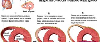
- acute left ventricular failure - a condition in which the function of the left ventricle is impaired, which leads to stagnation in the pulmonary circulation;
- decompensation of chronic heart failure - a syndrome that develops as a result of various heart diseases, appearing due to a violation of systole or diastole;
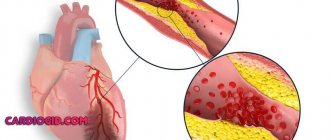
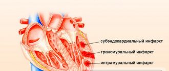
- acute myocardial infarction - a disease caused by blockage of the arteries of the heart, manifested by a violation of the contractile function of the heart;
- hypertensive crisis is an emergency condition characterized by a sharp and excessive rise in blood pressure;
- cardiosclerosis is a disease of the myocardium, manifested by the proliferation of connective tissue and the formation of scars;
- myocarditis is an inflammatory disease of the heart muscle;
- mitral stenosis is a heart defect in which there is narrowing and fusion of the cusps of the mitral valve, which is located between the left ventricle and the left atrium;
- severe aortic stenosis - a heart defect with a narrowing of the aortic opening due to fusion of its valves, which leads to disruption of the normal flow of blood from the left ventricle to the aorta;
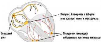
- acute arrhythmia - ventricular tachycardia, atrial fibrillation, atrial fibrillation;
- cardiac tamponade is an acute condition in which fluid accumulates between the layers of the heart sac, which leads to the impossibility of myocardial contractions due to pressure on it;
- rupture of a cardiac aneurysm is a formidable and very dangerous condition characterized by rupture of the thinned wall of the heart;
- neoplasms of the left side of the heart - various tumors;
- postpartum cardiomyopathy is causeless congestive heart failure that develops in the 3rd trimester of pregnancy or after childbirth.
- With damage to other organs:
- pneumonia - inflammation of the lungs;
- septicemia - blood poisoning;
- postoperative condition;
- bronchial asthma is a chronic lung disease in which attacks of suffocation of varying duration and frequency are observed;
- acute glomerulonephritis – immune inflammation of the kidneys with damage to the nephron, their main structural unit;
- acute cerebrovascular accident - damage to the brain due to the cessation of blood flow to one or another part of the brain;
- dependence on psychoactive substances – drug addiction, substance abuse, etc.
Reasons that increase the risk of developing cardiac asthma
- excessive physical and mental stress;
- an increase in blood volume during pregnancy, fever, or iatrogenic causes - caused by medical procedures (excessive amounts of intravenous infusions of medicinal solutions, too rapid removal of effusion in the lungs or abdominal cavity, uncontrolled therapy with glycosides, beta-blockers);
- fullness of the gastrointestinal tract (drinking and eating a lot), especially before bedtime;
- long stay in a horizontal position.
Causes
Cardiac asthma can develop against the background of cardiogenic and non-cardiogenic diseases. The cause of the development of cardiac asthma can be primary acute left ventricular failure or a chronic form in the acute stage. Cardiac asthma can complicate the course of diseases such as:
- IHD (including unstable angina , acute myocardial infarction );
- aneurysms ;
- postpartum cardiomyopathy ;
- acute myocarditis ;
- atherosclerotic cardiosclerosis ;
- post-infarction cardiosclerosis.
Potentially dangerous diseases in terms of the development of cardiac asthma are:
- arterial hypertension with congestion;
- atrial fibrillation (atrial fibrillation).
Asthma can develop against the background of decompensated cardiac defects of the mitral and aortic valves (stenosis, insufficiency), which is associated with difficulty in the outflow of blood. Changes in blood supply in the left parts of the heart can be facilitated by intracavitary neoplasms of the heart - myxomas , as well as the presence of a large intra-atrial thrombus in diameter.
Cardiac asthma can also develop against the background of acute cerebrovascular accident , damage to the renal system (acute glomerulonephritis ), infectious diseases ( pneumonia ).
Factors that can trigger an attack of cardiac asthma:
- severe emotional overload;
- inadequate physical activity;
- drinking plenty of food and fluids before bedtime;
- hypervolemia (during pregnancy , fever , fluid retention, or with intravenous administration of a large volume of fluid).
Pathogenesis
Cardiac asthma develops with an acute decrease in the contractility of the left chambers of the heart, in particular the left ventricle. At the same time, the return of the previous volumes of venous blood to the heart is maintained, but they are no longer pumped to the lungs due to damage to the ventricles. This leads to an increase in pressure in the pulmonary circulation, which, in turn, leads to the release of plasma into the surrounding lung tissue - interstitial edema.
Thus, the main function of the lungs is disrupted - the blood is less enriched with oxygen. Hypoxia occurs, the carbon dioxide content in the blood increases, which leads to excessive activity of the respiratory center in the brain. As a result, shortness of breath occurs. Cardiac asthma is the initial stage of pulmonary edema.
The second mechanism for the development of cardiac asthma is associated with an increase in the total volume of blood in the body (for example, physiological hypervolemia during pregnancy or fluid retention due to impaired renal excretory function, etc.). This increases the venous flow of blood to the heart, which leads to congestion of the pulmonary vessels, which can also lead to interstitial edema.
General information, what is cardiac asthma?
Cardiac asthma is a clinical syndrome characterized by sudden attacks of inspiratory dyspnea that progress to suffocation. Cardiac asthma refers to severe manifestations of acute left ventricular heart failure , which is a complication of diseases of the cardiovascular and other systems.
Cardiac asthma is characterized by a sharp decrease in the pumping function of the myocardium and congestion in the pulmonary circulation, which leads to acute disturbances in breathing and blood supply. Often, cardiac asthma develops before alveolar pulmonary edema , which is characterized by a fulminant course and death.
Clinical picture
Patients usually experience shortness of breath at rest, the onset or worsening of a cough several days before the attack.
Cardiac asthma itself occurs suddenly and is felt by lack of air and suffocation. The main period of manifestation is most often at night. The attack can last from several minutes to several hours.
Cardiac Asthma Clinic:
- The main and earliest symptom in patients is shortness of breath. The mechanisms of its occurrence may be:
- increased work of the respiratory muscles, because with interstitial edema of the tissue, the compliance of the lungs decreases;
- stimulation of the respiratory center in the brain due to irritation of stretch receptors, which are located in the lungs, by increasing edema;
- In addition to the lung tissue, the bronchi also swell, which leads to a decrease in their lumen and respiratory impairment.
Shortness of breath that occurs while lying down and disappears when sitting or standing is called orthopnea. The patient, who slept very peacefully, wakes up from a feeling of suffocation and strives to sit down and lower his legs from the bed or stand up to relieve the symptoms. It is this condition that is called an attack of cardiac asthma, which in medicine is designated by the term “paroxysmal nocturnal dyspnea.”
- The cough may be dry or with mucus that is difficult to clear.
- General weakness and fatigue occur due to decreased blood flow to the skeletal muscles.
- Frequent urination at night.
- Increased sweating, while the skin feels cold to the touch.
- Blue color of the skin and mucous membranes.
- Rapid pulse.
- Intense excitement, even to the point of fear of death.
- Swelling of neck veins.
- Respiration rate is more than 30 per minute.
Primary and secondary symptoms
An attack of cardiac asthma does not always occur suddenly: doctors distinguish between primary and secondary symptoms. The initial symptoms of cardiac suffocation include: difficulty breathing; feeling of heaviness in the chest; dyspnea; dry cough after physical activity (coughing attacks most often worsen when a person is in a horizontal position). Primary symptoms last from 2 to 4 days.
Secondary symptoms include:
- lack of air, suffocation;
- insomnia;
- increased sweating;
- cough with pink sputum;
- pale skin;
- gallop rhythm;
- dry wheezing;
- tachycardia.
An attack of cardiac asthma lasts from 2-3 minutes to 2-4 hours. At this time, people around should provide first aid to alleviate the suffering of the patient. If a person’s skin turns gray or dark blue, the veins in the neck are swollen, the pulse is weak during palpation, blood pressure has dropped, weakness, there is orthopnea and blood stagnation in the veins of the arms and legs, an ambulance should be called to provide qualified medical care.
Due to prolonged symptoms, the victim may develop pulmonary edema, so the patient should, after an attack and symptomatic therapy, consult a cardiologist and undergo a full examination of the body.
Risk factors and groups
- age over 60 years;
- repeated myocardial infarction with an asthmatic variant of its development, that is, the main complaint is attacks of suffocation;
- previous acute cerebrovascular accident;
- overweight;
- various chronic diseases;
- frequent exacerbation of angina pectoris.
Cardiac asthma: symptoms and treatment
Most attacks occur at night. This is facilitated by placing the body on a flat surface, for example, during sleep.
The main symptom of cardiac asthma is rapid breathing with deep inhalation and exhalation, paroxysmal cough (dry or with little sputum production). The clinical picture of cardiac asthma is complemented by difficulty breathing up to attacks of suffocation, a panic state (fear of death), pallor or blue discoloration of the skin of the face, lips, and fingers. To ease breathing, a person sits on the bed, lowering his feet to the floor, or rests his hands on the windowsill.
When listening to the patient's breathing, the specialist will not hear any altered noises, which will directly indicate cardiac rather than bronchial asthma. When signs of cardiac asthma herald the onset of pulmonary edema, the patient's breathing becomes noisy and has a wet, wheezing accompaniment.
Diagnosis of cardiac asthma
A characteristic clinical picture allows one to identify cardiac asthma.
Instrumental diagnostics
- ECG – is necessary to analyze the heart rhythm and can identify the cause of cardiac asthma;
- X-ray of the chest organs helps to assess the size of the heart and detect blood stagnation in the lungs;
- EchoCG is an ultrasound examination that allows you to analyze the function of the heart muscle.
Additional examination methods are
- coronary angiography is a research method in which a patient is injected intravenously with a radiopaque contrast agent, and under the control of an angiograph (special X-ray machine), the condition of the coronary vessels and the degree of their damage are assessed;
- catheterization of the left and right parts of the heart with measurement of pressure in the chambers and pulmonary artery - this technique allows you to evaluate end-diastolic pressure in the left ventricle, central venous pressure, filling pressure of the left ventricle, as well as stroke volume, minute volume and other hemodynamic parameters of the heart;
- radionuclide ventriculography – a radioactive drug is injected into the patient’s blood and, using special equipment (gamma camera), an image is formed where the contractile function of the heart can be assessed;
- computed tomography of the chest with contrast.
Laboratory diagnostics
- general blood analysis;
- biochemical blood test - urea, creatinine, ALAT, ASAT, potassium, sodium, blood sugar, MB-fraction of creatine phosphokinase, cardiac troponins T and I;
- general urine analysis.
Diagnostics
In this case, it is quite difficult to establish a diagnosis, since the clinical picture also points to other diseases. For example, for bronchial asthma. At the same time, there may simply not be time for diagnosis.
If possible, after a personal examination and clarification of the medical history, the patient is sent for diagnosis. The standard program includes the following studies:
- ECG;
- Ultrasound of the heart and chest;
- duplex scanning;
- radiography in 3 projections.
If these research methods are not enough to make an accurate diagnosis, then differential diagnosis is used. No self-medication is acceptable in this case.
Cardiac asthma
First aid for cardiac asthma
- Place the patient in a semi-sitting position.
- Ensure complete rest.
- Call an ambulance.
- Provide fresh air.
- Measure blood pressure and count pulse rate.
- Give 1 tablet of nitroglycerin under the tongue or nitrospray. If necessary, repeat the procedure after 3 - 5 minutes.
To reduce the cardiac return of blood to the lungs, hot foot baths and tourniquets are used on the lower extremities. In this case, venous blood accumulates in the extremities and thereby reduces blood pressure in the lungs.
Method of applying venous tourniquets:
- the patient needs to stand for 10 minutes, after which tourniquets are applied to the legs and one arm (they can be made from improvised means - tights, bandages);
- on the lower extremities it should be applied 20 centimeters from the inguinal fold. You need to place a towel or the patient’s clothing under the rubber;
- it should be placed on the arm in the upper third of the shoulder, also with a lining under the tourniquet;
- the tension force should be such that the pulse in the arteries is maintained;
- every 20 minutes you should change the position of the tourniquets on the arms and legs clockwise;
- It is important to monitor the condition of the skin below the applied tourniquet - if the skin is very pale and bluish, then the tension should be loosened.
Treatment
Cardiac asthma is a life-threatening condition and requires immediate hospitalization!
Drug treatment
- Oxygen therapy is the delivery of humidified oxygen through a nasal catheter.
- Narcotic analgesics - morphine is administered to relieve pain and shortness of breath. It causes dilation of the venous vessels and, to a lesser extent, dilation of the arteries, which leads to a decrease in pulmonary edema. Morphine also leads to a decrease in heart rate, which also has an important role in the treatment of cardiac asthma.
- Vasodilators are a group of drugs whose action is aimed at reducing congestion in the lungs.
- Nitrates cause dilation of venous vessels, but do not affect the contractile function of the heart. Nitroglycerin, nitrospray, isosorbide dinitrate are used. These drugs should be taken under strict control of blood pressure, as they can cause a pronounced decrease in blood pressure.
- Sodium nitroprusside is recommended for use in patients with severe heart failure or mitral heart disease. In case of myocardial infarction, their use is undesirable, since the manifestation of coronary “steal” syndrome is possible - this is a phenomenon when blood supply in the affected areas decreases, and in healthy areas it increases, as a result of which myocardial necrosis worsens.
- Nesiritide is a drug that causes powerful dilatation (expansion) of veins and arteries and gently increases the contractility of the heart. Used for acute decompensation of chronic heart failure.
Drugs that improve myocardial metabolism. Cardiac glycosides:
- strophanthin;
- korglucon;
- digoxin;
- Digitoxin.
Contraindications to the use of glycosides are:
- severe bradycardia - a condition characterized by a decrease in heart rate less than 50 beats per minute;
- ventricular arrhythmias - paroxysmal ventricular tachycardia - as there may be ventricular asystole (cardiac arrest);
- atrioventricular block is a violation of the conduction of electrical impulses from the atria to the ventricles, leading to heart rhythm disturbances.
Diuretics – used to treat fluid retention in the body. They simultaneously have a vasodilating effect and reduce vascular resistance in the lungs. The effect of diuretics is achieved very quickly - 5 - 30 minutes. From this group the following applies:
- furosemide - it must be used under the control of potassium and magnesium, as it flushes them out of the body;
- Torsemide is the most effective and modern drug, promoting less potassium loss than with furosemide.
Anticoagulants - indicated for patients with myocardial infarction, atrial fibrillation, etc. This group of drugs improves blood flow and prevents the formation of recurrent blood clots. Apply:
- heparin;
- low molecular weight heparins – enoxaparin, fraxiparin, dalteparin.
Surgery
Diseases that cause cardiac asthma and require surgical intervention:
- cardiogenic shock during myocardial infarction;
- ventricular septal defect after myocardial infarction;
- rupture of the wall of the left ventricle;
- aortic aneurysm or its dissection and rupture;
- acute mitral insufficiency;
- endocarditis – inflammation of the inner lining of the heart;
- acute aortic insufficiency;
- acute decompensation of cardiomyopathy.
Mechanical means of maintaining the myocardium:
- IABP - intra-aortic balloon counterpulsation - is a technique based on the mechanical injection of blood into the aorta using a special pump during diastole, which provides temporary replacement of cardiac function. Used for cardiogenic shock.
- Ventricular support devices are special pumps that are used for a short time before operations to restore heart function.
Heart transplantation is a surgical operation, the essence of which is to replace the heart of a sick person with a healthy heart.
Indications for transplantation usually arise:
- with severe acute myocarditis;
- postpartum cardiomyopathy;
- extensive myocardial infarction, when there is a poor prognosis after manipulations to restore blood supply.
Basic drugs for relieving cardiac asthma
| Group of drugs | Operating principle | Representatives | Mode of application |
| Nitrates | Dilate small vessels, reduce peripheral resistance, reduce blood flow to the heart |
| Under the tongue in the form of tablets, capsules, spray Intravenous drip |
| Sedatives | Reduce excitability Reduce tachycardia | Sibazon, Relanium | Intramuscular or intravenous |
| Narcotic analgesics | Reduces the excitability of the respiratory center, relieve pain, reduce adrenaline release |
| Subcutaneously, intramuscularly or intravenously |
| Diuretics | Reduce circulating blood volume, lower blood pressure |
| Orally, intramuscularly or intravenously |
| Antihypertensive drugs | Reduce blood pressure reduce resistance |
| Under the tongue Intramuscular or intravenous IV drip |
| Oxygen | Reduces hypoxia, reduces the formation of foamy sputum | Oxygen-air mixture Or a mixture of oxygen and alcohol vapor | Inhalation through nasal catheters |
| Cardiac glycosides | Strengthen myocardial contractility, increase stroke volume, reduce tachysystole |
| Intravenously slowly |
| Antiarrhythmic facilities | Affect polarization processes in the myocardium, relieve arrhythmia |
| Intravenous drip |
Differential diagnosis
First of all, cardiac asthma must be differentiated from bronchial asthma. An attack of bronchial asthma is characterized by difficulty in exhaling, and wheezing is also observed, which can be heard even at a distance. The sputum is viscous and difficult to separate.
Cardiac asthma is preceded by cardiovascular diseases, and with bronchial asthma - chronic bronchitis and repeated pneumonia.
Attacks in bronchial asthma are relieved by inhalers with bronchospasmodics, but in cardiac asthma they have no effect.
Tests and diagnostics
Correctly organized differential diagnosis of cardiac asthma with shortness of breath due to uremia , acute laryngeal stenosis, asthma attack due to bronchial asthma, hysterical attack and mediastinal syndrome allows for timely and high-quality medical care. Objective examination data, assessment of clinical manifestations, ECG and chest x-ray results allow us to establish an accurate diagnosis.
During an attack of cardiac asthma, auscultation is difficult due to the abundance of wheezing and respiratory sounds. However, it is possible to identify the gallop rhythm , muffled heart sounds, the accent of the second tone over the pulmonary trunk and the main signs of the disease (incompetence of the heart valves, rhythm disturbances , etc.). An increase and then a decrease in blood pressure is recorded, the pulse becomes frequent and has weak filling. When auscultating the lungs, attention is drawn to the presence of single, scattered dry or moist rales.
Prognosis for cardiac asthma
Sometimes an attack of cardiac asthma can go away on its own without any medical treatment. But even despite this, there is a high risk of cardiac asthma developing into pulmonary edema. Emergency hospitalization to a medical facility is required.
The prognosis for cardiac asthma depends on the disease that led to the development of this syndrome. In most cases, it is unfavorable, but, in general, timely treatment and compliance with medical recommendations makes it possible to prevent repeated attacks and maintain the patient’s satisfactory condition.
Resuscitation actions
There is a good chance that you may need emergency medical attention for cardiac asthma. Urgent medical measures in this case are as follows:
Place your feet in hot water
- move the patient to a sitting position, legs should hang from the bed, chair, etc.;
- place your feet in hot water, as this will ensure blood flow to the limbs;
- apply a tourniquet, but not longer than 20 minutes. The tourniquet should be 15 centimeters below the inguinal fold and always on top of the fabric.
Such assistance for cardiac asthma can save a person’s life and give doctors time to carry out the necessary medical measures.
Prevention
Prevention of cardiac asthma consists in the treatment and prevention of diseases leading to the development of this condition: treatment of coronary heart disease, chronic heart failure, arterial hypertension, kidney disease.
General recommendations:
- normalization of work and rest regimes;
- moderate amount of physical activity - daily walking for 30 minutes is best;
- limitation from stress and nervous fatigue;
- to give up smoking;
- daily monitoring of blood pressure and pulse;
- following a diet aimed at reducing salt intake, as well as limiting fluid intake and heavy foods.