© Author: A. Olesya Valerievna, candidate of medical sciences, practicing physician, teacher at a medical university, especially for SosudInfo.ru (about the authors)
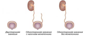
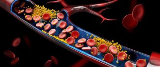
Brain angioma is a benign tumor-like process consisting of blood or lymphatic vessels. In the first case it will be called hemangioma, in the second - lymphangioma. Hemangiomas are much more common than tumors from lymphatic vessels, and the consequences from them can be fatal, so tumors from blood vessels are much more important clinically.
Externally, angioma looks like an interlacing of vessels of various diameters and structures, usually spherical in shape. The source of its formation can be both arteries and veins with capillaries, but almost always they have an abnormal wall structure, making the tumor dangerous in terms of rupture of its components.
The size of hemangiomas of the right or left hemisphere of the brain or cerebellum varies from a few millimeters to a centimeter or more. The larger the tumor, the more dangerous it is for its carrier, since it contributes to compression of the nervous tissue, and when it ruptures, a large focus of hemorrhage occurs, which can lead to death in a matter of days and even hours.
An angioma of the brain can be detected in any part of the brain - on the surface of the soft shell of the convexital (outer) surfaces of the brain, at its base, inside in the deep parts of the hemispheres, in the cerebellum. The deeper the tumor is located, the more difficult it is to reach it for surgical removal, however, modern methods of endovascular interventions make it possible to eliminate even the most inaccessible tumors.
Brain angioma often does not give any symptoms, and then its owner has no reason to look for pathology. In such cases, a sudden tumor rupture literally takes you by surprise, and the diagnosis of the disease occurs after the fact, when the patient requires intensive care or urgent surgery.
In other cases, vascular neoplasia may be accompanied by headaches and other symptoms that should prompt you to see a doctor for diagnosis. Timely detection and elimination of the tumor reduces the risk of its rupture, severe complications and death to zero.
Open operations with craniotomy to remove hemangiomas are becoming a thing of the past, gradually giving way to minimally invasive, gentle techniques that minimize the risk of brain injury, shorten rehabilitation and are well tolerated by patients. If the surgeon insists on such treatment, then it should not be postponed or abandoned altogether , however, it is important that the specialist performing the operation is sufficiently experienced and qualified.
What is cerebral venous angioma
The disease is not aggressive, but its presence should not be ignored. The interweaving of blood vessels are fused balls that expand over time, provoking the development of inflammatory processes in the brain.
The more the brain is affected, the greater the health risk caused by the presence of the formation. This pathology is dangerous for the body due to the rapid growth of angioma, which, affecting the tissues of nearby organs, leads to disruptions in the functioning of the entire body.
It is the brain that is responsible for the quality functioning of all systems. Pathological changes occur and, if not treated in a timely manner, destruction occurs. A patient with a tumor is at high risk of cerebral hemorrhage, which can be fatal.
At the first symptoms of the disease, it is necessary to begin treatment immediately. Therapy in the early stages of tumor development has a favorable outcome. At an advanced stage of the disease, drug and surgical interventions will not give positive results.
What it is. Reasons for appearance
The causes of brain cavernoma have not been fully elucidated by scientists and doctors. Most experts are inclined to believe that this disease is congenital. The impetus for the formation of vascular malformation in the fetus is a viral infection in the mother in the first trimester of pregnancy.
At this time, the circulatory system of the embryo is formed; viruses can provoke necrosis of vascular tissue and the appearance of cavities and vascular tangles.
The following are also considered unfavorable factors:
- premature or difficult birth with injuries;
- late or multiple pregnancy;
- violation of the integrity of the placenta;
- bad habits and illnesses of the mother during pregnancy.
Cavernomas are not exclusively a congenital pathology. Such formations can occur at any period of life. Sometimes they are caused by traumatic brain injuries; there are also suggestions that their development is facilitated by radiation, infections, inflammatory processes, and impaired immune status.
The term "angioma" refers to all vascular tumors consisting of lymphatic spaces or blood vessels. Hemangioma is considered a benign neoplasm consisting of blood endothelial cells.
Cavernoma or cavernous hemangioma is one of the types of disease characterized by a lumpy, bluish-colored surface with cavities (cavities) of a regular round shape. The cavities can be empty or filled with blood, blood clots, or scars. Along with single cavities, multiple ones occur in 10-15% of cases.
Such abnormal formations are prone to frequent hemorrhages, which is why the brain tissue located next to them has a yellow tint. The partitions between the cavernous cavities consist of dense fibrous tissue.
Cavernous hemangioma of the brain begins due to vascular pathologies in the womb, when for some reason the formation of areas of the nervous system is disrupted. Provoking factors can be:
- Difficult, protracted labor.
- Multiple pregnancy.
- Birth injuries.
- Premature birth.
- Severe intoxication.
- Late pregnancy.
- Placental pathologies.
- Infectious diseases of the mother during pregnancy.
- Smoking, addiction to alcohol or drugs during pregnancy.
- Difficult environmental situation.
Research has shown that the disease is not hereditary.
Kinds
Venous angioma is an enlarged venous column of vessels collected in a tangle. Depending on the location of the formations, the disease is divided into several types.
Frontal lobe angioma
She is responsible for initiative, correct decision-making, responsibility and showing interest. Therefore, when education is localized in this area, the character of a person changes greatly.
He becomes apathetic, a change in self-esteem occurs, speech is impaired, and inadequacy appears in his behavior and actions.
It is difficult for a person to think logically; he often commits unconscious actions. The patient cannot take responsibility and is unable to independently solve various problems.
Performance decreases, the emotional state is unstable, and over time the person suffers from depression. The patient does not feel cold or heat when touched.
Cerebellar angioma
If there is a tumor in this area of the brain, functional disturbances occur in the body. The patient's muscle movements are not coordinated, he cannot perform smooth movements, so all actions are abrupt. A person’s motor ability is greatly slowed down, all actions are performed in the form of jerks.
There are disturbances in speech, there are serious problems when writing words and sentences. Breathing is impaired, resulting in problems with the respiratory system and skeletal muscles.
It is impossible to cure the disease using medications. With this form of angioma, surgical intervention is indicated.
Angioma of the left hemisphere
There is inconsistency in the work of the muscles of the arms and legs. The patient has speech impairment and changes in gait.
On this topic
- central nervous system
How does a headache with a brain tumor hurt?
- Natalya Gennadievna Butsyk
- December 3, 2020
Taste preferences change completely. Vision decreases, nystagmus of the extraocular muscles is observed.
Due to impaired blood flow, a person is susceptible to epileptic seizures and convulsions. At an advanced stage of the disease, partial paralysis may occur.
Angioma of the right hemisphere
With this type of disease, the patient experiences a movement disorder. It becomes slow and sharp. Speech becomes slow, writing style changes. A person is bothered by tremors of the arms and legs.
Angioma of the parietal lobe
The patient's coordination of movements is impaired, tactile sensations and pain threshold change, and speech abilities may deteriorate.
Origin of cavernous angioma
Cavernous hemangioma is a type of infantile hemangioma. It occurs in the vast majority of cases in children and is congenital, but not hereditary. It is a pathological proliferation of blood vessels that form cavities (cavities) filled with blood. Under a microscope, overgrown vessels are visible, chaotically woven into knots.
The disease is benign in nature, although it grows rapidly and grows into surrounding tissues. It is rare - it accounts for 7% of vascular tumors according to WHO observations. The ICD-10 code is D18.0.
The exact causes of the disease are unknown. It is assumed that in the first trimester of pregnancy there is a disruption in the formation of the fetal vascular system. Predisposing factors: prematurity, pregnancy pathology, multiple pregnancies, viral diseases, bad habits and other factors that negatively affect the condition of the unborn child.
This type of tumor also occurs in adults, but is extremely rare. Reasons: disruption of blood vessels due to diseases of internal organs, toxic effects of certain drugs, radiation, excessive exposure to UV rays, poor ecology.
Liver hemangioma is a mysterious disease; the reasons for the development and growth of tumors have not yet been precisely established. Cavernous hemangioma occurs in approximately 2% of the population, with men affected less frequently than women. The disease can occur at absolutely any age; as a rule, it is diagnosed in patients 30-50 years old.
The disease is characterized by the proliferation of vascular walls with the formation of cavities (cavities). In fact, hemangioma is a benign formation, but it necessarily requires observation and, if necessary, timely and high-quality treatment.
Cavernous hemangioma does not pose a threat to life; if it is formed near physiological openings and on the mucous membranes, it can cause a lot of discomfort. When a hemangioma grows near the external auditory canal or eyes, there is a risk of deafness and blindness.
Cavernous hemangioma is a benign tumor in the liver. Symptoms are absent in most patients, often becoming obvious only when the tumor increases significantly. The incidence of the disease is approximately 7% of the population. More often, the pathological process is found in women.
The liver is an organ that lacks nerve endings.
What then worries people who, when they come to see a doctor, complain of pain under the ribs on the right side? Often one of the causes of pain in the right hypochondrium is cavernous hemangioma of the liver, which poses a serious threat not only to the organ, but also to health in general. Everything about the causes, symptoms and features of therapy is in this material.
There are two types of this disease: hereditary or sporadic.
- The mechanism of the congenital type of the disease is considered to be more studied, but there is also evidence of an autosomal dominant nature of inheritance; characteristic genes of the seventh chromosome have been identified and established, which affect the formation of pathological vascular ligaments
- The causes of sporadic tumors are still unknown. There are three scientific theories: the result of radiation, inflammation of the immune system, infection.
Localization of angiomas:
- 80% of angiomas are located in the upper lobes of the central brain. 65% of their component falls on the frontal, temporal and posterior parts. 15% on vascular occurrences of optical convexity.
- 8% are neocerebellum hemangioma.
- 2.5% vascular configurations in the dorsal medulla.
Detection is determined by the location of the tumor. The first signs are: migraine, not eliminated by medications; convulsions; sounds in the ears; disorganization of coordination; bouts of vomiting and nausea; weakness of the limbs; degradation of hearing and vision; memory distortion; disorder in speech. It happens that the angioma is “silent”. People don't even know about the disease. The tumor is detected when tested for other diseases or when it manifests itself in other family members.
Treatment of cavernoma should begin with a comprehensive diagnosis according to a regimen prescribed by doctors, which is created personally for the patient.
Symptoms
In addition to the differences between different types of angiomas, there are common symptoms that are characteristic of all types of cerebral venous angioma:
- frequent fainting;
- dizziness;
- paralysis;
- noise in the head;
- tremor of hands and feet;
- disorders ;
- headache ;
- imbalance and coordination of movements ;
- disturbances in the functioning of the cardiovascular system;
- epilepsy attacks
- problems ;
- convulsions;
- disorders ;
- blurred vision;
- decreased mental activity;
- noise in the head;
- change in taste preferences;
- impairment ;
- increased fatigue;
- nausea and vomiting;
- loss of ability to work.
How does a vascular tumor form?
More often, angiomas are formed by purely venous channels. But the process can also affect the arterial network. A normal vasculature connects arteries and veins through extensive branching of capillaries.
In 5% of cases of disease, the arterial part is directly connected to the vein, it turns out that the affected vessel “steals” other branches and disrupts the general blood flow in the area of location, and each tumor is an arteriovenous fistula
Pathology occurs in the late stage of formation of cerebral vessels in the fetus, from 40 to 90 days of pregnancy.
Forming angiomas have rays, so they are compared to a bicycle wheel. Due to increased filling, thinning of the vein walls occurs. The pressure in them increases and bleeding occurs (red blood cells leak out) if the tumor is located on the surface of the body. But with a deep location in the substance of the brain, hemorrhage occurs in the surrounding tissues.
Diagnostics
To perform an accurate analysis, a specialist carries out diagnostic measures.
First of all, laboratory tests are prescribed. The patient must undergo a general and biochemical blood test, as well as a general and biochemical urine test.
If the norm deviates, the patient is then prescribed an ultrasound examination to confirm the diagnosis. Magnetic resonance imaging will determine the presence of formations, identify the location and size of the angioma.
It is also possible to perform a computed tomography, angiography or x-ray. Only after all diagnostic procedures have been completed can treatment be prescribed.
Diagnostic methods and criteria
An appeal to a neurologist with complaints of systemic headaches, nausea, sensory disturbances is the reason for MRI and contrast angiography.
Sometimes we are talking about a neoplasm that is significant in size and significantly affects the performance of the brain. A tumor of this type can be detected in the middle and late stages by performing a non-routine CT or MRI of the head with contrast.
Modern diagnostic methods make it possible to accurately determine the location, size of the tumor, and type of vascular formation.
Venous angioma on MRI
Venous angioma of the right frontal lobe is indicated in the photo by an arrow
Treatment
Treatment of education includes taking medications, using traditional medicine, and in advanced cases, surgery.
Drug treatment
It is impossible to cure venous angioma by taking medications. This type of therapy is used only to alleviate the patient’s condition.
Drug therapy is prescribed if there is no threat of hemorrhage. To improve blood circulation, vascular drugs are indicated: Cerebrolysin, Cavinton, Bravinton, Vinpocetine, Telektol.
To improve the functions of cerebral vessels, the following drugs are effective: Actovegin, Mexidol, Emoxipin. Aspirin and Heparin are used to thin the blood. To increase blood supply to the brain, Tanakan and Bilobil are indicated.
On this topic
- central nervous system
What are the dangers of a porencephalic cyst of the brain?
- Olga Vladimirovna Khazova
- May 27, 2020
Sedatives occupy an important place. The most effective are: Persil, Sedavit, Fenozepam, valerian tincture, Persen, Tenoten, Deprim.
To eliminate headaches, drugs such as Nurofen, Tramadol, Ketanov, Diclofenac are indicated.
All drugs will only alleviate the patient’s condition, but will not contribute to recovery. A benign formation can only be removed surgically.
Surgical intervention
There are several methods of surgical treatment of the disease.
In severe cases of the disease, surgical removal of the accumulation of blood vessels is performed. A plastic spiral is inserted into the patient's lumen.
If the sclerotherapy method is chosen, a special substance is injected using a special catheter, which clogs the lumens of the vascular bundle and promotes scarring of the tumor. The operation is painful and lengthy.
It is possible to apply a superficial radiation beam to the tumor using a gamma knife. With its help, blood vessels are blocked. Education stops growing and developing, no longer presenting a danger to human life and health.
Liquid embolism can be injected into the collection of vessels, which easily penetrates into the smallest cavities and prevents the growth of angioma. Over time, the area where the tumor grew is replaced by connective tissue.
Diet
Diet plays a big role in treating the disease. The patient should exclude fatty and fried foods from his diet. Minimize the consumption of salt and sugar, coffee, chocolate and baked goods.
Avoid eating butter, pork, full-fat milk, liver and kidneys. It is necessary to eat vegetables and fruits, fish and seafood, cereals, dried fruits, and herbs.
Brain angioma is a serious disease. Therefore, it is strictly forbidden to self-medicate.
Drug therapy
It is impossible to get rid of angioma with the help of medications. Medicines can only eliminate the unpleasant symptoms of the disease. Treatment can be carried out using the following means:
- hormonal and painkillers;
- sedative and homeopathic;
- cytostatic and glucocorticosteroids.
They must be used if the tumor rapidly increases in size or if several tumors have been detected at once, affecting different parts of the brain. To normalize the patient's condition and prevent complications, Prednisolone, Sherizolon, Busulfan, Epirubicin and other drugs are prescribed. They can only be taken under the supervision of a specialist.
Attacks can be stopped with the help of homeopathic medicines, such as Lycopodin.
Can it develop into cancer?
Venous angioma of the brain is a benign formation. Therefore, it does not degenerate into a cancerous tumor.
On this topic
- central nervous system
Everything you need to know about pituitary adenoma
- Olga Vladimirovna Khazova
- February 28, 2020
Over time, it can lead to negative consequences and threaten human life. Therefore, it is important to consult a doctor in a timely manner for diagnosis and adequate treatment.
If the tumor does not manifest itself in any way, the person does not have behavioral disturbances or deterioration in health, you can live with this type of disease until old age. But in most cases, when diagnosing a tumor, surgical intervention is indicated.
Causes and types of vascular formations of the brain
The reasons why brain angiomas appear have not been thoroughly studied. A certain role is given to external adverse influences, the influence of genetic mutations, hereditary predisposition to angiodysplasia and disruption of the formation of connective tissue is high.
Most often, angiomas begin to form at the stage of intrauterine development of the vascular system of the brain, that is, they are congenital in nature, but the pathology can become known after years and decades of life. Congenital angiomas of the brain can be combined with other malformations of internal organs and genetic syndromes.
It is believed that the basis for the formation of angioma is a violation of the maturation of the vascular bed of the brain, when the arteries do not branch into smaller vessels, but form a tangle from which blood flows directly into the veins. Blood shunting leads to local hypoxia and a kind of theft of certain areas of nervous tissue due to a lack of capillary network. Thus, not only brain compression, but also local ischemia underlie the manifestations of the pathology.
Acquired vascular brain tumors account for only about 5% of cases. Among their causes are infectious pathology, in particular HIV infection, as well as severe craniocerebral injuries, the healing of which is accompanied by the formation of new blood vessels, the excessive growth of which can give rise to neoplasia.
The classification of brain angiomas can be based on a causative factor:
- Tumors as congenital defects;
- Acquired neoplasms.
Based on the number of tumor nodes, they are distinguished:
- Isolated solitary tumor of the left or right half of the brain, its base, cerebellum;
- Multiple angiomas (angiomatosis).
The structural features of the vascular walls of the neoplasm suggest the identification of types of hemangioma:
- Capillary - tumor vessels are similar in structure to the smallest blood vessels;
- Venous - consists of vein-like vessels with a relatively thin wall and wide lumens;
- Arterial - a tangle of vessels of an arterial structure with a well-defined muscular component;
- Cavernous - formed by many vascular thin-walled cavities of significant volume, where thrombus formation often occurs.
different types of angiomas
Abnormally developed vascular walls carry signs of tissue atypism - a violation of the ratio of cellular and stromal components, changes in the diameter of blood vessels, uneven thickness of layers, underdevelopment of structural elements, etc.
The presence of angioma changes blood flow towards its deficiency, resulting in local hypoxia and neurological symptoms. Compression of nervous tissue by a large tumor is manifested by focal symptoms, depending on the location of the hemangioma.
The most dangerous is considered to be cavernous angioma of the brain, the risk of hemorrhage in which is quite high. Cavernous vascular formations have thin walls that are easily torn from a variety of influences - increased blood pressure, excessive anxiety, physical overload, alcohol consumption. According to some reports, every third cavernous angioma ruptures sooner or later, and mortality and disability from complications remain high.
Venous angioma of the brain is represented by vessels of a venous structure with a poorly formed middle muscle layer and a relatively large diameter. It is not prone to rupture, since the blood pressure in it is not as high as in the arterial bed, but it can manifest itself as persistent cranialgia, weakness, and fatigue.
Based on the location of brain angiomas, neoplasms of the cerebellum, left or right frontal, temporal lobe, vertex, and base of the brain are distinguished. Depending on the location, the symptoms manifest themselves in their own way, which is associated with compression of specific nerve structures, and the tumors share a number of common signs of intracranial formation.
Forecast
An intact angioma may not be detected throughout life and may not cause serious trouble to the patient.
But if a hemorrhagic stroke or vasospasm occurs, the person may become disabled for life or die. Often, rupture of the vascular bundle leads to irreversible processes in brain activity.
Depending on the patient’s age, his general health, and the presence of vascular diseases, a prognosis for the course of the disease is made. A properly prescribed treatment regimen and timely surgical intervention will allow the patient to lead a healthy and fulfilling life for a long time.
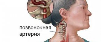
Symptoms
Symptoms are often caused by overload of the venous system. Impaired blood outflow occurs due to obstruction (blockage) of the duct or narrowing of the lumen as a result of mechanical compression, for example, with hydrocephalus. An angioma formed from the vessels of the venous system of the brain usually manifests itself with symptoms:
- Pain, noise and heaviness in the head area, dizziness.
- Attacks of nausea, often ending in vomiting.
- Epileptic, convulsive seizures.
- Darkness and loss of consciousness.
- Impaired motor coordination.
- Visual dysfunction.
- Deterioration of cognitive abilities.
The deterioration of the neurological status can be provoked by factors: stress, physical and mental fatigue, arterial and cerebral hypertension, progressive atherosclerotic lesions of the vascular wall.