The heart begins to beat long before we are born. Surprising and even magical, right? However, this means that this organ works longer and harder than others. And not only the quality of our life, but also life itself depends on how carefully we pay attention to his condition!
More than 17 million people die every year from heart and vascular diseases. It has been proven that 80% of premature heart attacks and strokes could be prevented!
Fortunately, most cardiovascular diseases are successfully diagnosed thanks to modern innovative equipment and the professionalism of doctors. And as you know, establishing an accurate diagnosis means taking an important step towards healing.
One of the most informative and safe studies is cardiac echocardiography (EchoCG) or, in other words, ultrasound of the heart.
1 ECHO-KG in "MedicCity"
2 Echocardiography at MedicCity
3 Ultrasound of the heart at MedicCity
The essence and purpose of echocardiography
Echocardiography or echocardiography is a non-invasive method of examining the heart using ultrasound. The echocardiograph sensor emits a special high-frequency sound that passes through the tissues of the heart, is reflected from them, and is then recorded by the same sensor. The information is transmitted to a computer, which processes the received data and displays it on the monitor in the form of an image.
Content:
- The essence and purpose of echocardiography
- Benefits of EchoCG
- Indications and contraindications for Echo-CG
- Types of echocardiography
- Preparation for echo-CG
- Research methodology
- Decoding the results
- Where to get an echocardiogram
- Echocardiography in children
- Preparing and performing the procedure for children
- Fetal echocardiography
Echocardiography is considered a highly informative research method, since it makes it possible to assess the morphological and functional state of the heart. Using this procedure, it is possible to determine the size of the heart and the thickness of the myocardium, check their integrity and structure, determine the size of the cavities of the ventricles and atria, find out whether the contractility of the heart muscle is normal, find out about the condition of the valvular apparatus of the heart, examine the aorta and pulmonary artery. This procedure also allows you to check the level of pressure in the structures of the heart, find out the direction and speed of blood movement in the heart chambers and find out the condition of the outer lining of the heart muscle.
This cardiological examination allows you to diagnose both congenital and acquired heart defects, find out about the presence of free fluid in the heart sac, identify blood clots, changes in the size of the chambers, thickening or thinning of their walls, detect tumors and any disturbances in the direction and speed of blood flow.
What is the essence of the method
There is an innovative, more important and reliable method of examining the heart - transesophageal echocardiography. Emergency ECHO KG – what is it? This is a relatively new method in diagnosing diseases of the cardiovascular system. At its core, it is an invasive ultrasound method for detecting heart disease.
Invasive, that is, associated with passage into the internal environment of the body through external barriers or through natural means.
In this case, the sensor is located not on the surface of the chest, but inside the esophagus. This procedure can be performed on patients of any age category, since the examination does not involve any radiation exposure. By performing an ultrasound of the heart through the esophagus, it is possible to determine the size and thickness of the heart cavities and assess the condition of the valve apparatus.
Carrying out a transthoracic (through the chest) ultrasound examination is less dangerous and the patient does not experience discomfort. But due to the presence of certain interference when performing this type of ultrasound of the heart (excessively developed subcutaneous fat layer, muscles, ribs, lung tissue), the examination results are often distorted, which leads to diagnostic errors.
Transesophageal ECHO CG must be performed by a functional or ultrasound diagnostics physician with a high qualification category. The use of this examination method is associated with the availability of special equipment. Before the procedure is scheduled, the patient must undergo a routine ultrasound examination of the heart through the chest.
One of the advantages of transesophageal echocardiography is the ability to monitor organ function during heart surgery, which does not interfere with doctors during surgery. The presence of special handles on the endoscope sensor, which can be rotated in different directions, significantly increases the inspection area.
Benefits of EchoCG
Echocardiography has a number of advantages over other types of cardiac examination.
First of all, this is an absolutely painless and non-invasive procedure that does not cause any discomfort to the patient. It is performed like a regular ultrasound. No injections or any other similar manipulations are performed before the procedure.
In addition, the procedure is completely safe for patients of any age group. It can be performed on children, adolescents, and pregnant women, since ultrasound does not have any negative effect on the fetus.
EchoCG is accessible, since the equipment for its implementation is available in almost any medical institution. The cost of echocardiography is much lower compared to MRI.
And the most important advantage of this type of examination is its excellent information content, which will allow the doctor to obtain the maximum necessary information and choose the right therapy.
Advantages
The most common method for identifying cardiac problems is an electrocardiogram (ECG), but it only shows a graphical record of heart activity, while the use of transesophageal echocardiography allows you to study the structure of the organ.
Systematic electrocardiogram examination is recommended even for healthy people
The use of transthoracic echocardiography (Echo CG) also has good information content and is widely used in medical practice to determine most heart diseases. The examination is safe for the body and does not cause discomfort to the patient due to the location of the ultrasound sensor on the surface of the body during the examination.
However, the occurrence of obstacles to ultrasound waves in the form of subcutaneous fat, ribs, muscle tissue, and lungs can significantly distort the results of transthoracic echocardiography and lead to errors.
Studying the heart through the esophagus has some advantages compared to Echo CG:
- the location of the sensor in close proximity to the heart during transesophageal echocardiological examination leads to obtaining a much better image than with EchoCG and, as a result, to greater information content;
- the presence of anatomical features of the patient’s body (close proximity of the ribs, excess weight, etc.) does not cause obstacles to his examination;
- the use of transesophageal echocardiography allows you to monitor the work of the heart during surgical interventions without interfering with the actions of doctors.
Cardiological studies also include transesophageal pacing (TEPS), which allows specialists to record the biocurrents of the heart.
Indications and contraindications for EchoCG
Echocardiography may be recommended to patients both if the doctor suspects they have any cardiovascular pathology, and during therapy to assess the effectiveness of the drugs used.
Indications for echocardiography are:
- Hypertension.
- Suspicion of the presence of congenital or acquired heart disease, including with a hereditary predisposition to this disease.
- Frequent dizziness, fainting, shortness of breath and swelling.
- Complaints about a “fading” heart, about “interruptions” in its work.
- Pain behind the sternum, especially if it radiates to the area of the left shoulder blade or the left half of the neck.
- Myocardial infarction, diagnosis of angina and cardiomyopathy, suspected heart tumor.
- Preventive examination of patients who often experience emotional and physical overload.
- Changes on the ECG and chest x-ray that require clarification of morphological changes in the heart.
It is worth mentioning separately in what cases echocardiography is recommended for expectant mothers. Pregnant women should undergo echocardiography if:
- The expectant mother has pain in the precordial area.
- The patient has congenital or acquired heart defects.
- Weight gain has stopped or sudden weight loss has occurred.
- Unreasonable swelling of the lower extremities and shortness of breath appeared with a slight antiepileptic load.
- Hemodynamic disturbances during pregnancy.
It should be noted that there are practically no absolute contraindications to echocardiography. However, certain types of this research are not recommended in certain situations, which will be discussed below.
What is transesophageal cardiac electrophysiology (TEC)?
The TEE technique is a study of the electrical activity of the heart through the esophagus, carried out on the principle of an ECG, but instead of applying electrodes to the skin in various leads, an electrode is inserted through a probe into the lumen of the esophagus; it is located directly near the posterior surface of the left ventricle.
Advantages and disadvantages of PPEFI
The method has significant advantages over traditional ECG. It is much more accurate, allows you to diagnose disturbances in the electrical conductivity of the heart (blockade, arrhythmias) in 90-100% of cases, and identify hidden forms of arrhythmia. The study also reveals not only the fact, but also the degree of coronary circulatory insufficiency, and helps determine the dose of medications.
The method is non-invasive and well tolerated by patients, including children. The disadvantage is the impossibility of its use in persons with an increased gag reflex, diseases of the esophagus and allergies to local anesthetics, which are used to treat the mucous membranes when a probe is inserted.
Scope of application of TPEFI in cardiology
The technique is indispensable for more accurate diagnosis in identifying various disorders of cardiac conduction, myocardial contractility, and differential diagnosis of angina attacks from other pathologies not related to blood circulation. TEE makes it possible to determine the degree of coronary insufficiency and monitor the dynamics of the state of coronary circulation in patients after myocardial infarction.
Indications and contraindications for electrophysiological examination of the heart
The electrophysiological method has the following indications:
- attacks of palpitations that are not diagnosed on a regular or daily ECG;
- frequent attacks of paroxysmal tachycardia;
- bradycardia (slow pulse less than 60 beats per minute);
- attacks of chest pain;
- fainting, dizziness not associated with diseases of the nervous system;
- Wolff-Parkinson-White syndrome (congenital anomaly of the cardiac pathways);
- to assess the effectiveness of antiarrhythmic drugs taken by the patient and adjust treatment.
Contraindications to the study are diseases of the esophagus (cardiospasm, tumors, cicatricial constrictions), cardiac pathology (heart block 2-3 degrees, atrial fibrillation, the presence of blood clots in the cavities of the heart), all acute diseases.
Preparation for the procedure
In order for the study results to be as reliable as possible and reflect true cardiac function, antiarrhythmic drugs are discontinued 2 weeks before the procedure, and coronary lytics - drugs against angina - are stopped 2 days before. This issue is resolved individually. If the patient’s condition does not allow one to do without these medications, then the study is replaced by other methods (Holter ECG, coronary angiography, ultrasound).
The day before and on the day of the examination, you should not drink drinks containing alcohol, caffeine (coffee, tea, tonics), or smoke; come to the procedure on an empty stomach.
The patient should also have on hand or on a card the results of a recent ECG, Holter monitoring, echocardiography, and 24-hour blood pressure monitoring.
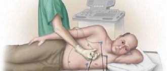
How the procedure is carried out, duration of the examination
The patient is placed on a flat surface without a pillow (couch), after anesthesia of the mucous membranes of the nasopharynx, a sterile thin electrode probe is inserted through the nasal passage or through the mouth, and is asked to make several swallowing movements.
The zone is lowered to a depth of 40 cm, and electrodes are applied to the skin of the chest. First, a regular electrocardiogram is recorded, then a small electrical impulse is applied to the inserted electrode to stimulate the heart. In this case, there may be some sensations behind the sternum - tingling or burning, which is quite acceptable. An ECG is recorded, the doctor observes the changes occurring on it.
The duration of the study usually does not exceed 30 minutes; sometimes attacks of tachycardia may occur during this time; they are assessed by the doctor, and the electrode can be removed if necessary.
Interpretation of results
When assessing the results, the appearance of arrhythmia as a result of electrical stimulation is taken into account. If it is absent, and the TEE data do not differ from the preliminary ECG, then such a study is considered normal. If rhythm deviations occur, the doctor gives them a detailed assessment: describes their type, source, time of occurrence, duration and other parameters. The patient is informed about the results obtained.
If, despite normal results, the patient continues to complain of heart failure, attacks of pain, dizziness, he is observed by a cardiologist, and after a while a repeat diagnosis is prescribed.
Types of echocardiography
Today, there are several types of echocardiography. What specific type of study to conduct is decided in each specific case by a cardiologist.
One-dimensional
At the moment, this type of echocardiography is rarely used independently, because it is considered less informative than others. No image of the heart is generated during the procedure. The data is displayed on the screen in the form of a graph. Using M-echocardiography, the doctor can determine the volume of the heart cavities and evaluate their functional activity.
B-echocardiography (two-dimensional)
During B-echocardiography, data from all structures of the heart enters the computer and is displayed on the monitor in the form of a black and white image. The doctor is able to determine the size of the heart, find out the volume of each of its chambers, the thickness of the walls, assess the mobility of the valve leaflets and how the ventricles contract.
Doppler echocardiography
As a rule, this study is performed simultaneously with B-echocardiography. It allows you to track blood flow in large vessels and on the heart valves, identify reverse blood flow and its degree, which may indicate the formation of pathological processes.
Contrast echocardiography
This test makes it possible to more clearly visualize the internal structures of the heart. The patient is injected intravenously with a special contrast agent, after which the procedure is carried out as usual. This procedure allows you to examine the inner surface of the chambers of the heart. Contraindications for this study are individual intolerance to contrast and chronic renal failure.
Stress echocardiography
To diagnose hidden heart pathologies that manifest themselves exclusively during physical activity, a special type of study is used - stress echocardiography. It makes it possible to identify diseases in the early stages, which do not remind of themselves if the patient is at rest.
Stress echocardiography is recommended to assess the condition of blood vessels and their patency, to find out how great the risk of complications is before performing surgical interventions on the heart and blood vessels. The procedure is also carried out in order to determine how effective the therapy for coronary heart disease is and to determine the further prognosis for this disease.
There are several contraindications to stress echocardiography. It should not be performed on patients suffering from severe respiratory, renal, hepatic or heart failure. It is also contraindicated in case of myocardial infarction, aortic aneurysm and a history of thromboembolism.
Transesophageal echocardiography
This is a special type of study, during which an ultrasound-generating sensor is lowered through the oropharynx through the esophagus to the required depth. Since the sensor has very small dimensions, it passes through the esophagus without problems. However, such a study is considered quite complex and is carried out exclusively in specialized medical centers. In addition, there are special indications for it. In particular, a transesophageal examination is performed in cases where a standard transthoracic examination does not allow assessing the condition of the heart and its structures. In particular, when doubts arise about the correct functioning of a previously prosthetic heart valve, if an aortic aneurysm and atrial septal defect are suspected, as well as if the patient has been diagnosed with infectious endocarditis and the doctor suspects an aortic root abscess.
At the same time, this type of study has contraindications from the upper digestive tract, namely, any tumor formations of the esophagus, bleeding from the upper gastrointestinal tract, the presence of a large diaphragmatic hernia or dilation of the esophageal veins. Transesophageal examination should not be performed in patients with severe osteochondrosis of the cervical spine, instability of the cervical vertebrae, or a history of esophageal perforation. Diagnosis may be complicated in patients with thyroid diseases.
Indications
Transesophageal echocardiography is mandatory for:
- the need to study the aorta in cases of aneurysm and dissection;
- infective endocarditis, its diagnosis and identification of consequences;
- examining a patient before surgery to replace a heart valve with a prosthesis;
- the need to diagnose problems with the prosthesis.
The study is recommended in the following cases:
- diagnosis of congenital heart pathologies;
- detection of tumor formations and blood clots in the heart;
- the need to assess the condition of the coronary sinus (venous canal);
- the ineffectiveness of using transthoracic echocardiography in cases of large patient weight, pulmonary emphysema, etc., as well as clarifying its results;
- suspicion of severe cardiac pathologies.
A significant advantage of the study is the ability to diagnose certain diseases in the early stages.
Transesophageal echocardiography is a semi-invasive method and, as a rule, is prescribed after a transthoracic examination.
Preparation for echo-CG
As a rule, when performing one- and two-dimensional echocardiography, as well as Doppler echocardiography, there is no need for any special preparation. If a transesophageal study is prescribed, there are a number of limitations.
So, the last meal should be no later than six hours before the procedure. Drinking is also not recommended. Immediately before the procedure, dentures should be removed.
On the eve of a transesophageal examination, persons with a labile nervous system are recommended to take a mild sedative. After the procedure, the patient will need some time to recover, so you should not overload yourself with work until the end of the day. It is also necessary to refrain from driving.
Research methodology
For transthoracic echocardiography, the patient is placed in the left lateral position. When a person lies in this position, the apex of the heart and the left side of the chest are brought closer together. This makes it possible to provide the most accurate visualization of the heart - as a result, all four of its chambers are visible on the monitor at once.
The doctor applies a gel to the sensor, which improves contact between the electrode and the body. After this, the sensor is alternately installed first in the jugular fossa, then in the area of the fifth intercostal space, where the apex beat of the heart can be monitored as clearly as possible, and then under the xiphoid process.
Of course, every doctor strives to ensure that the results of the study are as accurate as possible. It should be noted that how informative the procedure will be depends on three main factors.
First of all, the anatomical features of the patient should be taken into account. Serious obstacles to ultrasound are obesity, chest deformation and other similar factors. As a result, the resulting image may not be clear and cannot be interpreted properly. In order to clarify the diagnosis, doctors in such cases offer a transesophageal examination or MRI.
The quality of the equipment should also be taken into account. Of course, more modern equipment will provide the doctor with more opportunities to obtain sufficient information about the patient's heart.
Finally, the competence of the person conducting the examination should be taken into account. In this case, not only his technical skills are important (the ability to position the patient in the correct position and place the sensor at the right point), but also the ability to analyze the data obtained.
When performing stress echocardiography, the patient first undergoes a regular echocardiography, and then special sensors are applied that record parameters during physical activity. For this purpose, bicycle ergometers, treadmill test, transesophageal electrical stimulation or medications are used. In this case, the initial load is minimal, and then it is gradually increased, monitoring blood pressure and pulse indicators. If the patient's health worsens, the examination is stopped.
All this time, an electrocardiogram is continuously performed, which makes it possible to quickly respond if any extreme situations arise. During exercise, the patient may feel dizziness, increased heart rate, and discomfort in the heart area. After stopping the exercise, the heart rate slows down. Sometimes, in order for the heart to function completely back to normal, it is necessary to administer other medications. In this case, the patient's condition is carefully monitored until complete recovery.
Typically, the entire procedure lasts about an hour.
Transesophageal echocardiography begins with irrigating the patient's mouth and pharynx with lidocaine solution. This is intended to reduce the gag reflex during insertion of the endoscope. After this, the patient is asked to lie on his left side, a mouthpiece is inserted into his mouth and an endoscope is inserted through which ultrasound will be received and delivered.
Is cardiac echocardiography safe?
During this study, there is no radiation or other load on the organ. Therefore, if necessary, it can be prescribed even several times a week.
This study is characterized by the absence of complications and side effects.
EchoCG does not harm either the expectant mother or the fetus during pregnancy.
Limitations for the procedure may include inflammatory diseases of the skin of the chest, chest deformities and some other reasons.
Decoding the results
The doctor who conducted the study interprets the echocardiography results. He either transfers the received data to the attending physician, or gives it directly to the patient.
It should be borne in mind that a diagnosis cannot be made based solely on the result of echocardiography. The data obtained is compared with other information available to the attending physician: data from tests and other laboratory tests, as well as the patient’s existing clinical symptoms. Echocardiography cannot be considered as a completely independent diagnostic method.
Possible complications during the procedure
The most common problem that may arise when performing TEE is the urge to vomit, as well as discomfort in the throat.
However, such unpleasant symptoms are easily controlled with sedation and local anesthetics. After the study, the patient may have a sore throat for several days. Perforation of the esophagus occurs very rarely. This is an urgent situation that requires urgent surgical intervention.
There are also other risks associated with this procedure, for example:
- breathing problems;
- heart rhythm disturbance;
- infection of the esophagus;
- bleeding;
- trauma to the mouth, throat or esophagus.
If the patient has problems with the esophagus, for example, such as varicose veins, narrowing of the lumen of this organ resulting from radiation therapy, this may complicate the procedure. An allergic reaction to medications or latex is also possible.
There are other risks that are directly related to the patient’s health status, that is, the presence of a particular disease. You should definitely discuss all these issues with your doctor before the procedure.
Transesophageal echocardiography
Where to get an echocardiogram
Standard echocardiography is carried out both in public medical institutions (clinics and hospitals) and in private medical centers. To register for an examination, you must provide a referral from your attending physician or cardiologist.
More specific types of echocardiography - transesophageal examination or stress echocardiography - can only be performed in specialized medical institutions, since they require special equipment and personnel who have undergone special training.
Echocardiography in children
As noted above, the undeniable advantages of EchoCG are the non-invasiveness, painlessness and complete safety of this method of cardiac research. The manipulation is not associated with radiation exposure and does not provoke any complications. Therefore, if there are appropriate indications, the study can be recommended not only for adults, but also for children.
Diagnostics will help to timely detect congenital pathology in young children, which, in turn, will make it possible to select the most effective treatment. As a result, the child will be able to lead an absolutely fulfilling life in the future.
Indications for echocardiography in a child are:
- Heart murmurs.
- The appearance of shortness of breath either during physical activity or at rest.
- Blueness of the lips, nasolabial triangle, fingertips.
- Decreased or complete lack of appetite, too slow weight gain.
- Complaints of constant weakness and fatigue, sudden fainting.
- Complaints of frequent headaches.
- Discomfort behind the sternum.
- Decrease or increase in blood pressure.
- The appearance of swelling in the extremities.
Taking into account the fact that the method is safe, echocardiography can be performed on children more than once in order to track the development of the disease or assess how effective the treatment is. If any pathological changes have been identified, a study is carried out at least once every twelve months.
Best materials of the month
- Why you can't go on a diet on your own
- 21 tips on how to avoid buying stale food
- How to keep vegetables and fruits fresh: simple tricks
- How to curb your sweet cravings: 7 unexpected products
- Scientists say youth can be extended
Preparing and performing the procedure for children
Like adult patients, children do not need any preliminary preparation. It is advisable that the child does not eat anything for three hours before the test, because with a full stomach, the diaphragm is high, which can distort the result.
Parents should take with them the results of the electrocardiogram performed the day before, as well as the results of studies that were carried out previously. Without fail, the baby should be psychologically prepared for the procedure, explaining that no one is going to hurt him.
In order to carry out the procedure, the baby is undressed to the waist and placed on the left side on the couch. After moving the sensor across the chest, the doctor examines the resulting image.
How to do emergency echocardiography of the heart
Echocardiography using the transesophageal method is performed in a hospital setting or in clinics with intensive care equipment. The requirement must be observed to take urgent measures to save the patient’s life in case of sudden complications.
Transesophageal echocardiography is performed in a separate specialized room, the doctor is assisted by a nurse. For pain relief, anesthetic sprays or general anesthesia are used. The subject lies on his left side, and a mouthpiece is placed in the oral cavity. It will protect the patient's teeth and tongue, as well as the endoscope. The sensor is inserted into the throat and an image is displayed on the monitor.
Transesophageal echocardiography takes about 15–30 minutes. During this time, the clinic’s doctors and nursing staff monitor your condition. Next, the endoscope is removed, and the diagnostician begins decoding.
The person being examined observes his feelings and tells the nurse or doctor if he feels unwell. If there are no deviations in health within half an hour, the patient takes the results of transesophageal echocardiography of the heart and leaves.
Watch the master class on emergency ecoCG: