© Author: M. Vitaly Arkadyevich, resuscitator, especially for SosudInfo.ru (about the authors)
Pulmonary edema is a severe complication of many painful conditions, which is characterized by the release of an excessive amount of liquid blood from the lumen of the pulmonary vessels directly into the lung tissue (interstitium), and then into the lumens of the alveoli (the terminal sections of the bronchial tree in which gas exchange occurs).
Cardiogenic pulmonary edema is a serious complication of many pathologies of the heart itself, which without emergency qualified medical care often leads to death.
Cardiac asthma and pulmonary edema are essentially synonymous, since cardiac asthma is a complex of external manifestations (symptoms) that occur during the development of cardiogenic pulmonary edema at the initial stage - interstitial pulmonary edema.
Development mechanism
The development of cardiogenic pulmonary edema is associated with impaired pumping function of the heart muscle. This occurs under the influence of various pathologies. These include heart failure, acquired or congenital heart defects, myocardial infarction, and arrhythmias. Due to insufficient blood circulation, stagnation occurs in the body. As a result, fluid from the lymphatic vessels and capillaries begins to pass through the walls of the vessels into the pulmonary alveoli. This process is medically called fluid effusion. The liquid gradually displaces air from the lung tissue, which causes difficulty breathing. As the lungs fill with transudate, breathing becomes impossible, severe oxygen starvation of all tissues and organs develops, and death occurs.
Pathophysiology
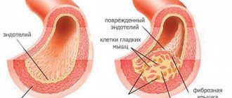
In normally functioning lungs, there is a small influx of fluid from the alveolar capillaries into the interstitial space of the lungs, facilitated by hydrostatic forces and the presence of microscopic gaps between capillary endothelial cells.
The rate of fluid entry is limited by the osmotic pressure gradient of the protein, which promotes the movement of fluid from the interstitial space back into the circulating plasma. As a result, the relatively small physiological movement of fluid from the vasculature into the lungs is usually compensated by the outflow of fluid from the interstitial space of the lung through the pulmonary lymphatic system, which ultimately returns to the systemic venous circulation. As fluid passes through the interstitial space of the lungs, it is delimited from the alveolar space by tight occlusive junctions between alveolar epithelial cells. In the absence of acute lung injury (eg, capillary injury), changes in the rate of fluid flow through the lungs are determined primarily by changes in hydrostatic pressure. Pulmonary capillary wedge pressure (PCWP), recorded from the catheter balloon at the moment of its wedging in the pulmonary artery segment, is a reflection of the filling pressure of the left atrium and is considered to most fully reflect the hydrostatic pressure of the microcirculation of the lung. Starling's equation for filtration mathematically demonstrates the fluid balance between the pulmonary vessels and the interstitial space, which "depends on the net difference in hydrostatic pressure and protein osmotic pressure, and the permeability of the capillary membrane."
Q = K*((Pmv - Ppmv) - (πmv - πpmv))
Where:
- Q = Net filtration of fluid through the vascular wall towards the interstitial space (flow of fluid filtered through the capillary wall into the interstitial space)
- K = Filtration coefficient
- Ppmv = Hydrostatic pressure in the perivascular interstitial space
- Pmv = Hydrostatic pressure inside the capillaries (HPLP)
- πmv = Osmotic pressure of protein in the vascular bed
- πpmv = Osmotic pressure of protein in the perivascular interstitial space
Although the Starling equation is useful for understanding the mechanisms that favor pulmonary edema, it is clinically impractical to accurately measure most of these parameters. However, a basic understanding of this equation is useful for clinicians working with patients with pulmonary edema.
Pathogenesis of cardiogenic pulmonary edema (increased hydrostatic pressure)
In cardiogenic pulmonary edema, hydrostatic pressure in the pulmonary capillaries leads to increased filtration of fluid through the vascular wall and is most often caused by volume overload or impaired left ventricular function leading to increased pulmonary vascular pressure. Moderate increase in pressure in the left atrium, expressed as PCWP 18-25 mm Hg. Art., causes the formation of edema in the perivascular and peribronchial interstitial spaces. With a further increase in left atrial pressure (PAPL > 25 mm Hg), the capacity of the lymphatic vessels and interstitial space (approximately 500 ml of fluid) is exceeded, and the fluid overcomes the pulmonary epithelial barrier, filling the alveoli with protein-containing fluid. The clinical picture of hypoxemia is caused by the accumulation of alveolar fluid, destabilization of the alveolar acini (impaired surfactant function) and, consequently, a violation of the ventilation-perfusion ratio (V/Q).
Pathogenesis of non-cardiogenic pulmonary edema (increased vascular permeability)
Non-cardiogenic pulmonary edema refers to any condition that promotes an abnormal increase in pulmonary vascular permeability, thereby promoting increased fluid and protein flow into the interstitial spaces and air spaces of the lungs. With regard to the Starling equation, pulmonary vascular damage equates to an increase in filtration coefficient and an increase in osmotic pressure in the interstitial space of the lung, which contributes to the formation of pulmonary edema. Another factor contributing to impaired gas exchange in noncardiogenic pulmonary edema is associated with disruption of the alveolar epithelial barrier, for example when the pressure in the interstitial space of the lungs is high enough to disrupt tight junctions, or when there is direct inflammatory or toxic damage to the alveolar epithelial lining. Damaged alveolar epithelium has a reduced ability to actively transfer fluid from the alveolar space to the interstitial space of the lung and causes impaired surfactant production (reduced active surface area), favoring alveolar collapse during normal breathing. An example is direct damage to the alveolar epithelium due to aspiration of gastric contents or pneumonia. Conditions that contribute to acute pulmonary capillary endothelial injury include systemic infections (sepsis), severe burns, trauma, and other systemic inflammatory conditions. Damage to the pulmonary capillary endothelium and/or alveolar epithelium is a hallmark of acute lung injury (ALI) and acute respiratory distress syndrome (ARDS), which are progressive non-cardiogenic lung injuries associated with impaired gas exchange (shunting, V/Q imbalance) and decreased pulmonary compliance (increased work of breathing).
Causes
Cardiogenic pulmonary edema is caused by heart disease in the decompensation stage. In addition to such common phenomena as myocardial infarction, arrhythmias, heart defects, the following conditions can cause pathology:
- cardiomyopathy;
- atrial tumor;
- endocarditis of bacterial origin;
- inflammatory process of the aorta;
- myocarditis;
- mitral valve stenosis;
- acute failure of the left ventricle of the heart;
- cardiosclerosis.
If cardiovascular diseases have not been treated, the development of cardiogenic pulmonary edema is possible with any pathology. Various intrauterine developmental disorders can provoke severe complications, for example, ventricular septal defect, coarctation of the aorta, patent ductus arteriosus.
Cardiogenic pulmonary edema always develops against the background of cardiac pathologies
Predisposing factors include bad habits, frequent emotional overload, increased blood pressure, lack of treatment for heart pathologies, non-compliance with doctor's orders, and more.
Mechanism and causes of development of pulmonary edema
The appearance of pulmonary edema (PE) can be caused by many reasons, but regardless of the factor that caused the complication, the mechanism of its development is the same - the accumulation of excess fluid in the interstitial tissues, the resulting thickening of the alveolo-capillary membrane and a decrease in the diffusion of gases (primarily oxygen). As a result, tissue hypoxia occurs (oxygen starvation of all tissues) and acidosis - a shift in the acid-base balance, leading to the inevitable death of the patient if he is not given emergency assistance.
There is no unified classification of pulmonary edema, but according to the pathogenetic mechanism it can be divided into:
- OB due to increased capillary pressure as a result of:
- acute myocardial infarction;
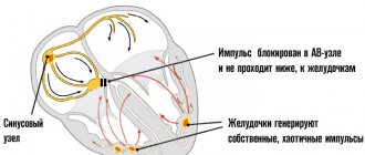
- cardiac arrhythmias;
- cardiomyopathies;
- myocarditis;
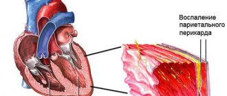
- exudative pericarditis;
- hypertensive crisis;
- pulmonary artery stenosis;
- heart defects;
- massive infusion of blood replacement solutions;
- renal failure in the anuric phase.
- OB due to increased permeability of the capillary wall with:
- acute respiratory distress syndrome;
- intoxications (for example, narcotic drugs);
- anticancer chemotherapy;
- use of X-ray contrast agents;
- inhalation of toxic substances;
- allergies.
- OB due to impaired lymphatic drainage due to cancerous lesions of the lymphatic vessels.
- OB due to changes in intrathoracic interstitial pressure during decompression sickness and evacuation (removal) of fluid from the pleural cavity.
- OL due to a decrease in protein content in the blood plasma.
- Mixed OL:
- neurogenic;
- postoperative;
- with eclampsia;
- with ovarian hyperstimulation syndrome;
- with altitude sickness.
Previously, a classification was used that included types of pulmonary edema such as interstitial and alveolar. Currently, it has been abandoned, since these two types of OA are actually only stages of the development of the syndrome. In addition, in terms of diagnosis and treatment, such a division does not provide any useful function.
Normally, only a small amount of fluid from the interstitium penetrates into the alveoli. Almost all of it is absorbed into the blood and lymphatic capillaries and removed from the alveolo-capillary membrane. However, if the permeability of the ACM is impaired, there is too much fluid and it does not have time to move all of it into the vessels. In this case, it permeates the interstitium, increasing its thickness, and in the most advanced situation it begins to enter the lumen of the alveoli, further worsening gas exchange.
Symptoms
Symptoms of pulmonary edema always depend on the stage of the pathology. Filling of the lung tissues with fluid occurs gradually, each stage is accompanied by its own manifestations. There are 4 stages in total:
- Dyspnoetic. Here the fluid just begins to enter the lung tissue, the patient’s breathing becomes difficult, a dry cough appears, accompanied by attacks of suffocation. When listening to the lungs, faint wheezing is heard.
- Orthopnoetic. Further filling of the lung with transudate provokes the transition of a dry cough into a wet one, breathing becomes more frequent, weakness, sometimes dizziness, and pale skin appear.
- Acute clinical stage. At this stage, a large amount of fluid accumulates in the lungs, the lung is filled by 50%, the cough intensifies, and mucous discharge mixed with blood appears. Whistles and wheezing are audible to others, and no special equipment is required to diagnose them. The patient can breathe in a vertical position of the body, but if he takes a horizontal position, the patient suffocates.
- Heavy. A dangerous condition that can cause death without medical help. At the last stage, the patient’s cough is intense and incessant, bloody foamy discharge appears from the mouth, and breathing is severely impaired. This condition indicates that the lungs are full of fluid and the patient may choke at any moment.
In addition to the stages and their characteristic signs, the following manifestations can be distinguished:
- difficulty, lack of air, especially in a lying position;
- open mouth, bulging eyes;
- panic, fear of death;
- profuse sweating;
- pallor and cyanosis of the skin, which intensifies with the development of complications;
- discomfort in the chest when trying to breathe;
- drop in blood pressure and pulse.
The accumulation of fluid in the lung tissues leads to breathing problems and coughing up blood.
In such a situation, it is necessary to call an ambulance as soon as possible. Even if such attacks had already been diagnosed earlier but went away on their own, the dangerous condition should not be ignored.
Important! Cardiogenic pulmonary edema is an extremely dangerous complication; death can be avoided in no more than 40% of cases.
Clinical manifestations and diagnosis
In the clinic, there is a division of the cascade of described pathological phenomena into stages, based on the existing symptoms:
- Dyspnea is a stage that is predominantly characterized by a subjective feeling of lack of air (shortness of breath).
- Orthopnea - shortness of breath progresses, forcing the patient to take a forced position, thanks to which the person feels better - the legs are lowered down, the upper body is raised (semi-sitting position).
- Stage of “expanded clinic” - the above symptoms are joined by distant wheezing (when wheezing is heard without a phonendoscope at a distance).
- The stage of severe clinical manifestations - the clinical manifestations of pulmonary edema include foaming from the mouth and nose, “bubbling” breathing (these manifestations are sometimes called the “boiling samovar” symptom), cyanosis (blueness) of the skin, and cool, sticky sweat.
Clinically, pulmonary edema in acute heart failure, quite conventionally, is divided into 2 types:
- Interstitial pulmonary edema is “subcompensated” pulmonary edema, the pathogenesis of which stops before fluid enters the lumen of the alveoli. This form of pulmonary edema is usually called “cardiac asthma”, the duration of which is measured in minutes or hours, but no more, then improvement occurs, often without specific assistance.
- Alveolar pulmonary edema - observed in cases where the alveoli fill with fluid and the above-described “vicious circle” is formed. An extremely life-threatening condition that requires emergency medical care, as it is the most common complication of heart disease, leading to death.
The symptoms of cardiogenic pulmonary edema are quite specific and allow it to be distinguished from other diseases that may also demonstrate clinical manifestations of asthma.
Clinical symptoms of cardiogenic pulmonary edema include:
manifestations of heart failure with pulmonary edema
- Shortness of breath, which is of a mixed nature, worsens when lying down and with physical activity; according to the wording of patients, “not to breathe in the air”;
- Tachypnea – increased frequency of respiratory movements;
- Orthopnea is a forced body posture that alleviates the feeling of shortness of breath;
- Pain in the heart area is caused by general hypoxia and myocardial ischemia;
- Wheezing in the lungs is initially dry, whistling, then the wheezing changes to moist, fine-bubble, and then large-bubble;
- Remote wheezing - heard at a distance when breathing in and out with the “naked” ear, so strong that it resembles the sound of boiling water;
- Discharge of finely bubbled white foam from the external openings of the mouth and nose, sometimes the foam is pink, since red blood cells can penetrate into the lumen of the respiratory tract;
- The cough at the beginning is dry, hacking, and as the disease progresses it becomes productive, with a large amount of foamy sputum;
- Tachycardia – increased heart rate;
- The pulse is thread-like, small in filling, frequent;
- Acrocyanosis – bluish discoloration of the skin of the hands, feet, nose;
- Cold clammy sweat;
- Pale skin of the body;
- Swelling of the veins of the head and neck;
- Emotional agitation is a panic fear of death, which, if medical care is not provided and the disease progresses, is replaced by apathy and drowsiness.
When diagnosing pulmonary edema, as well as in choosing treatment tactics, considerable importance is attached to the data of laboratory and instrumental research methods:
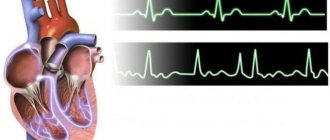
- Electrocardiography (ECG) - allows you to establish overload of the left side of the heart, acute coronary syndrome, arrhythmia, myocardial hypertrophy, myocardial dystrophy, etc.;
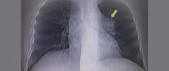
- pulmonary edema on x-ray
X-ray – allows you to confirm the presence of pulmonary edema and myocardial hypertrophy: expansion of the cardiac shadow, Kerley lines (B), the symptom of “butterfly wings”, however, it must be taken into account that in 20% of cases pulmonary edema is not reflected on the X-ray;
- Pulse oximetry - there is a decrease in the saturation of hemoglobin in the blood with oxygen (less than 90%);
- Measuring blood pressure - its indicators largely determine the tactics of providing medical care.
It is necessary to evaluate the manifestations of pulmonary edema not individually, but always in aggregate, taking into account the anamnesis, which will avoid diagnostic errors.
Difference between cardiogenic pulmonary edema
Before starting treatment for pulmonary edema, it is important to determine the nature of the pathology. In addition to cardiogenic (heart), there is non-cardiogenic edema. The clinical picture for these two conditions is very similar. Without the necessary knowledge, it is impossible to distinguish between them. Treatment of a particular condition begins only after confirmation of the diagnosis. If the therapeutic tactics are incorrect, death occurs.
Criteria by which cardiogenic edema can be distinguished from non-cardiogenic edema:
We advise you to read: Complications of myocardial infarction
- cardiogenic edema occurs against the background of cardiac pathologies, while the second option is provoked by pathologies not related to cardiac activity;
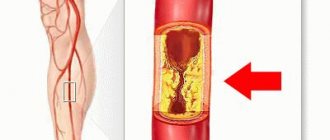
- cardiogenic edema is a consequence of the overflow of blood vessels and capillaries, the development of a stagnant process. With non-cardiogenic edema, pathology may appear due to damage to the vessels themselves, a violation of their integrity;
- Both pathologies differ in the nature of their course. Non-cardiogenic edema occurs suddenly and only a few hours pass before death occurs. With cardiogenic pathology, the course of the pathology takes longer;
- The differences on the x-ray are that with cardiogenic edema the heart is enlarged in size, and in the area of the lungs a darkened area is visible in the middle. In the noncardiogenic form, the heart is of normal size and areas of opacification are distributed throughout the lungs;
- echocardiography with cardiogenic edema shows impaired functioning of the left ventricle, in the second case the activity of the heart muscle is not impaired;
- the pressure in the pulmonary artery with cardiogenic exceeds 18 mmHg, with non-cardiogenic these levels are lower.
To accurately determine the nature of the pathology, it is necessary to conduct an examination in a hospital setting. Only an experienced specialist will be able to assess the patient’s condition and prescribe the necessary treatment.
Patient examination plan
Interstitial pulmonary edema can only be detected through instrumental and laboratory tests. It is important to establish the exact cause. Differential diagnosis is carried out with edema of non-cardiogenic origin. In the latter case, the causes are poisoning, kidney and liver pathology, and trauma.
The following studies are required:
- blood gas analysis;
- biochemical blood test;
- general clinical blood and urine tests;
- electrocardiography;
- determination of the acid-base state of the blood;
- echocardiography;
- plain radiography;
- catheterization of pulmonary arteries;
- physical examination;
- coagulogram.
Laboratory diagnostics allow you to assess blood oxygen saturation. With edema, the following disorders are detected:
- increase in carbon dioxide levels;
- increased fibrinogen (with thromboembolism);
- increase in transaminase concentration.
If cardiac asthma is suspected, then radiography is required. Cardiogenic edema often shows signs of pleural effusion. The heart is enlarged. A valuable diagnostic feature to exclude non-cardiogenic edema is the level of pressure in the pulmonary artery system. In case of heart pathology, it exceeds 18 mm Hg.
Who is at risk
In some patients, the risk of developing a dangerous condition increases due to certain provoking factors. These include:
- all heart diseases;
- frequent stress;
- age over 45 years;
- bad habits, namely drinking alcohol, smoking.
The conditions described above do not directly cause the development of edema, but they can increase the risk of its occurrence.
Elderly patients with cardiac dysfunction are at risk for developing cardiac pulmonary edema.
Manifestations of toxic edema
The clinical picture includes difficulty breathing as the main sign of toxic edema. Shortness of breath and cough appear, the skin and mucous membranes become bluish in color, the pulse increases, and blood pressure drops to critical levels - these are the indicators that the doctor will check first if the development of edema caused by toxins is suspected. It is possible that the patient will experience acute chest pain, as with cardiogenic edema. However, the difference between them is that toxic edema lasts longer, while the size of the heart muscle remains virtually unchanged.
If treatment for pulmonary edema is started in a timely manner, its manifestations can be reversed, wheezing, shortness of breath, cough can be eliminated, the skin can be restored to its previous appearance, and the blood count can be stabilized. An x-ray will show that the lesions are disappearing. Recovery time can take up to several weeks.
Classification
The condition is classified depending on the underlying factor. Pathology can develop against the background of insufficient activity of the left ventricle of the heart. This condition occurs due to myocardial infarction or coronary insufficiency. Another type is cardiogenic edema due to left ventricular overload. This occurs when the mitral or aortic valve is incompetent. A hypertensive crisis can provoke an increase in blood pressure in the systemic circulation. Another reason is the presence of hematomas, blood clots, and tumors in the vessels.
Symptoms and clinical picture
Symptoms of pulmonary edema of “cardiac” origin are specific and make it possible to make a diagnosis at the prehospital stage. Various respiratory disorders play a leading role:
- Orthopnea (taking a forced body position to facilitate breathing).
- Shortness of breath with difficulty breathing.
- Cough.
- Moist rales in the lungs, mainly in the lower sections.
- Discharge of foamy sputum.
Possible enlargement of the liver, swelling of the legs. In addition, there are signs of cardiac dysfunction. They vary depending on the primary pathology:
- Increased heart rate, decreased heart rate, or development of arrhythmias.
- Cold clammy sweat, pale skin.
- A decrease in blood pressure, less often an increase.
- With myocardial infarction: pressing, burning pain behind the sternum, radiating to the left shoulder blade, left arm.
Common symptoms include:
- Impaired consciousness.
- A sharp decrease in urine output.
There are three stages in the progression of fluid accumulation in the lungs during edema.
Stage 1. Characterized by the following symptoms:
- increased pressure in the left atrium;
- opening of small pulmonary vessels;
- gas exchange does not change.
Stage 2. Characterized by the following symptoms:
- accumulation of a small amount of fluid in the interstitial cavity;
- thickening of large pulmonary vessels;
- slight shortness of breath;
- dizziness;
- slight cough.
Stage 3. Symptoms:
- hypoxemia;
- decreased respiratory volume of the lungs;
- gas exchange disorders;
- severe shortness of breath;
- wheezing;
- cough with sputum.
Other common symptoms of pulmonary edema are:
- anxiety, restlessness;
- chest pain;
- tachycardia;
- cough with bloody sputum;
- low tolerance to physical activity;
- difficulty breathing when lying down;
- sudden awakening at night from lack of oxygen;
- swelling in the lower extremities;
- fatigue;
- fever;
- headache;
- high blood pressure.
Possible complications
Due to cardiogenic pulmonary edema, oxygen starvation of all organs develops. Hypoxia leads to inevitable death of brain cells, which without timely help causes death. In addition, the consequences may be as follows:
- ischemic disorders. Oxygen starvation leads to disruptions in the functioning of the kidneys and liver;
- scarring of lung tissue called pneumosclerosis;
- cardiogenic shock;
- pulmonary edema of a recurrent nature;
- deformation of the structure of the alveoli and bronchioles;
- Secondary pneumonia develops against the background of circulatory disorders.
Important! Complications can begin within 4–5 minutes after the development of cardiogenic pulmonary edema, so if signs of pathology appear, you should act immediately.
Research methods
A general blood test reveals leukocytosis. Sometimes, as a result of the death of neutrophils and increased consumption of platelets in the lungs, leukopenia and thrombocytopenia develop. Biochemical blood tests are usually within normal limits, although hypoalbuminemia and slight hyperglycemia are sometimes detected due to protein loss in the lungs.
X-ray examination of the chest organs is extremely necessary. At an early stage and with a mild course of NOL, an increase in the interstitial pattern of the lungs is determined. With moderate and severe NOL, infiltration appears in the lung tissue, indicating the involvement of the alveoli in the pathological process. Most patients have asymmetrical infiltration in the dorsocaudal regions of the lungs, predominantly on the right. When determining arterial blood gases, mild or severe hypoxia is detected, and with excessive hyperventilation, hypercapnia.
When examining a coagulogram, an increase in prothrombin and partial thromboplastin time is determined as a result of increased consumption of blood coagulation factors and disseminated intravascular coagulation. The severity of hypoxia is determined by oximetry data.
Diagnosis of the disease
In addition to the data obtained from a visual examination of the patient, laboratory tests are necessary for correct diagnosis. For this, the following tests are prescribed:
- blood gas examination;
- biochemical study of blood parameters;
- study of urea, creatinine, total protein;
- coagulogram;
- liver tests and more.
An electrocardiogram shows ischemic disorders, hypertrophy of the left ventricle of the heart, arrhythmias. Ultrasound examination reveals areas of myocardial hypokinesia, a decrease in fractional ejection, and an increase in diastolic volume.
An electrocardiogram and other diagnostic methods help to make the correct diagnosis and begin treatment
A chest x-ray shows expansion of the borders of the heart muscle and pulmonary roots. The lesion is most often visible on the picture as a darkened spot resembling a butterfly in shape. Focal opacities are diagnosed less frequently. To distinguish cardiogenic from non-cardiogenic edema, pulmonary artery catheterization is performed.
Diagnosis and differential diagnosis of pulmonary edema
The table above shows typical clinical symptoms that distinguish cardiogenic from non-cardiogenic pulmonary edema. Ironically, pulmonary artery catheterization, the most accurate diagnostic technique, is no longer used in routine practice due to frequent complications (eg, hemorrhage, pneumothorax, arrhythmias, infection, vascular injury) and its unreliability due to improper calibration or misinterpretation of data. Thus, less invasive techniques have largely replaced pulmonary artery catheters for routine evaluation of cardiogenic causes of pulmonary edema in the ICU setting.
Transesophageal echocardiography
Transesophageal echocardiography (TEE) is the most widely used technique for the evaluation of critically ill patients with suspected heart failure. In the context of pulmonary edema, the use of TEE can quickly detect serious cardiac disease associated with increased left ventricular filling pressure, including impaired left ventricular ejection fraction due to ischemic (usually causing abnormalities in wall motion) or non-ischemic (with diffuse wall motion disorders) myocardial diseases, significant valvular lesions or pericardial effusion causing the pathophysiological mechanism of tamponade.
PiCCO and KKT
Two alternative methods have emerged for the assessment of pulmonary edema.
- The Pulse Wave Continuous Cardiac Output (PiCCO) system assesses the pulmonary vascular permeability index in critically ill patients.
- Quantitative computed tomography (QCT) with thermodilution has also been used to diagnose pulmonary edema secondary to ARDS.
To date, no studies have demonstrated the superiority of PiCCO or QCT over traditional approaches (eg, CVP-based) for the assessment of pulmonary edema as a complication of ARDS.
Emergency care for a patient
Correctly provided emergency care for cardiogenic pulmonary edema helps to preserve the patient’s vital functions until the ambulance arrives. If symptoms characteristic of pathology develop, you must perform actions in the following order:
- seat the person in an upright position, legs and arms should be lowered;
- if necessary, remove foam and mucus from the mouth;
- it is important to ensure the flow of fresh air into the room; to do this, open the windows and door;
- pandemonium should not be allowed;
- the patient’s collar must be unbuttoned, nothing should restrict the neck;
- if the person is conscious, he needs to be given a Nitroglycerin tablet, as well as Furosemide.
When providing assistance, you should not panic. This will only aggravate the situation; the feeling of fear will be transmitted to the patient, which will negatively affect his condition.
First aid
According to clinical guidelines, prehospital emergency care in case of cardiogenic pulmonary edema includes the following prehospital measures:
- Inhalation of Nitrospray or taking nitroglycerin under the tongue.
- Administration (orally or intravenously) of diuretics, preferably Furosemide or Torasemide, 40 mg or more.
- For high blood pressure, use ACE inhibitors (Captopril under the tongue) or beta-blockers (Bisoprolol).
Among the non-drug effects, it is important to ensure access to fresh air and for the patient to assume a sitting position. As a last resort, foot baths with warm water are possible, however, this method is of a “field” nature.
In the case of a clinical picture typical of myocardial infarction, it is important to give the patient chewing Aspirin or Clopidogrel at a dosage of 300 mg, and ensure that he takes nitrates and ACE inhibitors or beta blockers.
The ambulance team is able to begin oxygen therapy, and they can also use narcotic analgesics. According to indications, cardioversion and reperfusion therapy can be performed.
Pulmonary edema due to acute left ventricular failure is an absolute indication for urgent hospitalization.
Treatment methods
Treatment of cardiogenic pulmonary edema is carried out exclusively in a hospital setting under the supervision of medical personnel. The main goals of therapy include:
- actions aimed at eliminating ischemia of body tissues and combating complications of the condition;
- normalization of blood pressure in the systemic and pulmonary circulation;
- establishing normal activity of the left ventricle of the heart;
- saturation of the blood with oxygen molecules.
Treatment for cardiogenic pulmonary edema is complex; its tactics are determined taking into account the patient’s symptoms and the characteristics of the course of the pathology. When severe pain develops, Morphine is used. It is possible to cope with the intense secretion of foam with blood using 30% alcohol inhalations. In case of severe cerebral hypoxia, the patient is transferred to artificial pulmonary inhalation. Urination is stimulated with diuretics. Nitroglycyrin helps strengthen the pumping function of the heart muscle. Antihypertensive medications are used to lower blood pressure. For cardiogenic pulmonary edema, which was not preceded by myocardial infarction, Eufillin is administered. This drug is prescribed to prevent bronchospasm.
The patient's serious condition requires the use of an oxygen mask
Therapy is considered effective when the following result is achieved:
- the patient can take a supine position without the risk of suffocation;
- moist wheezing in the lungs stops;
- the patient's dermis and mucous membrane acquire a normal pink tint;
- Severe coughing and secretion of mucus, foam, and blood stop;
- shortness of breath decreases, breathing normalizes.
Despite urgent and correct medical measures, cardiogenic pulmonary edema often causes serious consequences and death of the patient. To prevent a recurrence of the situation, the person must remain in a hospital until complete recovery.
Treatment
In a hospital setting, treatment of cardiogenic pulmonary edema begins with oxygen therapy or, if indicated, connection to a ventilator (artificial pulmonary ventilation). Oxygen is supplied through a nasal catheter or a mask in a humidified form (from 2 to 6 l/min); alcohol inhalations may be added for defoaming. Indications for intubation with mechanical ventilation:
- Dysfunction of the respiratory muscles.
- Unconscious state of the patient.
- In case of left ventricular failure due to ACS (acute coronary syndrome).
Among the medicines used are the following groups of drugs:
- Loop diuretics (“Furosemide” or “Torasemide” 40 mg or more intravenous bolus). Helps remove fluid from the body and reduce pulmonary edema.
- Vasodilators (“Nitroglycerin” from 20 to 200 mcg/min intravenously). They eliminate constriction of the small circle vessels, increase their capacitive function, thereby reducing the pressure in the pulmonary capillaries. Used only when systolic blood pressure is more than 90 mm Hg.
- Narcotic analgesics (“Morphine” from 10 mg). They eliminate unproductive shortness of breath, which additionally contributes to an increase in blood pressure in the pulmonary circulation, reduce psycho-emotional overexcitation, and cause slight dilatation of peripheral vessels.
- ACE inhibitors (Enalaprilat from 2.5 mg). Allows you to eliminate spasm of vessels resistant to other groups of drugs and reduce the load on the myocardium. Not suitable for routine use.
- Inotropic drugs (“Dobutamine” from 2 to 20 mg/kg/min). Necessary for increasing cardiac output and eliminating congestion in the pulmonary circle. Can be used for SBP below 90 mm Hg, but for heart rate up to 100 beats/min.
- Vasopressors (“Norepinephrine” 0.2–1.0 mg/kg/min). They are used exclusively when it is necessary to maintain blood perfusion in vital organs.
- Anticoagulants (“Fondaparinux” from 0.5 ml 2.5 mg, or “Enoxiparin” from 0.2 ml, or low molecular weight heparins from 5000 IU according to a regimen under the control of coagulogram parameters). They are used to prevent thromboembolic complications.
- Intravenous administration of 30% ethyl alcohol is used to suppress foaming.
Depending on the cause of the development of the threatening condition, etiotropic therapy should be carried out. Thus, when this condition develops against the background of a life-threatening arrhythmia, it is necessary to carry out electrocardioversion (cardioverter-defibrillator from 120 J) or drug cardioversion (Amiodarone up to 600 mg intravenously), it is possible to install a temporary or cardioverter/pacemaker.
Pulmonary edema due to acute left ventricular failure secondary to acute myocardial infarction requires thrombolytic therapy and/or percutaneous coronary intervention if urgently indicated. In addition, at the prehospital stage, Aspirin and/or Clopidogrel 300 mg and heparin therapy are used. In the event of the formation of acute mitral regurgitation, the question of the advisability of cardiac surgery is decided.
Exudative pericarditis requires urgent evacuation of fluid from the pericardial cavity.
For myocarditis, anti-inflammatory therapy is carried out.
Criteria for effective care for pulmonary edema:
- Normalization of respiratory rate (respiratory rate) and blood gas parameters.
- Stabilization of hemodynamics.
- The appearance of diuresis.
- General improvement in the patient's well-being.
Prognosis for patients
After the onset of cardiogenic pulmonary edema, the prognosis for the patient is extremely rarely favorable. Survival rate, according to various statistics, ranges from 30 to 50%. In this case, edema caused by myocardial infarction leads to death in 90% of cases.
After overcoming a dangerous pathology, many people experience various disorders of the respiratory, cardiac, and central nervous systems. After recovery, a person must regularly visit a doctor for 12 months, monitor his diet, and follow other preventive measures.
A favorable prognosis for the patient is possible only with timely assistance.
An important condition for complete recovery is intensive therapy for the disease that caused the swelling. Only after eliminating the root cause is the risk of a recurrent attack reduced to almost zero.
Important! Only timely diagnosis of cardiogenic pulmonary edema, correctly selected treatment tactics and strict implementation of all doctor’s recommendations provide a favorable prognosis for a person.
OL associated with disorders in the circulatory system
With cardiogenic pulmonary edema (caused by circulatory disorders), the first symptom is cardiac asthma, manifested by shortness of breath at rest, increased respiratory movements, a feeling of severe lack of air, and suffocation. Most often, the attack begins at night, the patient immediately wakes up and takes a sitting position in which it is easier for him to breathe. At the same time, he lowers his legs from the bed and rests his hands on its edge. This is the orthopneic position, which is accepted by almost every patient.
The onset of pulmonary edema is characterized by a desire to go to the window and breathe fresh air. In this state, the patient practically does not speak, but emotional stress is clearly visible on his face. As doctors put it, “the patient is completely devoted to the struggle for air.” The skin becomes pale, the nasolabial triangle becomes bluish (acrocyanosis). This indicates an increase in hypoxia. Cold, sticky sweat may appear - a sign of impending cardiogenic shock, which is an extremely severe complication of any cardiac pathology. With further development, the patient's breathing becomes noisy, bubbling in his chest can be heard even at a distance, and pink, foamy sputum may be released in large volumes. At this stage, the amount of liquid already far exceeds the capacity of the capillaries to remove it, and the liquid part of the blood begins to penetrate into the alveoli.
Prevention measures
Eliminating the causes that can provoke pulmonary edema is the prevention of the disease. To prevent a dangerous condition, you must adhere to the following rules:
- eat healthy and nutritious food. The diet should contain a large amount of minerals, vitamins and other nutrients. It is important to avoid foods that increase blood cholesterol;
- Each person is recommended to exercise and often walk in the fresh air;
- give up cigarettes, drugs and alcohol;
- promptly treat cardiovascular pathologies;
- avoid conflicts and stressful situations;
- normalize body weight;
- When working with harmful substances, you must strictly follow safety rules.
It should be remembered that it is impossible to completely insure against the development of pulmonary edema. The condition occurs suddenly, sometimes the patient is not even aware of problems with the heart or other organs. Compliance with prevention and a healthy lifestyle will help to significantly reduce the risk of a dangerous pathology. Timely assistance to a patient with developed pulmonary edema significantly increases the patient's chances of survival.
Diagnostics
Laboratory tests used in the evaluation of patients with cardiogenic pulmonary edema include the following:
- A complete blood count examines severe anemia and may cause sepsis or infection if a significantly elevated white blood cell count or bandemia is present
- Measurement of serum electrolyte levels. Patients with chronic CHF frequently use diuretics and are therefore predisposed to electrolyte disturbances, particularly hypokalemia and hypomagnesemia; persons with chronic renal failure are at high risk of hyperkalemia, particularly when not on hemodialysis
- Determination of urea content in the blood and creatinine - these research methods make it possible to study patients with renal failure and an expected response to diuretics; in low-output conditions such as systolic dysfunction, decreased BUN and creatinine levels may be secondary to renal hypoperfusion
- Pulse oximetry. Pulse oximetry is useful in assessing hypoxia and therefore the severity of cardiogenic pulmonary edema; it is also useful in monitoring the patient's response to supplemental oxygen and other treatments.
- Arterial blood gas test – This test is more accurate than pulse oximetry for measuring oxygen saturation; The decision to initiate mechanical ventilation is based primarily on clinical findings, but in rare cases, arterial blood gas results are taken into account.
Electrocardiography
Left atrial enlargement and left ventricular hypertrophy are sensitive, although nonspecific, signs of chronic left ventricular dysfunction. Electrocardiography may indicate acute tachydysrhythmia or bradydysrhythmia or acute myocardial ischemia or infarction as the cause of cardiogenic pulmonary edema.
CT scan
Chest CT can be a useful tool for differentiating cardiogenic pulmonary edema from acute respiratory distress syndrome in the emergency department, with an overall diagnostic accuracy of 88.5%. Features of chest CT with a high positive predictive value and moderate negative predictive value for cardiogenic pulmonary edema are likely to include the presence of ground-glass attenuation predominantly in the upper lobe or central region, as well as consolidation of the central air space.