Pathogenesis of the disease
Pulmonary artery aneurysm - what is it? This is a pathological expansion of the vessel that exits from the right ventricle into the pulmonary circulation, and whose task is to transport venous blood. The danger of AVA disease lies in its asymptomatic course (in most cases).
Presumably, the pathology is facilitated by a congenital anomaly in the structure of the wall of the pulmonary artery (certain areas are heterogeneous and defective). Its gradual stretching is facilitated by increased pressure in the small circle. At the site of wall expansion, blood flow moves according to the principle of turbulence.
Because of this, a number of hemodynamic processes are disrupted. The pressure in areas of the circulatory system changes; blood, according to physical principles, moves from areas with high pressure levels to those where they are low.
The pressure has a destructive effect on the thinned wall of the organ: it continues to stretch. Due to dystrophic transformation, the risk of rupture increases.
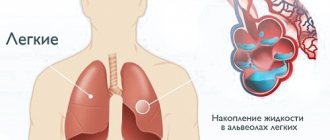
The developing pathology is visible in the image
AVA classification of lungs
In the literature, AVA of the lungs is also described under the names arteriovenous fistula (fistula), cavernous angioma, hemangioma, telangiectasia, cavernous sinus, and pulmonary arteriovenous malformation.
Arteriovenous pulmonary aneurysms can be single (60-70%) or multiple (30-40%). Three quarters of patients have unilateral lung damage, the rest - bilateral. In most cases (65-70%), the location of arteriovenous aneurysms is the lower lobes of the lungs, often on the right.
Anastomoses between arteries and veins can form at the level of segmental, subsegmental pulmonary vessels, arterioles and precapillaries. If vessels of medium and large caliber communicate with each other, then the pathology is classified as an arteriovenous fistula; Anomalies of smaller vessels that form saccular dilations are usually referred to as arteriovenous aneurysms.
In shape, pulmonary arteriovenous aneurysms can be round, oval, pear-shaped or grape-shaped, consisting of several cavities of various sizes (from 1 to 5-10 cm).
There are simple pulmonary AVAs (when one artery communicates with one vein) and complex arteriovenous malformations (when two or more feeding arteries communicate with several draining veins).
Anastomoses between arteries and veins can form at the level of segmental, subsegmental pulmonary vessels, arterioles and precapillaries. If vessels of medium and large caliber communicate with each other, then the pathology is classified as an arteriovenous fistula; anomalies of smaller vessels that form saccular extensions are usually classified as arteriovenous aneurysms.
In shape, pulmonary arteriovenous aneurysms can be round, oval, pear-shaped or grape-shaped, consisting of several cavities of various sizes (from 1 to 5-10 cm).
In vascular surgery, several classifications of aortic aneurysms have been proposed, taking into account their localization by segment, shape, wall structure, and etiology. In accordance with the segmental classification, there are: sinus of Valsalva aneurysm, ascending aortic aneurysm, aortic arch aneurysm, descending aortic aneurysm, abdominal aortic aneurysm, aneurysm of combined localization - thoracoabdominal aorta.
Assessment of the morphological structure of aortic aneurysms allows us to subdivide them into true and false (pseudoaneurysms). A true aneurysm is characterized by thinning and protrusion outward of all layers of the aorta.
According to etiology, true aortic aneurysms are usually atherosclerotic or syphilitic. The wall of the false aneurysm is represented by connective tissue formed as a result of the organization of a pulsating hematoma; the own walls of the aorta are not involved in the formation of a false aneurysm.
Pseudoaneurysms are more often traumatic and postoperative in origin.
In shape, there are saccular and fusiform aortic aneurysms: the former are characterized by local protrusion of the wall, the latter by diffuse expansion of the entire diameter of the aorta. Normally, in adults, the diameter of the ascending aorta is about 3 cm, the descending thoracic aorta is 2.5 cm, and the abdominal aorta is 2 cm.
An aortic aneurysm is said to occur when the diameter of the vessel in a limited area increases by 2 or more times.
Risk group
Doctors are conflicted about which people are more susceptible to aneurysms. It is believed that AVA is a congenital pathology that begins in the womb. The disease is rare (what a pulmonary artery aneurysm is is known to 5-6 people among a population of 200 thousand).
Despite the congenital factor, the disease does not manifest itself at an early age (it is diagnosed in a small number of patients). People aged 20 to 45 years are at risk - they are admitted to hospitals with different manifestations.
Aneurysms affect women and men equally. A single form of AVA is common in patients (20% of cases are attributed to aneurysms).
The proper development of the cardiovascular system in the fetus is influenced by the mother’s way of life: her habits, the environment. At risk are children who were exposed to chemicals in the womb, both from the environment and drugs (the woman suffered from addiction during pregnancy).
There are cases when an aneurysm is preceded by hereditary factors. If there were similar precedents in the family, then the risk of pathology in younger relatives is higher than in others. After birth, AVA begins due to cirrhosis, stenosis and other dangerous pathologies. An arteriovenous aneurysm occurs.
A pulmonary aneurysm is preceded by a congenital heart defect. Patients with pathology should pay attention to their health, just like those who have had experience in treating syphilis - one reason for the appearance of vascular anomalies.
General information about the disease
A pulmonary artery aneurysm is a congenital structure of the pulmonary vessels, which is characterized by the presence of a direct connection between the pulmonary arteries and veins and the discharge of blood into the bed of the pulmonary arteries
A pulmonary artery aneurysm is a congenital structure of the pulmonary vessels, which is characterized by the presence of a direct connection between the pulmonary arteries and veins and the discharge of blood into the bed of the pulmonary arteries. Typically, patients with this pathology have signs of arterial hypoxemia, namely:
- shortness of breath;
- blue discoloration of the skin;
- general state of weakness;
- deformation of fingers and toes.
Pulmonary aneurysm in most cases forms while the child is still in the womb. But this pathology usually manifests itself not in childhood, but in adulthood, about 20-50 years. Cases of this disease are recorded extremely rarely, and the pathology occurs equally in both men and women.
Aneurysm means local dilation, that is, the vessels and arteries of the human body have the ability to expand briefly and then return to their original shape. But under the influence of some unfavorable factors, chronic changes occur in the tissue structure, which lead to disruption of the functioning of the body.
Types of pulmonary artery aneurysm
There are only 2 types of aneurysm:
- True ABA. This type is characterized by uniform expansion of all layers of the artery walls.
- Pseudoaneurysm. The disease proceeds in almost the same way as in its true form, however, not all layers of the vessel walls increase. This does not mean a reduced risk of rupture - the artery is injured more often against the background of a pseudoaneurysm.
In medical sources you can find alternative names for aneurysm (cavernous sinus, fistula, hemangioma).
In the case of the pulmonary artery, the AVA is divided into:
- Single type. It is believed that this is a common pathology, in which the lesion occurs mainly in the right part at the bottom of the lung.
- Multiple type. The lungs are affected in several places at the same time.
- Simple type. Contact between artery and vein occurs.
- Complex type. Veins and arteries communicate through many connections.
Sometimes anomalies reach critical values - up to 10 centimeters in diameter.
Reasons for development
The lesion may be congenital or acquired. Congenital causative diseases:
- Stenosis, atresia, hypoplasia of the pulmonary trunk;
- Congenital heart defects;
- Cystic fibrosis;
- Transposition of the great vessels;
- Pulmonary vein anomalies.
Acquired causative diseases:
- Acquired heart defects;
- Chronic obstructive pulmonary disease (COPD);
- Protracted pneumonia;
- Pulmonary fibrosis;
- Emphysema;
- Bronchial asthma.
Dilation of the pulmonary vessels is always a consequence of cardiopulmonary diseases, therefore the basis for recovery and maintaining a good prognosis is the treatment of the primary pathology.
Symptoms
In 80% of cases, a person will not understand that he is suffering from AVA, since there will be no symptoms of this pathology. Diagnosis of the disease in appropriate episodes is possible in the final stages, life-threatening, in which the risks of rupture in places of wall anomalies are high, and noticeable disruptions in the functioning of the cardiovascular system appear.
The symptom may be acute respiratory failure, which appears for no reason (if the person has not previously suffered from heart pathologies, has not abused smoking, alcohol, or has not been in contact with toxic substances).
The intensity of symptoms indicates the size of the lesion and the number of aneurysms in the body. Clinical symptoms are not noticeable if the AVA value does not exceed 2 centimeters, and its number is no more than 1.
Significant deterioration in vital signs occurs with multiple aneurysms and their large diameter. They are expressed like this:
- The respiratory process becomes difficult (it becomes difficult to breathe).
- A bluish tint appears on the skin.
- The patient feels intense and prolonged pain in the chest.
- A cough appears, and during attacks you can see traces of blood.
- The lungs fill with blood, indicating bleeding.
- Severe shortness of breath, fatigue (when moving), and a hoarse voice become a consequence of AVA.
As a note: bleeding in the lungs may indicate concomitant hereditary diseases.
Symptoms
In most patients, an aneurysm develops without causing any symptoms. The disease is detected in an advanced form, accompanied by rupture of the artery. The severity of symptoms depends on the number and size of protrusions on the walls of blood vessels. Defects with a diameter of up to 2 cm do not cause dangerous violations. As the size of the pulmonary artery aneurysm increases, the following symptoms appear:
- shortness of breath that occurs after physical exertion;
- cough with bloody sputum;
- swelling of the lower extremities;
- increased fatigue, decreased physical activity;
- pain in the left side of the chest;
- general weakness, increased body temperature, chills.
Diagnosis
To identify the disease, the following diagnostic procedures are used:
- Examination of the patient. The pulmonologist pays attention to the condition of the patient’s skin and the appearance of whistling when breathing. A cough accompanied by hemoptysis is a sign of the development of dangerous complications that require emergency hospitalization. Analysis of the symptoms of the pathology allows us to make a preliminary diagnosis.
- Anamnesis collection. Helps identify the causes of the disease. Determining the etiology of the aneurysm plays an important role in choosing a treatment method. The operation is contraindicated in pathologies caused by severe diseases of the cardiovascular system and the presence of malignant tumors.
- CT. The procedure helps detect signs of pulmonary hemorrhage and artery rupture. The method provides visualization of the defect, allowing one to determine the type, location and size of the aneurysm.
- X-ray examination of the chest. In the photograph, the defect appears as a darkening. Radiography also helps to detect accompanying disorders - rupture of an artery or its branches, necrosis of lung tissue.
Important information: How to treat an aneurysm of the apex of the left ventricle of the heart and what is the prognosis for life
Causes
As an independent pathology, pulmonary artery aneurysm occurs in isolated cases. The disease is associated with other abnormalities of the heart and lungs. In addition to the congenital factor, an aneurysm can be caused by other diseases associated with blood circulation and the work of the heart muscle.
Doctors name the reasons:
- Cardiovascular pathology.
- Infectious and parasitic diseases.
In the first case, the presence of defects in the interpremedian or interventricular membranes is implied, in the second - arteritis against the background of syphilis, pulmonary mycoses, as a result of which abnormalities develop in the walls of the artery.
Aneurysms affect people who have pathologies of the respiratory system, vasculitis, Hughes-Stovin syndrome, respiratory tract injuries, and vascular damage due to surgery.
Diagnostics can detect bleeding in the lungs
Causes of pulmonary aneurysms
The main causes of pulmonary aneurysms: congenital heart defects and vascular insufficiency
Pulmonary artery aneurysm in most cases is congenital and appears as a result of the negative effects of various chemicals on the mother’s body. The occurrence of this problem can be influenced by many factors and diseases, which are divided into congenital and acquired causes. Thus, congenital causes of the development of pathology include:
- congenital heart defects;
- vascular insufficiency;
- pulmonary hypertension in severe chronic form;
- calcification of arteries and thrombus;
- ventricular or atrium septal defects.
Secondary arteriovenous pulmonary aneurysms, which arise as a result of acquired causes, are much less common than primary ones, but still occur. They are formed as a result of other diseases, so acquired causes of pathology include:
- cirrhosis of the liver;
- increased hemodynamics;
- pulmonary valve stenosis;
- infectious lung lesions;
- accelerated left-sided blood flow.
Other causes of pulmonary aneurysm include:
- various injuries that provoke the development of pseudoaneurysm;
- pulmonary vasculitis;
- tuberculosis;
- Behçet's disease;
- left atrial myxoma;
- poor environmental conditions;
- frequent stress and anxiety;
- Hughes-Stovin syndrome.
Many hereditary diseases are accompanied by the development of a complex type of aneurysm, which is characterized by the appearance of small spider veins and tumors on the skin. Also, such patients often experience bleeding. In many cases, AVA is combined with congenital heart disease, which ultimately leads to the death of the patient.
Diagnosis of the disease
Computed tomography and x-rays are the main methods to detect abnormalities. In the first case, medical workers discover that bleeding has begun in the lungs.
X-ray allows you to diagnose where exactly the structural disorder of the pulmonary artery wall is localized, what its size is, and how best to eliminate the threat to the patient’s life.
Despite the complexity of diagnostic measures, it is better to identify an aneurysm in the early stages, which will help to exclude death. During the initial examination, the pulmonologist interviews the patient, establishes the cause of the pathology, and draws conclusions about the appropriateness of certain types of exposure.
Diagnosis and treatment of pulmonary aneurysm
The main methods by which a doctor can make the correct diagnosis in this case are computed tomography and x-rays.
Difficulties often arise in diagnosing a pulmonary artery aneurysm, since its symptoms are similar to some related pathologies. But it is very important to identify the disease at an early stage and begin treatment to avoid death, especially if it concerns elderly patients. During the first appointment, the pulmonologist conducts a survey and physical examination of the patient. It is very important to determine the cause of the pathology and, starting from it, begin treatment.
The main methods by which a doctor can make the correct diagnosis in this case are computed tomography and x-rays. They can detect the presence of internal bleeding and determine the location, number and size of the aneurysm. Treatment for a pulmonary aneurysm involves surgery. Surgical intervention may not be necessary only if the anomaly is asymptomatic. In this case, the patient's condition is monitored. The type of surgery will depend mainly on the size of the aneurysm.
For example, a small lesion can be removed through a catheter, larger abnormalities can be eliminated using vascular resection, and in the most advanced cases, part of the organ is removed. After removing the problem area, the artery is sutured and stenting is performed. This treatment method involves installing a stent, which strengthens the artery wall and minimizes the likelihood of developing a recurrent aneurysm.
Complications
Common complications associated with AVA include:
- heart disease;
- narrowing of the pulmonary artery resulting in difficulty breathing;
- infectious diseases;
- decreased immune system functions;
- atherosclerosis;
- incorrect functioning of blood vessels.
When an aneurysm is diagnosed in the later stages of development, it may cause the following complications:
- pulmonary edema;
- thrombosis;
- strokes;
- vascular rupture and hemorrhage.
The listed complications pose a threat to the patient’s life and can lead to lifelong disability.
Diagnosis of aortic aneurysm
To confirm or refute the diagnosis of pulmonary artery aneurysm, the result of an X-ray examination of the lungs, as well as the medical history, is taken into account. Angiography is often required to determine the presence of abnormal changes in the vessels.
Treatment of the pathology is possible only with the help of surgical techniques.
Surgical intervention is performed to normalize blood circulation in the respiratory system and prevent fatal complications. When a pulmonary artery aneurysm develops, one of the main surgical methods is used, which is selected individually in each individual case. The choice of treatment method depends on several factors:
- etiology of the disease;
- presence of complications;
- patient's health status.
Urgent surgery is required for patients who experience bleeding when coughing, whistling during breathing, or severe pain.
At the initial appointment with a pulmonologist, complaints and duration of the disease, its connection with concomitant pathology are clarified; a physical examination is performed. Auscultatory phenomena characteristic of pulmonary arteriovenous aneurysm include systodiastolic murmur that increases with inspiration, “cat purring,” and “spinning top” noise.
Objectively, cyanosis of the skin and visible mucous membranes, deformation of the fingers in the form of “drum sticks” and nails in the form of “watch glasses” are determined. Data from laboratory tests (hemograms, blood gas studies) reveal polycythemia, a decrease in blood oxygen saturation.
On radiographs of the lungs, arteriovenous aneurysms are revealed as single or multiple round, clearly defined shadows, most often in the projection of the lower lobe. The vascular nature of the pathology is confirmed by performing functional tests of Valsalva and Müller: in this case, fluctuations in intrathoracic pressure entail an increase or decrease in blood flow to the aneurysm and a change in its size.
More accurate information about the nature of the formation can be obtained using angiopulmonography and MSCT with contrast of the pulmonary vessels, and lung perfusion scintigraphy. X-ray contrast studies make it possible to visualize the feeding and draining vessels, pathological shunting of blood from the artery to the draining vein.
Differential diagnosis is carried out with lobar emphysema, tuberculoma, solitary air cysts of the lungs.
Therapeutic tactics for pulmonary arteriovenous aneurysms can be different. In the case of multiple small vascular malformations, one has to limit oneself to conservative symptomatic therapy.
For small single arteriovenous aneurysms, the method of choice is interventional surgery—transcatheter endovascular occlusion of the arteriovenous anastomosis. In other cases, depending on the level of the lesion and the caliber of the communicating vessels, resection interventions are indicated, the scope of which can vary from atypical and segmental lung resection to lobectomy or pneumonectomy.
At the initial appointment with a pulmonologist, the complaints and duration of the disease, its connection with concomitant pathology are clarified, and a physical examination is performed. Auscultatory phenomena characteristic of pulmonary arteriovenous aneurysm include systodiastolic murmur that increases with inspiration, “cat purring,” and “spinning top” noise.
Therapeutic tactics for pulmonary arteriovenous aneurysms can be different. In the case of multiple small vascular malformations, one has to limit oneself to conservative symptomatic therapy.
For small single arteriovenous aneurysms, the method of choice is interventional surgery—transcatheter endovascular occlusion of the arteriovenous anastomosis. In other cases, depending on the level of the lesion and the caliber of the communicating vessels, resection interventions are indicated, the scope of which can vary from atypical and segmental lung resection to lobectomy or pneumonectomy.
The diagnostic search for aortic aneurysm includes assessment of subjective and objective data, X-ray, ultrasound and tomographic studies. An auscultatory sign of an aneurysm is the presence of a systolic murmur in the projection of the aortic dilatation.
Abdominal aortic aneurysms are detected upon palpation of the abdomen in the form of a tumor-like pulsating formation.
The X-ray examination plan for patients with a thoracic or abdominal aortic aneurysm includes fluoroscopy and chest radiography, plain radiography of the abdominal cavity, radiography of the esophagus and stomach. When recognizing aneurysms of the ascending aorta, echocardiography is used; in other cases, ultrasound dopplerography (USD) of the thoracic/abdominal aorta is performed.
Computed tomography (MSCT) of the thoracic/abdominal aorta makes it possible to accurately and visually represent aneurysmal dilatation, identify the presence of dissection and thrombotic masses, para-aortic hematoma, and foci of calcification.
At the final stage of the examination, aortography is performed, according to which the location, size, extent of the aortic aneurysm and its relationship to neighboring anatomical structures are clarified.
Based on the results of a comprehensive instrumental examination, a decision is made on the indications for surgical treatment of an aortic aneurysm.
Aneurysm of the thoracic aorta should be differentiated from tumors of the lungs and mediastinum; abdominal aortic aneurysm - from space-occupying formations of the abdominal cavity, lesions of the mesenteric lymph nodes, retroperitoneal tumors.
Why does the disease occur?
As a rule, calcium deposits are found in aneurysms, so the root cause is considered to be atherosclerotic changes, although it cannot be ruled out that calcified deposits are a consequence of inadequate hemodynamics in the pathological protrusion of the vessel. Currently, they are increasingly inclined to the genetic nature of aneurysms, so a mutation in one of the loci of chromosome 9 is associated with the development of an aneurysm in 20% of cases; the locus of this chromosome has also been associated with a high probability of severe atherosclerosis of the coronary arteries. It is possible that the genetic mutation primarily contributes to atherosclerosis, and the development of an aneurysm is secondary.
The genetic mutation fully explains the high frequency of inheritance of aneurysms by first-degree relatives in the male line, the presence of pathology in every tenth person with Marfan syndrome and some hereditary “vascular” syndromes. Infectious or “mycotic” aneurysms can result from infection of an existing aneurysm, they are characterized by significant thickening of the vessel wall, suggesting that a powerful inflammatory shaft is formed in response to atherosclerotic change.
Before the era of antibiotics, the “main supplier” of aneurysms was syphilis, but more often it was syphilitic aortitis with inflammation of a large area and the formation of pathological expansion along the entire diameter over a significant extent. Today, staphylococci and salmonella become common causes of primary inflammatory aneurysm. It is believed that human aging can lead to degeneration and necrosis of the vessel lining, so aneurysms occur in adulthood, and the most important risk factors for aneurysm are male gender, age over 50 years, and smoking. The role of tobacco smoking is so significant that quitting the bad habit is the most important therapeutic strategy.
Possible consequences
The consequences are fatal and difficult to diagnose:
- Thromboembolism of the pulmonary artery and its branches is a sudden closure of the lumen of the vessel by a formed thrombus. The complication clinic can be very short - a person gets up and immediately falls dead. With a small size of the blood clot, the danger to life is less pronounced, the main symptom is cutting-compressive pain behind the sternum;
- Rupture with bleeding is the second fatal complication, manifested by rapidly increasing hypoxia and profuse hemorrhage. Patients lose consciousness and suffer collapse, turning into shock. Mortality varies from 70 to 95%;
- Purulent mediastinitis is inflammation of the mediastinum that occurs against the background of bleeding with infection;
- Pneumonia is inflammation of the lung. It occurs as a focal or lobar type.
Prevention of the development of lung-related aneurysms is aimed at treating congenital and acquired cardiopulmonary diseases. Symptoms include basic respiratory syndromes, which makes timely diagnosis and treatment difficult. If you notice shortness of breath, bluish skin, increased heart rate, or chest pain, seek help immediately. Specialists in this pathology are pulmonologist, vascular and thoracic surgeons.
A pulmonary aneurysm is a rare abnormality of the arteries. For every 14 thousand autopsies, there is 1 case of pulmonary artery aneurysm, and often the disease occurs without symptoms and does not cause concern to the person and does not require treatment.
Aneurysm of the pulmonary arteries, trunk, veins: symptoms, causes, treatment
Damage to the vascular wall, accompanied by its limited expansion or stretching, can be located at any level of the vascular system. One of the rare but clinically significant types of such lesions is aneurysms associated with the lungs.
The course of the pathology is complicated not only by damage to the vessel, but also by respiratory failure, which complicates diagnosis and worsens the prognosis for recovery.
What it is?
Aneurysms associated with the lungs are local dilations of the walls of the vessels that provide blood supply to and outflow from the lungs. Associated with the respiratory system:
- The pulmonary trunk is an artery that delivers venous blood to the lungs;
- Proper pulmonary arteries are small vessels in the lung tissue that are not anatomically connected to the pulmonary trunk. Transport arterial blood;
- Pulmonary veins - four veins that carry arterial blood;
- Proper pulmonary veins are small veins with venous blood that are not connected to the pulmonary veins.
The pulmonary veins and pulmonary trunk are pericardial structures that have been called pulmonary because of their association with gas exchange. Aneurysms can be located in any of the four groups of vessels associated with the lungs.
Distinctive features:
- Progressive course;
- Relationship with the underlying disease;
- Tendency to thrombosis;
- High risk of thromboembolism;
- Respiratory failure predominates in the clinic.
The pathology affects people of both sexes. Men and women suffer equally often.
Reasons for development
The lesion may be congenital or acquired. Congenital causative diseases:
- Stenosis, atresia, hypoplasia of the pulmonary trunk;
- Congenital heart defects;
- Cystic fibrosis;
- Transposition of the great vessels;
- Pulmonary vein anomalies.
Acquired causative diseases:
- Acquired heart defects;
- Chronic obstructive pulmonary disease (COPD);
- Protracted pneumonia;
- Pulmonary fibrosis;
- Emphysema;
- Bronchial asthma.
Dilation of the pulmonary vessels is always a consequence of cardiopulmonary diseases, therefore the basis for recovery and maintaining a good prognosis is the treatment of the primary pathology.
Pulmonary trunk aneurysm
The ICD-10 code is I28.1.
The clinic distinguishes three syndromes:
- Respiratory failure;
- Hypoxia;
- Compression of adjacent anatomical structures.
When an aneurysm is present, the blood flow becomes turbulent. Less and less venous blood passes through the lungs - less blood becomes arterial. Hypoxia (oxygen starvation) occurs.
When large, the aneurysm compresses the heart chambers or one of the lungs, simulating the clinical picture of cardialgia, pleurisy, and inflammation of the mediastinum.
The course is long and steadily progressing . Symptoms are determined by the primary disease.
Prevalence: 2.3 per 100,000 population.
Causes:
- Congenital anomalies of the pulmonary trunk;
- Vices of Fallot;
- Acquired heart defects.
Based on complaints and clinical presentation, it is impossible to make a diagnosis. Imaging methods are used to confirm the diagnosis:
- X-ray – reveals an additional arch of the pulmonary trunk;
- Ultrasound of the heart - turbulent blood flow and a round vascular protrusion associated with the pulmonary trunk;
- Angiography – determination of the exact location of the aneurysm, thrombosis and bleeding. The pathology is represented by a limited unilateral expansion of the vascular wall, usually filled with a thrombus;
- CT and MRI - identifying the exact size of the aneurysm, thrombosis and thromboembolism.
Treatment is surgical in 100% of cases due to the high risk of fatal complications. Types of operations:
Aneurysm of the branches of the pulmonary artery
The ICD-10 code is I28.1.
The pulmonary artery is a common second name for the pulmonary trunk. The concepts are interchangeable and completely synonymous. The pulmonary artery, as it approaches the lungs, is divided into branches, for which the location must be specified. There are two branches:
- Right (sometimes called the right pulmonary artery);
- Left (left pulmonary artery).
Aneurysms rarely exceed 0.5-0.8 cm in size. The clinical picture develops slowly, sometimes over years, and is determined primarily by the underlying disease.
Distinctive features are the predominance of symptoms from the lung to which the affected branch approaches.
Diagnostic criteria:
- Respiratory failure (increasing shortness of breath, bluish skin);
- Tachycardia (due to hypoxia);
- In case of complications – unilateral pain syndrome.
Prevalence: 0.8 per 100,000 population.
Causes:
- Congenital anomalies;
- Acquired heart defects;
- COPD and bronchial asthma;
- Emphysema.
Diagnosis is difficult due to nonspecific and mild symptoms . The disease is confirmed by imaging methods:
- X-ray – decreased intensity of the pulmonary field;
- Ultrasound – a unilateral vascular protrusion is detected at the point where the artery enters the lung;
- Angiography – confirmation of localization and possible complications;
- CT (MRI) – identifying the exact size of the formation and thrombosis.
Surgical treatment:
- Clipping of the pathological area;
- Stent installation;
- Removal of the affected branch with prosthetics (read more about aneurysm removal here).
Disease of other pulmonary vessels
ICD-10 code: I72.8.
Intrapulmonary vessels are rarely affected. Due to their small size, such aneurysms may remain undetected indefinitely. No complaints. Characterized by rapid thrombus formation followed by calcification, which can be incidentally detected on screening radiography.
With rupture and bleeding, the clinical picture of small focal pneumonia is observed:
- Unilateral lung pain;
- Cough;
- Fever;
- In case of secondary infection, purulent-hemorrhagic sputum appears.
The frequency of occurrence is 0.1-0.3 per 100,000 population.
Causes:
- Congenital vascular anomalies;
- Emphysema;
- COPD;
- Bronchial asthma;
- Cystic fibrosis.
Diagnostics:
- X-ray – detection of rounded calcification in the lung up to 0.5 cm in size;
- Cardiac ultrasound and angiography are not performed;
- CT and MRI (rarely used) - small round formation filled with thrombus or calcification.
Treatment is carried out in relation to the underlying disease . When focal pneumonia develops, antibiotics, mucolytics, and painkillers are used.
Possible consequences
The consequences are fatal and difficult to diagnose:
- Thromboembolism of the pulmonary artery and its branches is a sudden closure of the lumen of the vessel by a formed thrombus. The complication clinic can be very short - a person gets up and immediately falls dead. With a small size of the blood clot, the danger to life is less pronounced, the main symptom is cutting-compressive pain behind the sternum;
- Rupture with bleeding is the second fatal complication, manifested by rapidly increasing hypoxia and profuse hemorrhage. Patients lose consciousness and suffer collapse, turning into shock. Mortality varies from 70 to 95%;
- Purulent mediastinitis is inflammation of the mediastinum that occurs against the background of bleeding with infection;
- Pneumonia is inflammation of the lung. It occurs as a focal or lobar type.
All complications require immediate hospitalization in a vascular or thoracic (thoracic) surgery hospital, long-term treatment and monitoring of vital functions. Many patients require cardiopulmonary resuscitation with tracheal intubation.
Prevention of the development of aneurysms associated with the lungs is aimed at treating congenital and acquired cardiopulmonary diseases.
Symptoms include basic respiratory syndromes, which makes timely diagnosis and treatment difficult. If you notice shortness of breath, bluish skin, increased heart rate, or chest pain, seek help immediately.
Specialists in this pathology are pulmonologist, vascular and thoracic surgeons.
Source: //oserdce.com/sosudy/anevrizmy/legochnyx-arterij-i-ven.html
Treatment methods
The tactics are determined by the size and growth rate of the aneurysmal protrusion. For aneurysms with a diameter of up to 5 cm, dynamic observation with regular follow-up examinations is possible. The gold standard for diagnosis is magnetic resonance imaging (MRI) or CT; in addition, if the thoracic region is affected, and not always, ECHO CG is performed.
Treatment is only surgical, preferably as planned, when the likelihood of complications is less. When a rupture begins, a successful recovery period will be hampered by the blood loss that has already occurred. At the observation stage, it is extremely important to quit smoking - the main provocateur of the growth of the bulge, and maintain blood pressure at the target level of 130/80 mm Hg. and a stable heart rate. It is possible to perform vessel reconstruction or stent implantation.
The material was prepared by the deputy chief physician for medical work of the Medicine 24/7 clinic, candidate of medical sciences Sergeev Petr Sergeevich.