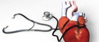
What is lung auscultation?
Auscultation is a method based on listening to changes in sounds that occur during the functioning of internal organs and systems. In the case of respiratory dysfunction, the doctor assesses the nature of the functioning of the lungs and bronchi.
A technique for studying breathing in a similar way was developed during the time of Hippocrates (IV-III centuries BC). To diagnose respiratory pathology, the doctor, during a standard examination of the patient, put his ear to the chest and listened for any extraneous or altered sounds.
The described method is called direct auscultation. In modern medicine, in 99% of cases an indirect version of the technique is used. Doctors use special instruments to auscultate the lungs - phonendoscopes (stethoscopes).
The device consists of a membrane and/or funnel that fits tightly against the area of the body being examined. The latter is connected by tubes (sound ducts) with rigid arms ending in ear olives. Due to the concentration of sound from the focus being studied, the doctor clearly hears what is happening under the membrane.
Auscultation of the lungs should be performed in all patients suffering from some form of respiratory pathology. The diagnostic method is simple, does not require the use of additional equipment and remains the basis for the initial assessment of the patient’s lung condition.
https://youtu.be/https://www.youtube.com/watch?v=38yYWg-aGlU
_
Noise classification
The main breathing noises include the following:
- Vesicular noise over the entire lung tissue.
- Bronchial noises that are heard above the larynx, trachea and large bronchi.
For pathological changes, for bronchitis and other similar pathologies. Associated with the work of the respiratory organs, various wheezing is added to the main noise or they stop being heard in the usual area. Listening makes it possible to establish the timbre, depth, duration and location of pathological noise.
Additional breath sounds include the following:
- Pleural friction noise characterizing dry pleurisy. It also occurs when metastases penetrate the pleura, with kidney failure or severe dehydration.
- Crepitation is a normal breathing noise that occurs when many alveoli are disconnected at the same time. The sound of this process is reminiscent of the crackling of cellophane or the rustling noise that appears as a result of rubbing your fingers against the hair near the ear.
- Moist wheezing occurs when a stream of air passes through the secretion, the low-viscosity liquid foams and air bubbles appear on its surface and immediately burst.
Bronchophony is a special type of auscultation when the doctor asks the patient to pronounce words in a whisper. If the words are defined, then we can talk about compaction of the lung tissue or the presence of resonating cavities that connect to the bronchus. This can happen with bronchial asthma.
In modern medicine, auscultation is gradually being replaced by the latest hardware diagnostic methods. The result of auscultation still retains some inaccuracies and only gives reason to assume the presence of one or another pathology. In this regard, the results obtained must necessarily be confirmed by other more objective diagnostic methods, for example, radiography, tomography or bronchoscopy.
Rate this article:
(No votes yet)
Loading...
Related posts:
- How and when is auscultation of the renal arteries performed?
- Auscultation method: when it is implemented, types and stages of diagnostics
- Types of auscultation and instruments for examination
- Diagnostic capabilities of arterial auscultation
- Location of cardiac auscultation points and assessment of examination results
- Purposes and techniques of fetal auscultation
Lung auscultation points
When using a phonendoscope, a certain sequence must be followed. Carrying out the methodology in accordance with generally known standards is the key to obtaining the most reliable results. An exception may be cases of dynamic monitoring of the condition of patients during long-term treatment. In such patients, the doctor specifically examines a specific pathological area.
Listen during auscultation of the lungs according to the scheme indicated below.
Alternately listening to sounds at the indicated points of auscultation of the lungs provides complete information about the work of the relevant organs.
The examination is carried out from top to bottom, from left to right (for the doctor). It is worth paying attention to the need for symmetrical application of the phonendoscope to the skin of the chest. You need to alternate between the left and right sides, as shown in the figure.
In the projection area of the heart, the lungs are not auscultated, which is due to the superimposition of the sound of the “body pump” on respiratory sounds, making it impossible to interpret them further.
Fact! Carrying out listening from behind provides the doctor with more space to work with a phonendoscope. Because of this, in the clinic, auscultation often begins from the back. From the point of view of propaedeutics, this approach does not provide a complete assessment of the patient’s condition. Therefore, it is recommended to start auscultation according to the scheme from the anterior surface of the chest.
Basic rules for implementing the methodology
The purpose of the examination is to identify and describe breathing sounds, as well as bronchophony over the entire area of the lungs. Auscultation of the lungs, listening points are determined with the patient sitting, standing, but with prolonged deep breathing, dizziness and fainting may occur due to hyperventilation in the lungs, as well as lying down for very weak patients.
It is important! The doctor sits or stands taking into account the patient’s position, the main thing is that it is comfortable and without much strain. Auscultation is carried out in the front, side or back. To obtain the necessary results, it is required that the patient breathe deeply during the procedure, so the specialist asks the patient to take deeper breaths before starting the examination.
When implementing the technique, it is carried out when specific points of auscultation of the lungs are identified:
- Auscultation ahead. In this case, the patient lowers his hands, and the doctor stands slightly to the right or in front of him. Auscultation begins from the upper part of the lungs, while the device is placed in the supraclavicular fossa so that the membrane touches the patient’s body over its entire area. The doctor focuses on the sounds heard, assessing them throughout the entire breathing cycle - inhalation and exhalation. Then the phonendoscope is installed in the symmetrical zone of the second supraclavicular fossa, listening to noise. Next, the examination consists of listening to noises in symmetrical areas of the anterior chest so that the midclavicular line intersects the installed sensor in the middle.
- Auscultation of the lateral sections. The patient should breathe deeply and evenly, clasping his hands and placing them behind his head. The phonendoscope is located on the side of the chest in the armpit. At the same time, breathing noise in this area is listened to and assessed. Then the examination continues, and the phonendoscope is successively shifted to symmetrical zones, descending to the lower part of the lungs.
- Auscultation of the posterior part. The patient should cross his arms over his chest. The phonendoscope gradually moves into the interscapular space, into the subscapular areas.
When auscultating the lungs, you need to listen very carefully to sounds. And after completing the diagnosis, the results are assessed:
- The main noise present at any point of auscultation.
- Identity of the main respiratory noise at symmetrically located points.
- The presence of collateral pathological respiratory noise with determination of its location.
Video auscultation of the lungs
A verbal description of the technique and localization of the main auscultation points in 80% of cases gives an approximate understanding of how the procedure is performed. To better understand the process, it is worth watching the video below. This guide demonstrates all listening points during lung auscultation, paying attention to important nuances.
A feature of the correct auscultation technique, which has not been mentioned before, is the need to listen to natural sounds from the healthy side to the sick side. Due to this technique, the localization of the pathological process and the severity of the problem become obvious. The doctor can compare the sound picture of the healthy and affected areas of the bronchopulmonary system.
Organization of decryption of received data
In medicine, there are some general rules for auscultation of the lungs. You can listen to the patient in any position, but it is most convenient when the patient sits on a chair and his hands are on his knees. This position allows you to obtain minimal background tones of the respiratory muscles.
It is important! Listening can also be done in a standing position, but then deep breathing can cause dizziness and even fainting. To prevent consequences and to press the sensor tightly to the body, the doctor always holds the patient with his free hand on the side opposite to listening.
During diagnosis, the sounds of the respiratory process during inhalation are initially compared, their characteristics are given, their volume and duration are assessed. Subsequently, the data obtained are correlated with the noises that were produced at a symmetrical point - this is comparative auscultation. Most attention is paid to the main breathing sounds - alveolar, heard above the lung area and disturbed during pneumonia. There are also bronchial sounds that can be heard above the larynx, above the large bronchi and above the trachea.
The propaedeutics of auscultation lies in the fact that the main noises are sound phenomena that complement the external breathing heard in a healthy person. The main noises are heard in both healthy and sick people. Pathological noises are special pathological manifestations that also accompany external breathing and are heard only in patients affected by any pathology. This phenomenon does not occur in healthy people.
Auscultation of the lungs in children
Auscultation of the lungs in children is an important diagnostic method that allows us to identify pathology of the respiratory system in young patients. The examination technology coincides with the principle of the procedure in adults.
Features of lung auscultation in children:
- The need to use smaller membranes or funnels;
- Poor development of the chest muscles, which leads to a significant increase in breathing sounds. This type of breathing is called puerile;
- The need for more careful control of the temperature of the phonendoscope applied to the child’s skin. Children react negatively to the touch of a membrane or funnel that is too cold.
The sequence of points and the principles of the procedure described above are relevant for small patients. Using auscultation, the presence and nature of wheezing, the localization of the inflammatory process, and the progression of organic or functional changes in the bronchopulmonary system are recorded.
Important! When using auscultation in children, the doctor always remembers that young patients are rarely distinguished by patience. Therefore, experienced pediatricians carry out diagnostics quickly, trying to turn the examination into a game.
Sometimes, for high-quality auscultation in a restless child, the doctor needs 2-3 attempts. Otherwise, the information obtained remains unreliable and may affect the choice of treatment method.
For what diseases
Over two thousand years of history of listening to the lungs, doctors have accumulated experience in diagnosing various diseases “by ear”. At medical universities, young doctors are taught how to recognize this or that pathology using a phonendoscope.
Diseases that are diagnosed using auscultation:
- Acute or chronic bronchitis;
- Pneumonia. Pneumonia is a serious pathology that changes the function of the relevant organs. Auscultation of the lungs during pneumonia is a method used additionally to monitor the quality of therapy;
- Bronchial asthma;
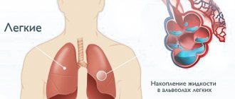
- Hydro- or pneumothorax - accumulation of fluid or air in the pleural cavity;
- Acute pulmonary edema is stagnation of blood in the tissues of the corresponding organ.
Using the described technique, tuberculosis or lung cancer can be suspected. However, these diagnoses cannot be established without the use of auxiliary methods.
Important! Auscultation is the primary diagnostic method, allowing the doctor to get a general picture of pulmonary dysfunction. To clarify the cause of the symptoms characteristic of a particular case, additional procedures are required. Otherwise, important details that affect the patient's outcome may be missed.
What is it used for?
Auscultation is used to detect a variety of diseases of the lungs, bronchi, heart and circulatory system. To do this, an assessment of the main and secondary breathing noises is carried out. Bronchophony over the entire surface is also assessed. These indicators are then supposed to be compared with normal ones, on the basis of which a conclusion is made about the presence or absence of diseases.
Thanks to auscultation, the following pathological conditions inherent in children and adults can be detected:
pneumonia;- pulmonary infarction;
- presence of a tumor in the lung;
- pulmonary edema;
- tuberculosis;
- pneumothorax;
- accumulation of fluid in the pleural cavity;
- heart failure.
Since the main signs by which such a diagnosis is carried out are noises, it is necessary to find out what kind of noises can be detected during auscultation. This:
- Vesicular respiration. This type of noise is soft and uniform and should be continuous when inhaling. The sound resembles the sound “v” or “f”.
- Bronchial breathing. It is observed in the phases of inhalation and exhalation, similar to the sound “x”. When exhaling, this noise is more sharp than when inhaling.
- Mixed breathing. It can be called intermediate between the first two, since it has the features of both of them.
In addition to the main ones, the doctor may hear additional noises during auscultation, which are signs of pathological phenomena. This:
- Wheezing. Can be dry or wet. They appear as a whistling, buzzing or buzzing sound (dry) or resemble the sound of bursting bubbles (wet).
- Crepitus. This phenomenon is a jerky squeaking sound.
- Pleural friction noise. When this noise is detected, it can be assumed that its source is very close to the surface. It sounds like the crunch of snow or the rustle of paper.
In order for the diagnosis to be correct, the doctor must take into account not only the existing extraneous noises, but also the characteristics of the main noises. In addition, it is necessary to take into account the symptoms that the patient names, his individual characteristics and much more.
Algorithm for performing lung auscultation
A feature of modern lung auscultation remains the presence of a phonendoscope. A few doctors use a stethoscope - a wooden tube without flexible elements and the usual ear tips.
Diagnostics can be carried out both in a hospital (clinic) and at the patient’s home. In extreme situations, listening to the lungs is carried out in the conditions in which the person finds himself. The main thing is to establish the presence of damage to the lung tissue and decide on the necessary treatment.
Algorithm for performing lung auscultation:
- The patient stands or sits during the examination;
- It is important that the room is warm and quiet;
- For high-quality auscultation, it is recommended to undress the patient from top to waist. The rustling of clothes can cause the sounds heard by the doctor to be misinterpreted;
- The doctor alternately applies the head of the phonendoscope to the corresponding points, according to the diagram indicated above.
Doctors are recommended to use one instrument, which helps them become accustomed to its operation. During diagnosis, the doctor pays attention to the volume of sounds occurring in the chest, height, symmetry, possible migration, homogeneity.
For differential diagnosis and comprehensive research, auscultation is carried out:
- during the patient’s normal breathing;
- during deep inhalations and exhalations;
- after the patient coughs;
- when changing the position of the body.
Due to these techniques, it is possible to distinguish some features of pathological processes.
Diagnosis in adults based on lung sounds
Even with auscultation, the first thing the doctor pays attention to is the clinical signs of the disease that appear in the patient.
If we talk directly about auscultation, when listening to the lungs, the development of pneumonia is indicated by the noises and sounds discussed in this chapter. Based on their nature, one can make assumptions regarding the form of pneumonia.
- Croupous pneumonia - when listening to the lungs, crepitation is clearly audible, which is often compared to a crunching sound. In addition, wheezing is classified as wet, occurring mainly at the time of deep inspiration. The occurrence of crepitations and moist rales is caused by the passage of air through accumulations of mucus on the walls of the alveoli and vesicular exudate in the bronchi.
- Focal pneumonia - this form of pneumonia is characterized by so-called rough breathing, accompanied by dry wheezing and clearly localized crepitus, which can be heard directly in the area of the inflammation. In this case, crepitations occur during inhalation, which is explained by the opening of the alveoli, which have stuck together due to the accumulation of mucus. Dry wheezing, on the contrary, occurs during exhalation, and as air escapes, it turns into fine bubble noise.
Regardless of the form or type of pneumonia, characteristic noises and wheezing occur both during inhalation and exhalation. After coughing, pathological sounds in the lungs become more distinct or change completely, as well as from a change in body position. It is for this reason that the doctor is obliged to conduct auscultation before and after coughing attacks, and also ask the patient to take a horizontal and vertical position.
Important! In addition to wheezing and noise, the doctor should pay attention to any deviations from the norm. Even weakened or heavy breathing can suggest pneumonia.
What you need to know and possible consequences
Auscultation of the lungs is the generally accepted standard for diagnosing diseases of the respiratory system. The procedure is safe for the patient. During the examination, the person does not feel any discomfort except for the touch of a cool phonendoscope. The duration of the examination depends on the severity of the pathology. On average, a doctor needs 2-5 minutes to fully carry out the corresponding procedure.
Undesirable consequences of auscultation are a myth. It is extremely difficult to harm a patient using the appropriate technique.
Stereophonendoscope
Human hearing is imperfect - we hear well mainly spatial vibrations. In auscultation, much depends on the stage of development of pneumonia and the hearing characteristics of the doctor who conducts the diagnosis. Because of this, some details may slip through the cracks, leading to a misdiagnosis or allowing the disease to progress until the doctor can hear certain sounds.
To prevent this from happening, you can resort to a more accurate method of auscultation, during which a special device is used - a stereostethophonendoscope. The device has a number of undeniable advantages :
- The ability to “hear” pneumonia at the earliest stages of the pathological process, that is, before the appearance of a bright auscultatory picture. The human ear cannot recognize such sounds.
- The ability to accurately determine the location of the source of inflammation.
- Thanks to the sensitivity of the device, it is possible to begin treatment in record time.
- The diagnostic process is significantly accelerated.
Normal indicators or normal auscultatory picture
The concept of normality during auscultation requires an understanding of the principles of the formation of sound vibrations during the passage of air through the respiratory tract.
There are two types of breathing:
- Vesicular (alveolar). When auscultating the lungs, this type is normally heard over the entire surface of the lungs. The formation of characteristic noise is due to the filling of the alveoli with air, which is accompanied by a turbulence of its flow with tension in the walls of the corresponding structures. During auscultation, a characteristic “f” sound is heard, mainly during inspiration. The exhalation is heard for a very short time;
- Bronchial. This type of sound is determined above the surface of the larynx and trachea. The peculiarity remains the same duration of the two phases of the respiratory cycle.
In children, vesicular breathing is heard as noisy with a higher amplitude. The reason is the poor development of the muscular corset and the adherence of the lungs to the inner wall of the chest.
Normally, the breathing pattern is the same for all locations. The severity of noise may decrease at the upper and lower points of auscultation, which is due to a decrease in the number of alveoli in these places due to the anatomical features of the lungs.
Auscultation rules
Correct performance of lung auscultation involves a number of aspects:
- maintaining silence during the procedure;
- comfort for the patient and the doctor;
- following the pattern of auscultation points;
- careful analysis of the information received.
By following these rules, the doctor receives the maximum amount of relevant information to assess the condition of the patient's respiratory tract.
Main breath sounds
During auscultation of the lungs, the doctor hears a variety of sounds. A variant of the norm is described above. The table below shows the most common diseases with characteristic changes in the auscultation pattern.
A description of the pathological changes will be presented below.
Vesicular respiration
The principle of the corresponding noise is the filling of the alveoli with air. Pathological changes are manifested by weakening of vesicular respiration. Possible pathogenetic causes of the corresponding situation:
- Narrowing of the respiratory tract. The result is a decrease in the amount of air that enters the lungs;
- The appearance of compaction foci in the tissues of the corresponding organs. The result is a decrease in the number of active alveolar conglomerates, which leads to weakened air exchange;
- Inflammatory or congestive process in the lungs. Pneumonia is a typical example of this pathology mechanism;
- Increased alveoli in size due to emphysema (increased pneumatization). The result is that the walls of the corresponding structures become inelastic, which interferes with the normal process of noise formation;
- Accumulation of fluid or air in the pleural cavity. The result is compression of the lung tissue leading to collapse of the organ and the inability to perform the function with a complete loss of vesicular respiration. Apnea (lack of lung function) is also accompanied by a corresponding auscultatory pattern.
Qualitatively, vesicular respiration can acquire a hard tint. The causes are predominantly bronchogenic in nature. Normally, the doctor hears a soft blowing sound. In the case of pathology, a hard, dry grinding sound is detected, which indicates the presence of narrowings or other changes in the respiratory tract. The corresponding picture is typical for smokers.
Scanning breathing may also occur. This pathological variant of vesicular noise is characterized by intermittency. There are long pauses between breathing cycles, and the patient feels unwell.
Bronchial breathing
Bronchial breathing under normal conditions is heard only in the area of the larynx and trachea. Its appearance in other parts of the chest indicates a dysfunction of the respiratory tract.
Interesting! You need to understand that bronchial breathing cannot occur in the lung tissue. If the doctor hears a corresponding sound in the middle part of the chest, then this means that the alveoli have become denser and are not filled with air while maintaining patency of the bronchi. The corresponding noise simply spreads through the respiratory tract, like along highways.
Pneumonia, lung cancer, pneumosclerosis and other pathologies accompanied by hardening of the lungs will cause a corresponding auscultatory picture.
Things to remember
Today there are general rules for auscultation of the lungs. There are only a little more than ten basic rules, and following them is not difficult.
So, it is important to remember:
- The fact that in the room in which this manipulation is carried out there must be peace and absolute silence.
- The fact is that for the procedure to be carried out correctly, the patient must be naked, since friction of the device against clothing causes side effects of sound.
- The need to pay attention to the hair of the actual area (in order to avoid side effects of sound, the hair on the chest needs to be soaped or wet).
- Maintaining the correct temperature in the room (if a person shivers from the cold, this will interfere with listening to the organs).
- The fact is that the position of the patient and the specialist listening to the organs should be as convenient and comfortable as possible.
- Applying the stethoscope evenly to the listening surface (this should be done as tightly as possible, but without pressure).
- The fact is that it is better not to touch a hard instrument during the procedure, as this may contribute to the appearance of side sound effects.
- The fact is that there is no need to put pressure on the instrument, because it will not only be uncomfortable for the patient, but also painful.
- Using the same tool.
- Controlling the patient's breathing.
- Systematic and persistent listening to relevant organs.
If the patient is being listened to in an upright position, the doctor should grasp him as tightly as possible. This will help you hear all sound phenomena.
In order to hear all the noise, a specialist must abstract from the surrounding reality and focus on the area of study.
If the patient has pneumonia
In case of pneumonia, manipulation is carried out using a special instrument called a stethoscope. This tool consists of:
- sound-amplifying membrane;
- tube systems made of high-quality plastic.
When a person’s lungs are normal, the specialist hears a clear sound, indicating that breathing has not undergone any abnormal changes. And with pneumonia, exudate prevents a person from breathing normally. In this case, the specialist clearly hears the sound of weak, labored breathing. Specific wheezing can also be heard.
If the patient has bronchitis
With bronchitis, the patient's breathing can be either uniform or uneven. It is harsh in places, and the exhalation is a little lengthened. The noises that accompany breathing are either continuous or intermittent.
Intermittent sound phenomena are:
- group;
- single;
- having inconsistent localization.
They are caused by specific secretions of a more liquid consistency.
Sometimes with bronchitis, sound phenomena that are not interrupted by respiratory phases are heard. They are formed against the background of the presence of a fairly viscous secretion.
This symptom indicates the development of dry bronchitis.
The sounds can sometimes be creaking and sometimes purring. Often the doctor hears a sound similar to the noise of a working spindle.
If the patient has bronchial asthma
In bronchial asthma, due to weakened breathing of both lungs during inhalation and exhalation of air, the specialist clearly hears whistling dry rales of a wide variety of shades.
In the event that attacks of suffocation occur for a long time, then symptoms of right heart failure are observed.
Additional breath sounds
The noises described above are the main ones. In addition to bronchial and vesicular breathing, additional sound phenomena can be recorded during auscultation, which affect the understanding of the pathology developing in the patient’s lungs.
Wheezing
Wheezing is an auxiliary breathing noise associated with the passage of air masses through the respiratory tract, in which additional obstacles are formed (sputum, pus, blood). During contact with a liquid, turbulence of the gas mixture occurs, which leads to the appearance of a corresponding phenomenon.
Wheezing occurs:
- dry;
- wet;
- mixed.
Dry wheezing is formed when the airways are blocked by thick and viscous mucus. Depending on the diameter of the section of the respiratory tract where the block occurs, the height, timbre and duration of the corresponding phenomenon changes. There are buzzing, whistling wheezing sounds. The latter are more common and are characteristic of bronchial asthma.
Moist rales differ in the mechanism of their occurrence. For the corresponding sound to appear, air must pass through a liquid medium with the formation of bubbles, which, when bursting, provide the appearance of the described phenomenon. Depending on the localization of the pathological process and the diameter of the area of the affected respiratory tract, wheezing can be small-, medium- and large-bubble. The cause of this sound is the accumulation of blood, pus, and liquid sputum in the bronchi.
Crepitus
Crepitation is a sound characteristic of the early and late stages of pneumonia. Unlike moist rales, the pathogenetic basis for the appearance of noise remains the penetration of fluid into the cavity of the alveoli. During exhalation, the corresponding structures decrease in size. The liquid envelops the walls of the bubbles, which leads to sticking together. During inhalation, air fills the alveoli, which is accompanied by the walls peeling off with a characteristic click.
This sound occurs simultaneously in all bubbles, which creates a corresponding auscultatory picture that resembles rubbing hair near the ear.
A characteristic feature of crepitus remains the need for deep inspiration to straighten the alveoli. With shallow breathing, the phenomenon is not recorded. Therefore, for differential diagnosis of early and late stages of pneumonia, it is imperative to ask the patient to breathe deeply.
Crepitus additionally occurs in all lung diseases that are accompanied by the penetration of fluid into the respiratory vesicles.
Pleural friction rub
Pleural friction noise is a pathological phenomenon that is not associated with dysfunction of the lung tissue specifically. The source of the problem becomes the pleural cavity, the visceral and parietal layer of the corresponding connective tissue structure. Normally, all of these elements are smooth and elastic.
In the presence of an inflammatory or infectious process, partial leakage of plasma into the specified space is observed. Quite quickly, excess liquid is absorbed back into the vessels, however, the dry part in the form of fibrin remains.
The result is the deposition of hard fibers on the surface of the pleura. During the next respiratory movements during auscultation, the doctor records noise that occurs due to friction of fibrin conglomerates. The sound phenomenon resembles the rustling of snow underfoot. The typical cause is dry (fibrinous) pleurisy.
At the same time, the patient is worried about increased body temperature, chest pain, and discomfort during deep breathing.
The pleural friction noise resembles crepitus or moist rales. For differential diagnosis, the patient is asked to close his mouth and nose with his hands and imitate the respiratory movements of the chest.
If the noise remains, then the pleura is affected. With wheezing and crepitus, the connection with the air flow is always maintained. Additionally, you can ask the patient to cough. Wheezing and crepitus change their character after the appropriate test, which is not typical for pleural friction noise.
Auscultation of the lungs. Technique, algorithm
Auscultation of the lungs is a research method based on the perception of sounds naturally occurring in the body through indirect or direct contact of the researcher’s ear with the surface of the body.
Auscultation of the lungs using a simple technique
At the same time, this is a very difficult to interpret research method, which in its significance and value in some cases is not inferior to x-ray examination. Listening requires experience, one must have a correct understanding of the sound impressions perceived by the ear, and most importantly, one must be able to find in these extremely diverse acoustic phenomena a reflection of the pathoanatomical processes taking place in the lungs according to the place of listening.
To correctly understand the auscultated lung sounds, it is necessary to pay attention to their nature, strength, relationship to the phases of breathing (i.e., inhalation and exhalation), localization and distribution. The same with percussion, at the beginning we carry out comparative auscultation. Listening at strictly symmetrical places in the chest, we compare the data obtained. It is necessary to mentally compare inhalation with exhalation on the same side, inhalation with exhalation and exhalation with exhalation on opposite sides.
The patient's position during auscultation, depending on the condition, can be any. However, the most comfortable position will be a standing or sitting position with your hands freely lowered or placed on your knees. You should not listen to severe, weak patients in a standing position; – when breathing deeply, they often experience dizziness and fainting. The most incorrect position is when the patient sits on the bed with his legs extended. The patient should be naked to the waist, as clothing often introduces extraneous sounds. It is necessary to teach the patient to breathe correctly: deeply,
calmly, evenly, through the nose and only at the special request of the doctor - through the mouth at an average pace, i.e. take about 25 breaths per minute.
According to the doctor’s sign, at the end of the exhalation, the patient should, without inhaling, cough briefly vigorously, but silently, only with residual air; Take a deep breath again immediately after coughing.
Failure to follow this rule is a big omission: in almost half of patients with tuberculosis, mild wheezing is heard only after coughing. And a doctor who does not instruct the patient how to breathe does not receive what auscultation can give. Proper installation of the stethoscope is also of great importance. If the stethoscope does not fit tightly to the skin, then you can easily hear noises and wheezing that are not really there.
When listening to the lungs, first of all, you need to listen to breathing sounds, determine the nature of breathing, its intensity, and establish the ratio of inhalation and exhalation.
After this, pay attention to possible side noises or wheezing. When listening to respiratory sounds, breathing through the mouth is undesirable (the patient breathes through the nose), while when wheezing, breathing through the mouth will promote stronger air movement in the bronchi and thereby easier formation, and therefore the perception of wheezing.
Then listen to the friction noise of the pleura, which can most often be heard in the inferolateral parts of the chest, where the excursion of the lungs is small, and, therefore, the conditions for listening to the friction noise are the best.
Finally, the voice is heard. Both loud speech and whispers are heard. Both through a stethoscope and directly by the ear. The order of listening places is the same as for percussion, i.e. apexes, anterior surface (top to bottom), lateral surfaces (from the axillary fossae downwards), posterior surface (above the shoulder blades, between them and under the shoulder blades) in symmetrical places alternately.
The sounds or noises that arise when listening to the respiratory organs are divided into three main groups: 1. Breathing noises. 2. Side noises or wheezing and crepitus. 3. Pleural friction noise.
Auscultation of the lungs. Algorithm
1. Preparation for auscultation, instructing the patient, identifying and eliminating factors that can cause the appearance of auscultation artifacts 2. Strict symmetry of the auscultation points on the right and left. If asymmetry of the main respiratory sounds is detected and the presence of side (localized) respiratory sounds is suspected, proceed to auscultation in zones 3. Determination of the presence (or absence) and differentiation of the main respiratory sounds A. Determination of the time ratio of the inhalation and exhalation phases B. Determination of the qualitative characteristics of the main respiratory sounds noise
| Vesicular a) Predominance of the inhalation phase over the exhalation phase (3: 1) b) Qualitatively resembles the sound “f” pronounced on exhalation c) Relatively soft | Bronchial a) The inhalation and exhalation phases are equal in duration and exhalation is somewhat predominant b) Qualitatively resembles the sound “x” c) Coarser d) As a rule, determined locally or unilaterally |
4. Assessment of the nature and changes in vesicular respiration a) Physiological vesicular respiration (puerile) b) Pathological vesicular respiration and its characteristics A. Symmetry 1. Its total change 2. Unilateral 3. Local B. Increased vesicular respiration (hard, rough) C. Weakening of vesicular respiration D. Saccaded vesicular respiration E. Unilateral (local) disappearance of vesicular respiration
5. Assessing the nature and changes in bronchial breathing a) Physiological (above the trachea and larynx) b) pathological - in the area where vesicular breathing is supposed to be heard A. Localization (one-sided, local) B. Volume 1. Normal 2. Loud (metallic) 3. Quiet (compressive stelectasis) B. Rare variants (amphoric)
6. Determination of the presence of adverse respiratory sounds A. Over the entire surface of the lungs B. Unilateral C. Local
7. Based on qualitative characteristics (duration, pitch, timbre, stability) and additional auscultatory techniques (forced breathing, coughing, tension and relaxation of the abdominal muscles with the upper respiratory tract closed), a specific respiratory noise is determined
8. Wheezing A. Timbre B. Pitch C. Duration Wet / Dry Small bubbles / Medium bubbles / Large bubbles Whistling / Humming Oral (bubbling breathing) / Oral (rooster crowing)
9. Crepitus Differentiated from fine-bubbly moist rales In contrast to them a) Does not change the caliber (only volume) b) Qualitatively resembles the crackling sound of hair when rubbed near the ear With pleuropneumonia: a) Initial b) Terminal
10. Pleural friction noise Differentiates from dry rales, in contrast to them a) It is heard as if “closer” to the ear b) Does not change in pitch after coughing c) Changes in volume when the phonendoscope is pressed on the chest d) Is usually accompanied by a typical pain syndrome Qualitatively reminiscent crunching snow, creaking door hinge
11. Rare auscultatory symptoms a) Noise of a “falling drop” b) Splashing noise
12. Assessment of the relationship between the main and secondary respiratory sounds 13. Assessment of the relationship of auscultatory symptoms with data obtained by other methods of studying the respiratory system.
Auscultation. Methods, rules
Auscultation (auscultare - listen, listen) is a method of research using the perception of sounds that naturally occur in the body, which are perceived through direct or indirect - with the help of some solid body - contact of our ear with the surface of the body. Listening to voices, coughing, sneezing, loud breathing, wheezing, rumbling in the intestines and other sounds heard at a distance does not apply to the auscultation method.
Auscultation. History of origin
Listening to sounds occurring inside our body was used for diagnostic purposes in ancient times. Thus, in the writings of Hippocrates there are references to the friction noise of the pleura, the sound of splashing in the pleural cavity, and moist rales in the lungs. At the beginning of our era, they undoubtedly knew how to listen to heart sounds. But then, for one and a half thousand years, listening did not play a role in the study of patients.
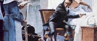
Listening became a diagnostic method only thanks to the French scientist Rene Laennec (1781-1826), who was a talented clinician, pathologist and teacher at a medical school in Paris.
In Russia, the development of the auscultation method is associated with the names of P.A. Charukovsky and M.Ya. Mudrova. The talented Russian professor Grigory Ivanovich Sokolsky, whose name is associated with the doctrine of rheumatism (Buyo-Sokolsky disease) in his works “On the study of diseases with hearing and a stethoscope” and “The doctrine of chest diseases”, described in detail the auscultatory phenomena heard in heart defects and respiratory diseases .
Auscultation deals with very faint sounds from our body that travel little or nothing through the air. Therefore, if there is at least the thinnest layer of air between the ear and the surface of the body, we do not hear sounds, but begin to perceive them as soon as a continuous communication through the solid body is established between the ear and the sounding body. This is achieved either by direct contact of the ear, for example, with the chest, or by connecting them with some kind of solid body capable of conducting vibrations (a stethoscope). On the same basis, the already inaudible tone of a tuning fork placed in front of the ear is again perceived well and for a long time if the tuning fork is placed on the head. Thus, it was proven that sound conduction in stethoscopes occurs not through the column of air inside them, but along their walls.
The name stethoscope was given by Laennec. His stethoscope originally resembled a paper parcel. It was a hollow wooden tube 33 cm long with the same diameter throughout, which was disassembled in the middle. Modifications to this original form went in different directions: thinning the tube, shortening it, making the ear end more convenient, and using various materials for making the tube.
The stethoscope is a cylindrical tube. In most cases, its wide part is funnel-shaped and is applied to the auricle, and the narrower part, the so-called bell of the stethoscope, is applied to the patient’s body. Stethoscopes are made from various materials: wood, metal, ivory, plastic. Subsequently, instead of rigid stethoscopes, flexible ones were proposed, first recommended by N.F. Filatov. In this case, two rubber tubes come from the bell of a conventional stethoscope, the ends of which are inserted into the researcher’s ears. Finally, the latest modifications of the stethoscope concerning its thoracic end were expressed in the addition of a device for resonance to it in order to enhance sound phenomena. This is how various forms of phonendoscopes arose. First, simple ones, when the chest end of the stethoscope was covered with a rubber membrane, and more complex, when the end part of the phonendoscope was a metal cavity covered with a membrane. Sound phenomena arising in a particular organ are transmitted to the membrane, which vibrates. The cavity covered with this membrane, according to the theory of resonance, amplifies the sound. Sometimes a multiphonendoscope is used in classes with students.
In principle, the term stethoscope given by Laennec does not accurately reflect the purpose of this device, since it comes from two words - stethos - chest and scopeo - look. The advantage of flexible stethoscopes is twofold: convenience of examination for the doctor and the patient and a more significant amplification of sound. They almost do not change the nature of natural sounds and produce little side noise. The disadvantage is a significant change in the natural character of sounds and a large number of easily occurring extraneous noises. They are inconvenient for the doctor and the patient and their use is tedious.
Auscultation of the lungs. Theory
Auscultation, as a research method, deals with very faint sounds from our body, which have very little or no propagation in the air.
We can perceive such sounds under the condition of continuous communication through a solid body between the ear and the sounding body. This is achieved either by direct contact of the ear, for example, with the chest, or by connecting them (the ear and the sounding body) with some kind of solid body capable of conducting vibrations (a stethoscope). If there is at least the thinnest layer of air between the ear and the surface of the body, we do not hear sounds. From this it is clear that in stethoscopes of any design, sound is transmitted not through the column of air inside them, but along their walls, just as in
In our ear, sound vibrations are conducted not through the air of the ear canal, but through the auricle, the wall of the ear canal, the eardrum, etc.
Full stethoscopes are preferred not because they conduct sounds through the air of their canal, but because they are lighter and more capable of vibration.
To maintain the maximum ability of the stethoscope to vibrate, you need to apply it to the body as lightly as possible and not touch it with your hand while listening. The size of the funnel of the chest part of the stethoscope matters: the narrower it is, the more accurately it is possible to localize the sounds being heard, but they are collected from a smaller surface and, therefore, weaker. The wider the funnel, the weaker sounds can be perceived.
Auscultation. Methods
In general, what matters is not the choice of stethoscope or phonendoscope, not the method of listening, but the ability to auscultate. You should always use the same device, since each stethoscope and phonendoscope has its own individual characteristics.
There are two main methods of auscultation:
- A method of immediate or direct auscultation, in which listening is performed directly with the ear attached to the patient’s body. This method is still widespread in France, the homeland of Laennec, the founder of mediocre auscultation. French doctors usually use direct auscultation through a thin cloth.
- The method of mediocre, indirect or instrumental auscultation is carried out using a stethoscope or phonendoscope.
| Advantages | Flaws | |
| Method of immediate or direct auscultation | – large surface of perception – natural character of the sounds heard | – localization of sounds is difficult, especially when listening to the heart; impossibility of use in some parts of the body (in the supraclavicular and axillary fossae); unhygienic, dangerous for infectious diseases. |
| Method of mediocre, indirect or instrumental auscultation | – the ability to localize sounds anywhere in the body and in any position of the patient, which is especially important in seriously ill patients – the hygiene of the method. | – smaller surface of perception – changed nature of sounds heard |
Auscultation. Choosing a Stethoscope Solid Stethoscopes
Advantages: they change little the character of natural sounds, produce little side noise, and at the same time convey tactile sensations along with sounds.
Flaws. The examination is inconvenient and tedious for the doctor and the patient, painful when pressed.
Flexible stethoscopes
Advantages: ease of examination for the doctor and the patient, the ability to see his facial expression and significant amplification of sound.
Flaws. Significant change in the natural character of sounds. For beginners in auscultation, it is better to use a stethoscope, and it makes no difference which is better - hard or soft. It is not the method or method of auscultation that decides the matter, but the ability to auscultate.
Auscultation. Rules
- Auscultation is carried out in a warm room; it is necessary that silence be maintained, since the appearance of muscle tremors will interfere with auscultation.
- The patient's body should be bare, as friction of clothing can cause collateral noise.
- You should pay attention to the hair on the body, since the friction of the instrument against the hair imitates typical pathological auscultatory phenomena. Moisten or soap the hair at the listening site to avoid unwanted noise.
- The bell of a stethoscope or phonendoscope should be tightly, but not tightly applied to the listening surface (apply evenly, tightly, but lightly to the listening surface).
- The position of the doctor and the patient should be comfortable.
- It is better not to touch the solid stethoscope with your hand while listening to avoid unwanted sounds and a decrease in sound conductivity.
- When listening to a patient in a standing or sitting position, the doctor should clasp (hug) the patient with his free hand so that they form a single whole.
- Do not press the stethoscope so as not to cause pain to the patient.
- Use the same stethoscope whenever possible.
- When listening to the respiratory system, control the breathing of the subject.
- Listen systematically and persistently.
You need to get used to being distracted from everything around you. For this purpose, it is useful to close your eyes and plug your free ear when listening (to eliminate unnecessary sound and visual irritation).
Usually comparative auscultation is performed, in which the listening order is the same as for comparative percussion.
Listen in symmetrical areas and alternately on one side or the other, comparing each time the auscultation data in the following order: apexes, anterior surface of the lungs from top to bottom in symmetrical areas, lateral surfaces (from the axillary fossae downwards, preferably with arms thrown behind the head) , posterior surface in the supra-, inter- and subscapular areas.
First, they listen to breathing sounds, which are called basic, that is, they determine the nature of breathing, its intensity, the ratio of inhalation and exhalation. Then they pay attention to side noises or wheezing, crepitus, pleural friction noise, and then listen to the voice.
Auscultation of the lungs
Auscultation of the lungs is performed according to a certain plan: first in front and above, moving down and to the sides. Then in the same sequence - from behind and along the side lines.
Places for listening to the lungs: a—in front; b - behind
Auscultation determines the nature of respiratory sounds that form auscultatory types of breathing:
vesicular - inhalation and ]/3 exhalation are heard in the form of the sound “f”; hard (puerile in children under six years of age) _ both inhalation and exhalation can be heard with the sound “f”; bronchial - you can hear inhalation and exhalation with the sound “x”; amphoric - inhalation is similar to the sound produced by forcefully blowing air into the narrow neck of an empty glass vessel. Indicates the presence of a smooth-walled cavity in the lung (abscess, Glossary Link cavity); saccaded - inhalation consists of two or more phases. It happens when there is a tumor or foreign body in the trachea or large bronchus. Normally, vesicular breathing is heard below the angles of the scapula in the back, on the lateral surfaces of the chest and in front below the 2nd rib and lateral periosternal lines; bronchial - in the interscapular space and behind the sternum - in front.
Types of breathing:
o - vesicular; 6 - weakened vesicular; c - enhanced vesicular; g - bronchial; d — weakened bronchial; e - enhanced bronchial; g - bronchovesicular; z - saccade
Adverse breath sounds:
crepitus - a small crackling sound when inhaling, evidence of pathology in the alveoli; pleural friction noise - inhalation and exhalation are similar to the crunch of snow underfoot or the friction of two sheets of paper - evidence of pathology of the pleural layers (they have become rough). The pleural friction noise is sometimes similar to crepitus or moist fine bubbling rales.
Features:
after coughing, wheezing changes its character or disappears; with strong pressure on the chest with a stethoscope, the pleural friction noise intensifies (wheezing does not); crepitus is heard only during the inhalation phase, and wheezing and pleural friction noise are heard in both phases of breathing; with the mouth and nose closed and the abdomen moving, the pleural friction noise is heard, but wheezing and crepitus are not; wheezing: dry - whistling, wheezing, buzzing - occurs in the bronchi due to narrowing of their lumen or due to the accumulation of viscous sputum; wet - small, medium, large bubbles, reminiscent of the sound of bursting air bubbles passed through water. They occur in the bronchi of the appropriate caliber when liquid sputum accumulates in them.