The quality of a person’s life and his health largely depend on the state of the cardiovascular system. The heart and blood vessels perform important functions - they pump blood, which supplies all organs and tissues with oxygen and nutrients, and removes carbon dioxide. The modern level of medical technology, innovations in treatment and diagnosis have significantly reduced mortality from cardiovascular diseases, but they still remain the main causes of death in all countries. One of the serious pathologies diagnosed by cardiologists in middle-aged and elderly people is myxomatous degeneration of the mitral valve.
What changes does the pathological process suggest?
This pathological condition of the human heart has other names. Doctors may diagnose the patient using the terms “mitral valve prolapse” or “endocardiosis.”
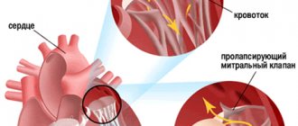
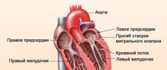
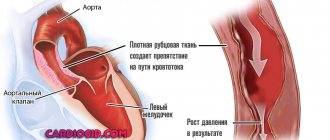
The mitral valve separates the left atrium from the left ventricle. Under normal conditions, it prevents the reverse flow of blood from the atrium into the ventricle of the heart. For some reasons, most often due to genetic predisposition or viral infections, a person experiences degeneration of the valve leaflets - their stretching and thickening.
This process is called MVP (mitral valve prolapse), its development in the patient causes disturbances in the functioning of the heart. A backflow of some blood from the atrium into the ventricle occurs - this phenomenon is called regurgitation. The development of the disease leads to a change in the patient’s condition and the appearance of murmurs during heart function.
The myxomatous process leads to further changes in the functioning of the organ. Its result is an increased size of the left ventricle, and subsequently the entire heart, arrhythmia, heart failure, and disturbances in the functioning of other valves.
What do degenerative changes in organ tissues look like?
Myxomatous degeneration of the mitral valve - signs, diagnosis, therapy
The quality of a person’s life and his health largely depend on the state of the cardiovascular system. The heart and blood vessels perform important functions - they pump blood, which supplies all organs and tissues with oxygen and nutrients, and removes carbon dioxide.
The modern level of medical technology, innovations in treatment and diagnosis have significantly reduced mortality from cardiovascular diseases, but they still remain the main causes of death in all countries.
One of the serious pathologies diagnosed by cardiologists in middle-aged and elderly people is myxomatous degeneration of the mitral valve.
What changes does the pathological process suggest?
This pathological condition of the human heart has other names. Doctors can announce the diagnosis to the patient using the terms mitral valve prolapse or endocardiosis.
The mitral valve separates the left atrium from the left ventricle. Under normal conditions, it prevents the reverse flow of blood from the atrium into the ventricle of the heart. For some reasons, most often due to genetic predisposition or viral infections, a person experiences degeneration of the valve leaflets - their stretching and thickening.
This process is called MVP (mitral valve prolapse), its development in the patient causes disturbances in the functioning of the heart. A backflow of some blood from the atrium into the ventricle occurs - this phenomenon is called regurgitation. The development of the disease leads to a change in the patient’s condition and the appearance of murmurs during heart function.
The myxomatous process leads to further changes in the functioning of the organ. Its result is an increased size of the left ventricle, and subsequently the entire heart, arrhythmia, heart failure, and disturbances in the functioning of other valves.
Changes depending on the degree of disease
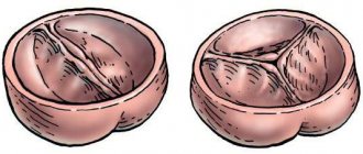
Myxomatosis of the mitral valve goes through three stages of development (degrees). Each of them has its own characteristics and requires a different approach to therapy.
Myxomatous valve degeneration of the first degree is expressed in a slight thickening of its leaflets - less than 5 millimeters. In this case, the valves close completely, there are no manifestations of the disease at all. This condition requires observation by a cardiologist and a change in the usual lifestyle with a review of attitudes towards bad habits, nutrition, and physical activity.
The second stage of the disease is diagnosed when the valve thickens in the range from 5 to 8 millimeters, and there is a stretching of its valves, a violation of their closure, and a change in the outline of the hole between them.
Thickening of the valve leaflets by more than 8 millimeters indicates the third stage of the disease. With it, the valve leaflets do not close, and there is a pronounced deformation of the mitral ring.
Manifestation of symptoms of the disease
Any suspicion of heart disease should be a reason to urgently contact a cardiologist. Myxomatous degeneration of the mitral valve leaflets manifests itself depending on the degree of progression of the pathology.
Its first degree develops asymptomatically, subsequent stages of the disease are manifested by characteristic symptoms:
- a person’s performance and endurance decrease, persistent fatigue appears,
- there is pain in the chest,
- cardiac arrhythmia appears - the heartbeat may increase without physical activity, noticeable interruptions in the functioning of the heart are observed,
- possible fainting, dizziness, nausea,
- there is a feeling of lack of air, accompanied by shortness of breath and cough.
Methods for confirming the diagnosis and conducting examinations
The diagnosis of myxomatous degeneration can be made using several types of diagnostics. During the initial examination of the patient, the doctor may suspect the presence of the disease by listening to the heart tone with a stethoscope.
Systolic murmur becomes an important reason for referring the patient for a detailed examination.
It is carried out using:
- ultrasound examination,
- chest x-ray,
- deciphering the data obtained when taking an electrocardiogram,
- laboratory tests.
Such diagnostic methods make it possible to study the changes that have occurred in the valve, identify possible threats to the further development of the pathology, and prescribe treatment.
Treatment and preventative actions
The patient's condition at the initial stage of the disease does not require drug treatment. It is prescribed by a cardiologist if the pathology begins to progress.
Drug therapy in this case is aimed at achieving the following results:
- restoration of cardiac activity,
- relief of pain symptoms,
- preventing the occurrence of blood clots.
This treatment is indicated for the second degree of the disease. The subsequent, third degree involves surgical intervention. Its goal is to replace the mitral valve with a prosthesis. When carrying out the intervention, high-tech techniques are used that have a gentle effect on the health of the person being operated on.
During and after treatment, it is important to follow preventive measures.
A patient with myximatous mitral valve degeneration requires:
- forget about drinking alcohol, smoking,
- use of physical activity, its types and intensity must be agreed upon by a doctor,
- switch to a diet using healthy foods prepared in a certain way.
It should be remembered that symptoms of myxomatous condition of the mitral valve require mandatory medical attention. Joint actions between the doctor and the patient will help improve the patient’s condition and avoid serious complications of the pathology.
Loading…
Source: https://dlja-pohudenija.ru/serdcze/prichiny-miksomatoznoj-degeneraczii-mitralnogo-klapana-i-ee-simptomy
Changes depending on the degree of disease
Myxomatosis of the mitral valve goes through three stages of development (degrees). Each of them has its own characteristics and requires a different approach to therapy.
Myxomatous valve degeneration of the first degree is expressed in a slight thickening of its leaflets - less than 5 millimeters. In this case, the valves close completely, there are no manifestations of the disease at all. This condition requires observation by a cardiologist and a change in the usual lifestyle with a review of attitudes towards bad habits, nutrition, and physical activity.
The second stage of the disease is diagnosed when the valve thickens in the range from 5 to 8 millimeters, and there is a stretching of its valves, a violation of their closure, and a change in the outline of the hole between them.
Thickening of the valve leaflets by more than 8 millimeters indicates the third stage of the disease. With it, the valve leaflets do not close, and there is a pronounced deformation of the mitral ring.
Manifestation of symptoms of the disease
Any suspicion of heart disease should be a reason to urgently contact a cardiologist. Myxomatous degeneration of the mitral valve leaflets manifests itself depending on the degree of progression of the pathology. Its first degree develops asymptomatically, subsequent stages of the disease are manifested by characteristic symptoms:
- a person’s performance and endurance decrease, and persistent fatigue appears;
- there is pain in the chest;
- cardiac arrhythmia appears - the heartbeat can increase without physical activity, noticeable interruptions in the functioning of the heart are observed;
- possible fainting, dizziness, nausea;
- there is a feeling of lack of air, accompanied by shortness of breath and cough.
Methods for confirming the diagnosis and conducting examinations
The diagnosis of myxomatous degeneration can be made using several types of diagnostics. During the initial examination of the patient, the doctor may suspect the presence of the disease by listening to the heart tone with a stethoscope. Systolic murmur becomes an important reason for referring the patient for a detailed examination. It is carried out using:
- ultrasound examination;
- chest x-ray;
- deciphering the data obtained when taking an electrocardiogram;
- laboratory tests.
Such diagnostic methods make it possible to study the changes that have occurred in the valve, identify possible threats to the further development of the pathology, and prescribe treatment.
How pathology is displayed on ultrasound images
Treatment and preventative actions
The patient's condition at the initial stage of the disease does not require drug treatment. It is prescribed by a cardiologist if the pathology begins to progress.
Drug therapy in this case is aimed at achieving the following results:
- restoration of cardiac activity;
- relief of pain symptoms;
- preventing the occurrence of blood clots.
This treatment is indicated for the second degree of the disease. The subsequent, third degree involves surgical intervention. Its goal is to replace the mitral valve with a prosthesis. When carrying out the intervention, high-tech techniques are used that have a gentle effect on the health of the person being operated on.
During and after treatment, it is important to follow preventive measures. A patient with myximatous mitral valve degeneration requires:
- forget about drinking alcohol, smoking;
- use physical activity - its types and intensity must be agreed upon by a doctor;
- switch to a diet using healthy foods prepared in a certain way.
It should be remembered that symptoms of myxomatous condition of the mitral valve require mandatory medical attention. Joint actions between the doctor and the patient will help improve the patient’s condition and avoid serious complications of the pathology.
More:
Is it possible to play sports with stage 1 mitral valve prolapse? What are the restrictions? Degrees of mitral valve insufficiency, disease characteristics and treatment
From the large number of reasons for the transformation of normally elastic valves into myxomatous ones, the following main factors can be identified: - hereditary determined myxomatous transformation of the valves;
Congenital microanomalies of the architecture of the leaflets, chords and atrioventricular ring, leading to myxomatous transformation of the valve;
Myxomatosis as an acquired process.
In some cases, myxomatous changes in the valve may be due to genetic reasons. With autosomal dominant inheritance, the syndrome genes are mapped to chromosomes 16p12.1 (OMIM 157700), p11.2 (OMIM 607829) and 13. Another locus was found on the X chromosome and causes a rare form of MVP, which is designated “X-linked myxomatous valvular dystrophy” .
Some authors consider the following to be the histological manifestations of hereditarily determined myxomatosis.
The fact is presented that the thickness of the spongiosis zone in the mitral valves (the main structure producing mucopolysaccharides) is regulated by genotype. Thickening of the spongiosis zone (over 60% of the total thickness of the valve) predisposes to MVP syndrome. The presence of blanc-B loci of lymphocyte surface antigens (HLA antigens) is associated with a 50-fold increase in the likelihood of myxomatous degeneration of the mitral valve leaflets.
Features of the architecture of skin capillaries (according to capillaroscopy and laser Doppler flowmetry) in patients with primary mitral valve prolapse are similar to those in hereditary connective tissue diseases (Marfan's disease). This allowed the authors to believe that there is a phenotypic continuum between primary mitral valve prolapse and Marfan disease, and the MVP syndrome itself is, in fact, a frustrated (incomplete) form of hereditary connective tissue disease. The familial nature of MVP was confirmed in 20% of cases, and it was usually observed in mothers of probands. In 1/3 of family cases, relatives of the proband exhibit signs characteristic of connective tissue deficiency: varicose veins, pectus excavatum, scoliosis, hernia.
It is known that fibrillin is one of the structural components of elastin-associated microfibrils that are found in the mitral valve. Using polymerase chain reaction, C. Yosefy and A. Ben Barak (2007) identified polymorphism fibrillin - 1 gene in exon 15 TT and exon 27 GG. This polymorphism was significantly associated with MVP.
The pathogenesis of myxomatous degeneration of the mitral valve may involve polymorphism of the T4065C gene, responsible for the production of urokinase-plasminogen activator.
Immunohistochemical analysis of the leaflets of myxomatous valves removed during surgery revealed a disturbed distribution of fibrillin, elastin, collagen I and III compared to normal valves.
Experimental studies have established an increase in NADPH-diaphorase activity in myxomatous valves.
An association has been established between polymorphism of the angiotensin-converting enzyme gene and mitral valve prolapse, especially the M235T gene.
Myxomatosis can also occur due to congenital microanomalies in the architecture of the leaflets, chords and atrioventricular ring, which over time, due to repeated microtrauma against the background of hemodynamic influences, become more pronounced, accompanied by excess production of predominantly type III collagen in the valve stroma.
There is a hypothesis of a primary defect in the development of the connective tissue apparatus of the mitral valve, the latter being combined with an increase in the number of stigmas of dysembryogenesis.
The hypothesis of congenital microanomalies of the mitral valve is confirmed by the high frequency of detection of impaired distribution of chordae tendineae to the mitral valves and abnormal chordae in the left ventricle.
These anomalies were also found in the control group of healthy children without mitral valve prolapse syndrome. However, such microanomalies as dilatation of the right atrioventricular orifice, trunk of the pulmonary artery, sinuses of Valsalva and improper distribution of the chordae of the anterior mitral leaflet were observed significantly more often in primary MVP than in the control.
Most of the listed microanomalies are related to the connective tissue structures of the heart. Some minor anomalies, such as impaired distribution of the chordae, may be directly related to the MVP syndrome, being a causative factor. Other anomalies, such as dilatation of the great vessels, coronary sinus and others, reflect the inferiority of connective tissue structures.
Of particular importance are abnormally attached tendon chords of the subvalvular apparatus. A number of authors consider them to be the cause of mitral valve prolapse.
Congenital microanomalies of the heart, according to our data, occurred significantly more often in children with primary MVP, whose mothers worked in chemical production (hydrogen sulfide, carbon disulfide) during pregnancy. Mitral valve prolapse occurs much more often in children born and living in environmentally unfavorable areas (Aral region, Ust-Kamenogorsk). In an era of environmental distress, this fact is of great importance in understanding and the origin of dysembryogenesis of the heart and its connective tissue elements.
Some congenital anomalies lead to prolapse of the mitral leaflets, accompanied by mitral regurgitation. For example, severe mitral valve prolapse, with holosystolic murmur and mitral regurgitation, is observed in the absence of commissural tendon filaments of the mitral valve. This anomaly is detected by two-dimensional Doppler echocardiography and occurs in 0.25%, according to autopsy data. Congenital mitral insufficiency with large prolapse is observed with annular ectasia.
A number of authors consider myxomatosis as an acquired process. It is known that myxomatous stroma is represented in small quantities in the leaflets of intact valves. Its local or diffuse spread is detected in various valve lesions, for example: rheumatic heart disease, congenital mitral regurgitation, infective endocarditis. In this regard, myxomatous transformation is associated with a nonspecific reaction of the connective tissue structures of the valve to any pathological process.
Proponents of the “embryonic myxomatosis” hypothesis consider myxomatosis as a result of incomplete differentiation of valve tissue, when at the early embryonic stage the influence of factors stimulating its development weakens. However, this hypothesis is not supported by data from epidemiological studies of the frequency of prolapse during ontogenetic development. According to this theory, MVP should occur more often in young children, which is not confirmed by population studies.
Along with the “myxomatous” causes of primary MVP, there is a “myocardial” hypothesis, based on the fact that in patients with prolapsed valves, angiographic studies reveal changes in left ventricular contraction and relaxation of the following types: 1) “hourglass”; 2) lower basal hypokinesia; 3) inadequate shortening of the long axis of the left ventricle; 4) abnormal contraction of the left ventricle of the “ballerina’s leg” type; 5) hyperkinetic contraction; 6) premature relaxation of the anterior wall of the left ventricle.
Such variants of asynergic contraction and relaxation can lead to dysfunction of the mitral valve, its sagging into the left atrium during systole. However, impaired contractility and relaxation of the left ventricular myocardium is not found in all patients; it is mainly documented in congenital anomalies of the coronary arteries in children and in coronary heart disease in adults.
Many authors attach particular importance to disturbances in the metabolism of microelements in the etiopathogenesis of mitral valve prolapse. Magnesium deficiency is considered as the main etiopathogenetic factor leading to valve prolapse.
Some authors consider the occurrence of mitral valve prolapse in connection with a violation of the valve innervation that occurs with various autonomic and psycho-emotional disorders. A close relationship has been established between mitral valve prolapse and panic disorders and anorexia nervosa. However, the etiopathogenesis of leaflet prolapse due to impaired valve innervation is more complex. Thus, with anorexia nervosa, along with innervation anomalies, metabolic disorders and microelements are determined, mainly hyponatremia, hypokalemia, hypochloremia, hypophosphatemia, hypoglycemia and hyperazotemia.
In recent years, a large number of publications have appeared on the high incidence of MVP in patients with coronary artery anomalies, for example, the common origin of the coronary arteries from the right sinus of Valsalva. According to autopsies, congenital anomalies of the coronary arteries are found in 0.61% of cases and in 30% are accompanied by mitral valve prolapse. MVP syndrome is most often found when the right coronary artery arises anomalously from the left or non-coronary aortic sinus. Probably, minor anomalies of the coronary arteries cause local dyskinesia of the left ventricular segments, mainly in the area of the papillary muscles, which leads to their dysfunction and valve prolapse. Thus, left ventricular ischemia causes predominantly prolapse of the posterior mitral leaflet, its central and posteromedial lobes.
The occurrence of secondary mitral valve prolapse is observed in many conditions and diseases. MVP is observed in patients with hereditary pathology of connective tissue (Marfan syndrome, Ehlers-Danlos syndrome, elastic pseudoxanthoma, etc.), valvular-ventricular disproportion, neuroendocrine abnormalities (hyperthyroidism).
In hereditary connective tissue pathology, there is a genetically determined defect in the synthesis of collagen and elastic structures, and the deposition of glycosaminoglycans in the valve stroma.
Many authors associate the occurrence of MVP with valvular-ventricular disproportion, when the mitral valve is too large for the ventricle or the ventricle is too small for the valve. This reason causes the occurrence of MVP in most congenital heart defects, accompanied by “underload” of the left heart: Ebstein’s anomaly, atrioventricular communication and atrial septal defect, abnormal drainage of the pulmonary veins, etc.
Thus, mitral valve prolapse is a polyetiological disease, in the genesis of which both genetic and environmental factors are of great importance. Each of the above hypotheses for the occurrence of mitral valve prolapse is confirmed in the clinical picture, which determines the phenotypic polymorphism of the syndrome.
The results of a comprehensive examination of children with primary MVP suggest that several factors simultaneously play a role in the occurrence of leaflet prolapse in these children, the main ones being the inferiority of the connective tissue structures of the valve, minor anomalies of the valve apparatus and psycho-vegetative dysfunction, which contributes to hemodynamic dysregulation.
Many cardiovascular diseases debut in adulthood, or are discovered by chance during preventive examinations.
Myxomatous mitral valve degeneration is one example of such scenarios.
The pathology requires dynamic monitoring and conservative therapy to prevent complications.
Conducted research
This is a set of morphological changes that occur in the mitral valve leaflets. They correspond to weakening of the connective tissue and are described by morphologists when examining materials acquired during cardiac surgery (in people with mitral valve prolapse and severe, hemodynamically significant mitral regurgitation). Japanese authors in the early 1990s developed echocardiographic indicators of myxomatous degeneration, the specificity and sensitivity of which is approximately 75 percent.
They include thickening of the leaflet over 4 mm and reduced echogenicity. Identifying patients with myxomatous leaflet degeneration is very important, since all complications from mitral valve prolapse (strokes, bacterial endocarditis, severe valve regurgitation requiring surgical treatment, or sudden death) have been noted in 95-100 percent of cases in the presence of myxomatous degeneration.
Some experts believe that such patients should undergo antibiotic prophylaxis for bacterial endocarditis (for example, during tooth extraction).
Mitral valve prolapse, together with myxomatous degeneration, is also considered the cause of stroke in young patients with the absence of generally established risk factors for stroke development (primarily arterial hypertension).
The frequency of transient ischemic attacks and ischemic strokes in people aged no more than 40 years was studied based on archival information from 4 capital hospitals over a five-year period. The incidence of such conditions in people under 40 years of age was about 1.4%. The causes of strokes in young patients include hypertension (20% of situations), but 2/3 of young patients did not have any established risk factors for the development of ischemic damage in the brain.
Some of these patients underwent echocardiography, and in 93 percent of cases, mitral valve prolapse was detected along with myxomatous degeneration of the leaflets.
Myxomatously modified leaflets in the mitral valve can become the basis for the development of macro and microthrombi, so the loss of the endothelial layer with the occurrence of small ulcerations due to increased mechanical stress is associated with the deposition of platelets and fibrin on them. As a result, strokes in these people are of thromboembolic origin. Therefore, for people with myxomatous degeneration and mitral valve prolapse, some experts recommend taking small doses of acetylsalicylic acid every day.
According to the definition of A.I. Martynova et al. (2000), myxomatous degeneration is a process of destruction and loss of the normal architecture of fibrillar structures of connective tissue with the accumulation of acidic mucopolysaccharides without signs of inflammation.
In the early 90s of the last century, Japanese authors developed echocardiographic criteria for myxomatous degeneration. Their sensitivity and specificity are about 75%. These include leaflet thickening greater than 4 mm and decreased echogenicity.
The experience of ultrasound examinations in patients with MVP allowed us to use the EchoCG classification of myxomatous degeneration (MD) proposed by G.I. Storozhakov, G.S. Vereshchagina, N.V.
Malysheva (2001) for practical work: MD 0 - there are no signs of myxomatous lesions of the valve apparatus.
MD degree I - minimally expressed MD: slight thickening of the mitral leaflets (from 3 to 5 mm), arched deformation of the mitral orifice within 1-2 segments, closure of the leaflets, as a rule, is not disturbed.
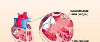
MD degree II - moderately severe MD: significant thickening (5-8 mm) and elongation of the leaflets, deformation of the contour of the mitral orifice over several segments, there are signs of stretching of the chords, less often their single ruptures. Possible moderate stretching of the mitral ring, disruption of closure of the valves.
MD III degree - pronounced MD: the mitral leaflets are sharply thickened (more than 8 mm) and elongated, the maximum depth of prolapse is noted, there are multiple ruptures of the chords, a significant expansion of the mitral ring, there is no closure of the leaflets, their significant systolic separation is noted, multi-valve prolapse is also possible and dilatation of the aortic root. As the authors show, the presence of stage II-III MD is always accompanied by the development of moderate or severe mitral regurgitation. Examples of myxomatous degeneration are presented in the figure.
Identification of individuals with myxomatous degeneration of the leaflets seems very important for the following reasons: - all complications of MVP (sudden death, severe mitral valve insufficiency requiring surgical treatment, bacterial endocarditis and strokes) in 95-100% of cases were noted only in the presence of myxomatous degeneration of the leaflets;
Myxomatous valve degeneration progresses with age.
Thus, with myxomatous valve degeneration, mitral regurgitation progresses with age. According to YB Deng et al. (1990), among patients with myxomatous MVP, mitral regurgitation was found in 29% of patients, its frequency almost doubled over a 4-year observation period.
16 children (10 boys, 6 girls) with myxomatous degeneration of the mitral valve were observed. In these children, mitral valve prolapse was combined in 5 cases with tricuspid valve prolapse and in one case with aortic valve prolapse. Myxomatous degeneration and mitral regurgitation were determined in all children during Doppler examination.
It is characteristic that with myxomatous valve degeneration, external phenotypic signs of connective tissue dysplasia are absent or weakly expressed. For this reason, we consider MVP with myxomatous degeneration as an independent nosological group, probably due to genetic mechanisms.
Myxomatous degeneration of the mitral valve is not as serious a pathology as it might seem at first glance. It has several alternative names that a person may hear from a cardiologist after undergoing an examination: endocardiosis, valve prolapse.
To be able to learn more about such a defect as myxomatous degeneration of the mitral valve leaflets, what it is and how to deal with it, we will consider further. We are talking about thickening and stretching of the valve flaps, which contributes to their loose closure. As a result, the formation of reverse blood flow directed towards the ventricles.
There are several degrees of pathology, depending on which the prognosis for a person changes, and a decision will be made about prescribing effective treatment:
- 1st degree – here there is a thickening of the valve leaflets up to 3-5 mm. Such changes do not disrupt their closure, therefore, a person usually has no symptoms of pathology. Doctors do not sound the alarm when diagnosing this stage of the disease; they recommend reconsidering your lifestyle and conducting preventive examinations twice a year;
- 2nd degree - the valves are stretched, thicker, their indicators reach 5-8 mm. In addition, deformation of the mitral orifice contour is observed. Sprains and isolated ruptures of the chordae are diagnosed. The closure of the valves is disrupted;
- Grade 3 – thickening of the mitral valves is very noticeable, their thickness exceeds 8 mm. There is deformation of the mitral ring, stretching and rupture of the chordae. There is no closure of the valves at all.
Conclusion: stage 1 of the pathology is considered safe, there are no disturbances in the functioning of the heart, and there is no regurgitation (reverse blood flow). At stages 2 and 3, blood returns in a certain volume, because the closure of the valves is disrupted or completely absent. This state of affairs should not be ignored, because there is a high risk of developing serious complications.
Important! In order for the left ventricle to cope with the increased blood volumes due to reverse blood flow, it will begin to increase in size. The result is LV hypertrophy. But this is only one of the negative consequences of the vice we are considering.
It is known that myxomatous degeneration of the mitral valve leaflets only progresses with age. There is a risk of additional complications: sUA insufficiency, bacterial endocarditis, stroke, sudden death. The prognosis is not reassuring, so it is worth taking care of the possibility of timely detection of this pathology, allowing effective measures to be taken to treat it and prevent complications as soon as possible.
What is the problem
Myxomatosis of the mitral valve is a disease based on an increase in the volume of its valves due to the spongy layer located between the ventricular and atrial surfaces of the valve. This process occurs due to a change in the chemical composition of cells, when the content of mucopolysaccharides in them increases significantly.
The outcome of all such deviations is valve prolapse, which gradually leads to a number of pathological processes:
- phenomena of fibrosis on the surface of the valves;
- thinning and lengthening of the chordae tendineae;
- damage to the left ventricle, its dystrophy.
The changes are irreversible, leading to aggressive patient management tactics.
The distinctive properties of the pathology are:
The severity of the disease is determined by the degree of prolapse (sagging) of one or two leaflets into the cavity of the left ventricle. The severity of myxomatous degeneration is determined by cardiac ultrasound.
Myxomatous mitral valve degeneration
Many cardiovascular diseases debut in adulthood, or are discovered by chance during preventive examinations.
Myxomatous mitral valve degeneration is one example of such scenarios.
The pathology requires dynamic monitoring and conservative therapy to prevent complications.
What is the problem
Myxomatosis of the mitral valve is a disease based on an increase in the volume of its valves due to the spongy layer located between the ventricular and atrial surfaces of the valve. This process occurs due to a change in the chemical composition of cells, when the content of mucopolysaccharides in them increases significantly.
The outcome of all such deviations is valve prolapse, which gradually leads to a number of pathological processes:
- phenomena of fibrosis on the surface of the valves;
- thinning and lengthening of the chordae tendineae;
- damage to the left ventricle, its dystrophy.
The changes are irreversible, leading to aggressive patient management tactics.
The distinctive properties of the pathology are:
- Affects persons over 40 years of age.
- More often diagnosed in men.
- The presence of mitral regurgitation (reverse blood flow when the heart muscle relaxes after contraction).
- Progressive course of the disease.
- Formation of heart failure.
The severity of the disease is determined by the degree of prolapse (sagging) of one or two leaflets into the cavity of the left ventricle. The severity of myxomatous degeneration is determined by cardiac ultrasound.
Degree Criteria
0 Signs of myxomatous degeneration are absent on ultrasound scanning, but initial changes can be detected by examining histological materials
I Unpronounced thickening of the valves – no more than 0.03–0.05 cm; the opening of the mitral valve takes the form of an arch
II Marked enlargement of the valves up to 0.08 cm with a violation of their full closure, involvement of the chords in the process
III Sharp thickening - more than 0.08 cm, accompanied by rupture of the chordae, expansion of the aortic root
Thus, myxomatous degeneration has a similar picture to mitral valve prolapse, but different causes.
Why do pathological changes form?
Not many reasons are known for the development of myxomatous degeneration of the mitral valve leaflets. The most common:
- rheumatism;
- chronic rheumatic heart disease;
- secondary atrial septal defect;
- birth defects;
- hypertrophic cardiomyopathy;
- cardiac ischemia.
Pathology always develops secondary. A hereditary predisposition to the occurrence of myxomatous degeneration plays an important role.
Symptoms of the disease
In the early stages of the development of pathological changes, the patient does not complain, or they are caused by the main problem. As it progresses, the following are noted:
- increased fatigue;
- heartbeat;
- sharp fluctuations in blood pressure;
- anxiety;
- panic attacks;
- pain in the apex of the heart not associated with physical activity;
- increased shortness of breath;
- decreased resistance to physical and everyday stress;
- heart rhythm disturbances;
- the appearance of edema in the lower 1/3 of the lower leg and feet.
The severity of symptoms increases as the degree of valve prolapse increases.
Diagnostics
Mitral valve myxomatosis is determined by the results of several studies:
- assessment of patient complaints;
- anamnesis data;
- objective examination;
- additional examination methods.
During the examination, characteristic auscultatory signs of pathology are:
- systolic click;
- midsystolic murmur;
- holosystolic murmur.
A distinctive feature of the auscultatory picture in myxomatous degeneration is its variability (the ability to change from visit to visit).
From an additional examination, the doctor prescribes:
- ECG;
- Holter monitoring;
- Ultrasound of the heart (transthoracic, transesophageal) is the only method that allows visualization of pathological changes;
- tests with dosed physical activity;
- X-ray of the lungs;
- MSCT;
- electrophysiological study.
Such extensive diagnostics are necessary to determine further tactics for patient management and monitor therapy.
Treatment options
Myxomatous degeneration of the mitral valve leaflets, grade 0-I, does not require aggressive measures. Doctors choose a wait-and-see approach, regularly assessing the patient’s condition. No specific treatment is carried out. The patient is given a number of general recommendations:
- eliminate heavy physical activity;
- normalization of body weight;
- treatment of concomitant diseases;
- healthy sleep;
- physiotherapy;
- proper nutrition.
For patients with a higher degree, symptomatic treatment is indicated:
- β-blockers;
- calcium antagonists;
- ACE inhibitors;
- antiarrhythmic drugs.
With the development of infective endocarditis or thromboembolism, the following is prescribed:
- antibiotics;
- disaggregants;
- anticoagulants.
The influence on the mental status of the patient is of great importance. For these purposes, magnesium preparations and sedatives are used.
Surgical correction is carried out when the clinical picture is severe and the degree of myxomatosis increases.
Patient management tactics are determined individually by the cardiologist.
Source: https://VseOSerdce.ru/valve/mitral/miksomatoznaya-degeneraciya-mitralnogo-klapana.html
Degree Criteria
0 Signs of myxomatous degeneration are absent on ultrasound scanning, but initial changes can be detected by examining histological materials
I Unpronounced thickening of the valves – no more than 0.03–0.05 cm; the opening of the mitral valve takes the form of an arch
II Marked enlargement of the valves up to 0.08 cm with a violation of their full closure, involvement of the chords in the process
III Sharp thickening - more than 0.08 cm, accompanied by rupture of the chordae, expansion of the aortic root
Thus, myxomatous degeneration has a similar picture to mitral valve prolapse, but different causes.
Why do pathological changes form?
Not many reasons are known for the development of myxomatous degeneration of the mitral valve leaflets. The most common:
- rheumatism;
- chronic rheumatic heart disease;
- secondary atrial septal defect;
- birth defects;
- hypertrophic cardiomyopathy;
- cardiac ischemia.
Pathology always develops secondary. A hereditary predisposition to the occurrence of myxomatous degeneration plays an important role.
Diagnostics
Mitral valve myxomatosis is determined by the results of several studies:
- assessment of patient complaints;
- anamnesis data;
- objective examination;
- additional examination methods.
During the examination, characteristic auscultatory signs of pathology are:
- systolic click;
- midsystolic murmur;
- holosystolic murmur.
A distinctive feature of the auscultatory picture in myxomatous degeneration is its variability (the ability to change from visit to visit).
From an additional examination, the doctor prescribes:
Such extensive diagnostics are necessary to determine further tactics for patient management and monitor therapy.
Forecasting
Prognosis for myxomatous mitral valve degeneration is related to the stage of the disease at the time of diagnosis, including the speed of development of the disease. This pathology manifests itself in some patients at an early age, and in some patients the disease progresses rapidly. Mostly, the development of myxomatous valve degeneration occurs very slowly and quietly, over several years, which is why some patients may be asymptomatic for a long period after the detection of systolic murmurs. If heart failure develops, the average life expectancy with optimal treatment is 6-18 months.
Treatment options
Myxomatous degeneration of the mitral valve leaflets, grade 0-I, does not require aggressive measures. Doctors choose a wait-and-see approach, regularly assessing the patient’s condition. No specific treatment is carried out. The patient is given a number of general recommendations:
For patients with a higher degree, symptomatic treatment is indicated:
- β-blockers;
- calcium antagonists;
- ACE inhibitors;
- antiarrhythmic drugs.
With the development of infective endocarditis or thromboembolism, the following is prescribed:
The influence on the mental status of the patient is of great importance. For these purposes, magnesium preparations and sedatives are used.
Surgical correction is carried out when the clinical picture is severe and the degree of myxomatosis increases.
Patient management tactics are determined individually by the cardiologist.
What is myxomatous degeneration of the mitral valve leaflets? This is a pathological condition where the heart valves change due to the expansion and thickening of the valves. The closure of the valves is disrupted and blood flows in the opposite direction, producing noise when listening. Parts of the organ are stretched due to degenerative changes and increased blood flow in the opposite direction.
Causes and symptoms of manifestation
The symptoms of the pathology directly depend on the degree of degeneration. In the initial stages, the cardiologist listens to systolic murmurs. As the disease progresses, the size of the heart and blood circulation increase, so signs of a pronounced nature begin to appear:
- endurance decreases;
- shortness of breath appears;
- appetite worsens;
- possible fainting;
- coughing begins.
Additional complaints include:
- pain in the chest area;
- paroxysmal heartbeat (can be observed at rest or with minor exertion);
- interruptions in heart function due to extrasystole;
- shortness of breath (lack of air);
- feeling tired for no apparent reason.
Important! Any signs of heart problems require immediate consultation with a cardiologist. Timely identification of pathology increases the chance of complete recovery.
Pain in the heart area varies in nature and depends on the development of the disease. Rupture of the hypertrophic left atrium or valve flaps can be fatal.
Myxomatous degeneration of the mitral valve is considered a fairly common pathology. But today the true reason for its development has not been determined. Some people may have a natural or genetic defect.
People with problems with growth and the formation of cartilage tissue are more susceptible to the disease. This is the connecting thread between this pathology and non-standard development, degeneration of connective tissues in the valve flaps.
Doctors are conducting research to identify the influence of hormonal factors on the progression of this disease.
Signs of MD
At the initial stage, any signs will be absent, because in this case the blood circulation is not impaired, regurgitation is completely absent. But, as the disease progresses to a more serious stage, the person will experience the following symptoms:
- decreased performance, general weakness, fatigue even with minimal loads;
- shortness of breath that appears with minimal physical or emotional stress, a feeling of lack of air;
- pain in the heart, which most often manifests itself in the form of tingling. Occur periodically and are short-lived;
- dizziness accompanied by arrhythmia, often leading a person to a pre-fainting state;
- cough, it should be considered as an additional symptom that may not appear. At first it is dry, then accompanied by the release of sputum, which may contain streaks of blood.
When visiting a doctor, signs of disruption of the cardiac system will first be noticed when listening to the heart. The doctor will hear noises that accompany the return flow of blood to the ventricle. This alone can become a reason for a more detailed examination, including anamnesis, laboratory tests, electrocardiography, and echocardiography.
If electrocardiography shows only the presence of a disorder, its stage, then ultrasound of the heart will be able to provide more complete information, because it will allow one to determine the size of the valves, the features of their deformation, in other words, all the pathological changes that occur in this case.
Methods for diagnosing the disease
Pathology is determined by listening to the heart. The doctor hears a systolic murmur in the mitral valve.
For the final diagnosis, the physiological state of the person is examined and echocardiography (ultrasound diagnostics of the heart) is prescribed. An echocardiogram allows you to determine the maneuvering of the valves, their structure and the functioning of the heart muscle. For examination, one-dimensional and two-dimensional modes are used. This research method allows us to determine the following pathological factors:
- the anterior, posterior or both valves thicken by more than five millimeters in relation to the mitral annulus;
- the left atrium and ventricle are enlarged;
- contraction of the left ventricle is accompanied by sagging of the valve leaflets towards the atrium;
- the mitral ring expands;
- tendon threads lengthen.
An electrocardiogram is required. The ECG records all kinds of irregularities in heart rhythm.
Additional diagnostic methods include chest x-ray.
Methods of treating the disease
To date, doctors have not identified effective methods of prevention that can prevent or stop the progression of this pathology. If your doctor finds systolic murmurs and only minor changes in the structure of the heart, he may recommend regular medical examinations without prescribing drug therapy. In this way, you can monitor the development of the disease and its possible progression.
- give up bad habits: alcohol, nicotine, caffeine drinks;
- stick to a balanced diet: less fatty and salty, more fresh vegetables and fruits. It is worth reducing the consumption of cholesterol-containing foods. It is better to steam or boil food; it is better to avoid fried foods;
- moderate physical activity;
- spend more time outdoors;
- fully relax after a working day.
In more complex forms of pathology, the doctor prescribes medications to minimize the progression of severe symptoms caused by hypertrophy and changes in the structural parts of the heart.
If heart failure is detected, the patient is prescribed medications that will remove excess fluid from the body, help maintain the functionality of the heart muscle, and increase the speed of blood flow.
As a rule, drugs are combined. This allows you to reduce symptoms and improve the patient’s well-being. Treatment of the pathology directly depends on the presence of concomitant diseases (especially for liver and kidney pathologies).
Important! You should not take medications without a doctor’s prescription, since they may be personal intolerance and negatively affect the development of pathology.
Myxomatous degeneration of the mitral valve leaflets has a favorable prognosis if the pathology was detected in the early stages and does not have pronounced symptoms. The disease can develop at a fairly early age, and manifests itself quite rapidly. This manifestation requires early diagnosis and surgical therapy.
But, as a rule, the valve degenerates slowly and moderately over several years. Even if a systolic murmur is detected, the patient may have an asymptomatic period.
Once heart failure develops, the average life expectancy is about a year. But this is only an approximate figure, which is influenced by many factors. Therefore, after making a diagnosis, it is necessary to fully follow the doctor’s recommendations and instructions. This will prolong life and improve its quality, and in most cases completely get rid of pathology.
In the work of the heart, the function of the mitral valve cannot be underestimated: it is a partition of 2 valves between the left ventricle and the atrium, which open and allow blood into the ventricular cavity. After which they close and stop its supply, throwing blood into the aorta, thus organizing blood circulation. The valves must be thin and elastic, and changes in their structure can disrupt the quality of the valve and the organ as a whole. Pathological processes are not uncommon here, myxomatous degeneration of the mitral valve is one of them.
For information! This defect has an alternative name - endocardiosis, according to ICD 10 it does not have a separate designation, but refers to mitral valve prolapse (under code 134.1).
Myxomatous degeneration of the mitral valve: causes of development
What is myxomatous degeneration of the mitral valve leaflets? This is a pathological condition where the heart valves change due to the expansion and thickening of the valves. The closure of the valves is disrupted and blood flows in the opposite direction, producing noise when listening. Parts of the organ are stretched due to degenerative changes and increased blood flow in the opposite direction.
Causes and symptoms of manifestation
The symptoms of the pathology directly depend on the degree of degeneration. In the initial stages, the cardiologist listens to systolic murmurs. As the disease progresses, the size of the heart and blood circulation increase, so signs of a pronounced nature begin to appear:
- endurance decreases;
- shortness of breath appears;
- appetite worsens;
- possible fainting;
- coughing begins.
Additional complaints include:
- pain in the chest area;
- paroxysmal heartbeat (can be observed at rest or with minor exertion);
- interruptions in heart function due to extrasystole;
- shortness of breath (lack of air);
- feeling tired for no apparent reason.
Important! Any signs of heart problems require immediate consultation with a cardiologist. Timely identification of pathology increases the chance of complete recovery.
Myxomatous mitral valve degeneration
Pain in the heart area varies in nature and depends on the development of the disease. Rupture of the hypertrophic left atrium or valve flaps can be fatal.
Myxomatous degeneration of the mitral valve is considered a fairly common pathology. But today the true reason for its development has not been determined. Some people may have a natural or genetic defect.
People with problems with growth and the formation of cartilage tissue are more susceptible to the disease. This is the connecting thread between this pathology and non-standard development, degeneration of connective tissues in the valve flaps.
Doctors are conducting research to identify the influence of hormonal factors on the progression of this disease.
Methods for diagnosing the disease
Diagnosis of myxomatous mitral valve degeneration
Pathology is determined by listening to the heart. The doctor hears a systolic murmur in the mitral valve.
For the final diagnosis, the physiological state of the person is examined and echocardiography (ultrasound diagnostics of the heart) is prescribed. An echocardiogram allows you to determine the maneuvering of the valves, their structure and the functioning of the heart muscle. For examination, one-dimensional and two-dimensional modes are used. This research method allows us to determine the following pathological factors:
- the anterior, posterior or both valves thicken by more than five millimeters in relation to the mitral annulus;
- the left atrium and ventricle are enlarged;
- contraction of the left ventricle is accompanied by sagging of the valve leaflets towards the atrium;
- the mitral ring expands;
- tendon threads lengthen.
An electrocardiogram is required. The ECG records all kinds of irregularities in heart rhythm.
Additional diagnostic methods include chest x-ray.
Methods of treating the disease
To date, doctors have not identified effective methods of prevention that can prevent or stop the progression of this pathology.
If your doctor finds systolic murmurs and only minor changes in the structure of the heart, he may recommend regular medical examinations without prescribing drug therapy.
In this way, you can monitor the development of the disease and its possible progression.
At this stage, it is recommended to change your lifestyle:
- give up bad habits: alcohol, nicotine, caffeine drinks;
- stick to a balanced diet: less fatty and salty, more fresh vegetables and fruits. It is worth reducing the consumption of cholesterol-containing foods. It is better to steam or boil food; it is better to avoid fried foods;
- moderate physical activity;
- spend more time outdoors;
- fully relax after a working day.
Treatment of myxomatous mitral valve degeneration
In more complex forms of pathology, the doctor prescribes medications to minimize the progression of severe symptoms caused by hypertrophy and changes in the structural parts of the heart.
If heart failure is detected, the patient is prescribed medications that will remove excess fluid from the body, help maintain the functionality of the heart muscle, and increase the speed of blood flow.
As a rule, drugs are combined. This allows you to reduce symptoms and improve the patient’s well-being. Treatment of the pathology directly depends on the presence of concomitant diseases (especially for liver and kidney pathologies).
Important! You should not take medications without a doctor’s prescription, since they may be personal intolerance and negatively affect the development of pathology.
Myxomatous degeneration of the mitral valve leaflets has a favorable prognosis if the pathology was detected in the early stages and does not have pronounced symptoms. The disease can develop at a fairly early age, and manifests itself quite rapidly. This manifestation requires early diagnosis and surgical therapy.
But, as a rule, the valve degenerates slowly and moderately over several years. Even if a systolic murmur is detected, the patient may have an asymptomatic period.
Once heart failure develops, the average life expectancy is about a year. But this is only an approximate figure, which is influenced by many factors. Therefore, after making a diagnosis, it is necessary to fully follow the doctor’s recommendations and instructions. This will prolong life and improve its quality, and in most cases completely get rid of pathology.
Source: https://medsosud.ru/aritmiya/miksomatoznaya-degeneratsiya-stvorok-mitralnogo-klapana-borba-s-zabolevaniem.html
General information about the defect and causes
When considering myxomatous degeneration of the mitral valve leaflets, the question arises: what is it? So, this is a pathological condition, which is not the most dangerous for the body: if the defect is detected in a timely manner, there are measures to take and preventive programs are recommended.
Myxomatous degeneration of the valve leaflets is a stretching or increase in their thickness, which, as the disease progresses, begins to interfere with the complete closure of the valve at the time of systole and cannot resist reverse blood flow. Most often, this defect is diagnosed in older and middle-aged people.
In total, there are three degrees of development of the pathological process:
- the first degree is characterized by an increase in the thickness of the valves ranging from 3 mm to 5 mm, which does not interfere with closure;
- on the second, the thickening reaches 8 mm, which leads to valve deformation, single ruptures of the chords and impaired closure;
- at the third stage, when the thickness of the leaflets increases above 8 mm, the valve does not close and regurgitation of blood occurs (reverse flow), in which part of it returns to the atrium.
The causes of pathology can be many factors
The initial stage is not life-threatening, but progression of myxomatous degeneration and transition to later stages can lead to mitral valve insufficiency, stroke, infective endocarditis, and death.
To date, no specific causes that can lead to this defect have been identified. In some cases, heredity is a dangerous factor. A pattern has been identified in which patients with this pathology have problems with growth. Doctors do not rule out the influence of hormonal imbalances, but this factor is still being studied.
Myxomatous degeneration of the mitral valve: signs, diagnosis
In the work of the heart, the function of the mitral valve cannot be underestimated: it is a partition of 2 valves between the left ventricle and the atrium, which open and allow blood into the ventricular cavity.
After which they close and stop its supply, throwing blood into the aorta, thus organizing blood circulation. The valves must be thin and elastic, and changes in their structure can disrupt the quality of the valve and the organ as a whole.
Pathological processes are not uncommon here, myxomatous degeneration of the mitral valve is one of them.
For information! This defect has an alternative name - endocardiosis, according to ICD 10 it does not have a separate designation, but refers to mitral valve prolapse (under code 134.1).
General information about the defect and causes
When considering myxomatous degeneration of the mitral valve leaflets, the question arises: what is it? So, this is a pathological condition, which is not the most dangerous for the body: if the defect is detected in a timely manner, there are measures to take and preventive programs are recommended.
Myxomatous degeneration of the valve leaflets is a stretching or increase in their thickness, which, as the disease progresses, begins to interfere with the complete closure of the valve at the time of systole and cannot resist reverse blood flow. Most often, this defect is diagnosed in older and middle-aged people.
In total, there are three degrees of development of the pathological process:
- the first degree is characterized by an increase in the thickness of the valves ranging from 3 mm to 5 mm, which does not interfere with closure;
- on the second, the thickening reaches 8 mm, which leads to valve deformation, single ruptures of the chords and impaired closure;
- at the third stage, when the thickness of the leaflets increases above 8 mm, the valve does not close and regurgitation of blood occurs (reverse flow), in which part of it returns to the atrium.
The causes of pathology can be many factors
The initial stage is not life-threatening, but progression of myxomatous degeneration and transition to later stages can lead to mitral valve insufficiency, stroke, infective endocarditis, and death.
Read it! Supraventricular extrasystole: what is it?
To date, no specific causes that can lead to this defect have been identified. In some cases, heredity is a dangerous factor. A pattern has been identified in which patients with this pathology have problems with growth. Doctors do not rule out the influence of hormonal imbalances, but this factor is still being studied.
At the beginning of its appearance, the pathological process may not be accompanied by certain symptoms due to the fact that no disturbances in the functioning of the organ occur.
With the development of the defect and the transition to the second and third degrees, myxomatous degeneration of the mitral valve is accompanied by quite characteristic signs:
- periodic pain in the left side of the chest, stabbing in nature and short-term manifestation;
- deterioration of general condition (increased fatigue, decreased physical activity, weakness, decreased appetite);
- the appearance of shortness of breath even with little physical activity;
- feeling of lack of air;
- dizziness, lightheadedness and fainting.
In some cases, an additional symptom may be a cough. At first it is dry, and then with phlegm and splashes of blood.
Diagnosis and treatment methods
The presence of pathology is indicated by systolic heart murmurs, which the doctor can hear during auscultation (listening). To confirm the diagnosis, prescribe:
- electrocardiogram;
- echocardiography (a type of ultrasound of the heart);
- X-ray of the chest area.
Attention! Genetic tests and blood tests are currently not required to identify this defect.
At the initial stage, when myxomatous degeneration of the mitral valve leaflets does not interfere with the functioning of the heart and does not affect the general condition of the body, active treatment, much less surgical intervention, is not required. However, the patient must be registered with a cardiologist and undergo regular examinations.
There are currently no effective drugs that could completely stop and eliminate this pathological disease.
Therefore, as the pathology progresses, medications are prescribed that help eliminate symptoms and significantly slow down the dangerous process.
Such medications include those that remove excess accumulated fluid from the body, are aimed at maintaining the performance of the heart muscle and improving blood circulation, and regulating the heart rate.
Read it! Mitral valve prolapse
In cases where the pathology has led to mitral regurgitation and blood regurgitation, surgical intervention may be indicated (you can watch the video on the Internet resource), in which it is possible:
- preservation of the valve with plastic surgery of the valves or their replacement;
- prosthetics (the affected mitral valve is removed and a biological or artificial prosthesis is placed in its place).
Quitting bad habits will help you stay healthy
Despite the fact that the causes of the development of myxomatous mitral valve degeneration have not been fully established, and it is difficult to talk about specific prevention, there are some important recommendations.
- You should definitely be under the supervision of a doctor and undergo regular preventive examinations.
- Lead a completely healthy lifestyle (excluding all bad habits).
- Create and adhere to a work and rest schedule.
- Review your diet, include only healthy foods (more vegetables and fruits, quail eggs). Focus on foods that contain heart-healthy components (for example, potassium-rich foods - dried apricots, prunes, cabbage, rose hips). Avoid strong black tea and coffee.
Source: https://SosudiVeny.ru/aritmiya/miksomatoznaya-degeneratsiya-mitralnogo-klapana.html
What symptoms does it manifest?
At the beginning of its appearance, the pathological process may not be accompanied by certain symptoms due to the fact that no disturbances in the functioning of the organ occur.
With the development of the defect and the transition to the second and third degrees, myxomatous degeneration of the mitral valve is accompanied by quite characteristic signs:
- periodic pain in the left side of the chest, stabbing in nature and short-term manifestation;
- deterioration of general condition (increased fatigue, decreased physical activity, weakness, decreased appetite);
- the appearance of shortness of breath even with little physical activity;
- feeling of lack of air;
- dizziness, lightheadedness and fainting.
In some cases, an additional symptom may be a cough. At first it is dry, and then with phlegm and splashes of blood.
Symptoms of the disease
Symptoms of mitral degeneration vary depending on the stage of valve leaflet degeneration. At an early stage of development, systolic murmurs are detected in the heart. According to the degree of increase in changes, enlargement of the heart and blood circulation, other symptoms arise, consisting of decreased physical activity, shortness of breath, loss of appetite, fainting, and cough. In some cases, in the later stages of the disease, death is possible (the reason for this is rupture of the hypertrophied left atrium or rupture of the valve leaflets).