0
Author of the article: Marina Dmitrievna
2017.10.23
328
Vessels
Rupture of the aorta of the heart is the most severe consequence of negative processes on the walls of blood vessels, which is fatal in 90% of cases. Sometimes surgical treatment is not always optimal.
In recent decades, this pathology has become common and has increased 7 times. It is much more difficult to save a patient from death due to a rupture of the main artery than from other emergency situations associated with problems of the cardiovascular system - myocardial infarction, hemorrhagic and ischemic attacks, etc.
A rupture diagnosed by surgeons
With modern medical capabilities, it is possible to identify transformations of the aortic wall that threaten the integrity of the aorta, but it is not possible to protect against rupture. The aorta is the largest vessel of the human body. A large volume of blood passes through it, and it is this that provides nutrition and oxygen to organs and tissues. Functioning without interruption and with sufficient tension, the aorta becomes vulnerable to many negative transformations. However, atherosclerotic growths inside have recently become a greater danger to the integrity of the walls of the main artery.
The term “aortic rupture” implies a section of the surface of the epithelium of the vessel due to an aneurysm, which was the result of atherosclerotic deposits, dystrophic metamorphosis, and inflammation.
How is the aorta structured and what is an aneurysm?
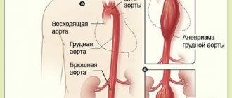
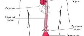
The aorta, due to its size, was divided into several conventional sections:
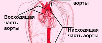
- The ascending aorta starts from the left ventricle of the heart.
- The arc from which branches emerge that feed the chest, arms and head.
- The thoracic section of the aorta, which continues to the diaphragm (also called the descending section).
- The abdominal part, extending below the diaphragm, passes through an opening in it into the abdominal cavity.
The very structure of this vessel includes:
- The internal cavity through which blood flows.
- The inner wall is made of dense flat cells, ensuring the unhindered passage of blood.
- The middle wall, consisting of connective and a small amount of smooth muscle tissue, is strong and elastic, allowing blood to move through the internal cavity in a normal manner.
- An outer wall consisting of fibrous tissue, which allows you to fix the position of the entire vessel in space and maintain its integrity.
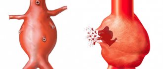
Damage can occur at any “depth” of the aorta and in any section, and most often takes the form of an aneurysm.
An aneurysm is a pathological change in the walls of a vessel in which they expand due to inflammation, atherosclerosis or mechanical damage.
Blood accumulates in this kind of sac, the affected area grows until the walls of the vessels can no longer withstand the load.
After this, the aorta ruptures - blood freely splashes out, blood flow quickly deteriorates or is interrupted altogether, and irreversible internal damage occurs due to bleeding.
First aid
It is important not only to know what causes aortic rupture, but also how to provide first aid to the patient so that his life can be saved. The subsequent prognosis for a person will largely depend on the quality and timeliness of first aid provided. You definitely need to call an ambulance, and take certain measures before it arrives. In particular, you need to give the patient a horizontal position and try to raise his head. The person must remain motionless all this time.
You need to try to calm the patient so as not to further aggravate the condition or provoke attacks of shock. In addition, it is forbidden to give food or drink, or use laxatives. If you have nitroglycerin at home, then you need to put one tablet under the patient’s tongue to somewhat relieve the pain.
After this, you need to wait for an ambulance, since a traumatic aortic rupture can only be treated through surgery. In addition, additional drugs are prescribed to treat the underlying disease, which can cause rupture of the vessel.
Why might a rupture occur?
The reasons that cause an artery to grow and rupture are:
- In old age – deterioration of vascular elasticity.
- Death of tissue in the walls of blood vessels due to infections or deterioration of the immune system.
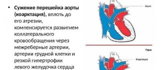
- Congenital pathology of vessel narrowing - coarctation.
- Atherosclerosis.
- Underdevelopment of connective tissues caused by genetic predisposition - Marfan syndrome.
- Hypoplasia.
- Aneurysm.
- Tumor damage to the aortic wall, including tumors of the esophagus, stomach, intestines, and pancreas.
- Injuries in the chest and abdomen.
If any of these factors are present, the patient's condition can deteriorate very quickly.
The causes of death are massive blood loss and shock.
According to statistics, without emergency assistance, and if the vessel rupture is large, the person dies in 90 percent of cases.
If symptoms are detected early in the development of wall damage, the chances of survival increase.
Provoking factors and types of ruptures
In a number of diseases, a person’s aorta can dilate, which can subsequently lead to an aneurysm. These include a disease called Marfan syndrome.
This is a severe hereditary disease manifested in connective tissue pathologies. Without proper treatment, patients suffering from this disease usually live no more than 35 years.
The cause of death in such patients is vascular rupture or heart failure.
The first signs of an advanced process of aortic rupture are a violation of the integrity of the inner layer of the vascular wall. In the patient's condition, this is reflected by strong unpleasant sensations: pain on the left side of the chest and left arm.
The person turns pale and feels dizzy. Vomiting may occur.
In this case, pressure surges are inevitable, followed by a strong increase in indicators and a faint or semi-fainting state of the patient. It happens that death occurs instantly.
What causes the rupture of the aorta of the heart as a result of an aneurysm? Why is this happening? First of all, the explanation is provided by the very understanding of the structure of the heart muscle.
The fact is that the myocardium constantly, continuously pulsates. A huge volume of blood, flowing through the heart muscle in an endless stream, puts force on the thin bridges inside the heart, protruding and stretching them.
This is a normal process and, with a naturally normal structure of the arteries, in the presence of healthy, elastic vascular walls and myocardium, this process does not have negative consequences.
However, a rupture of the aorta of the heart jeopardizes the entire functional functioning of the cardiovascular system. An aneurysm that already exists only expands, capturing more and more new areas and significantly delaying blood flow due to improper distribution of blood inside the vessel.
Delamination stages and possible forms
There are three stages in the development of pathology:
- There is a tear in the inner layer of the walls - the intima.
- Due to the rupture, the middle shell (media) begins to separate - exfoliation occurs.
- The outer wall (adventitia) is stretched, followed by rupture due to high pressure.
In its normal state, the adventitia is strong enough that rupture does not occur spontaneously under the influence of the blood flow itself or changes in it (for example, due to a rapid heartbeat). However, the deterioration of the condition of the vessels due to their chronic damage or immediate severe damage (for example, due to injuries) leads to stratification and divergence of the layers.
The forms of delamination depend on the time of the process:
- Acute - up to two days. Possible instant death.
- Subacute - from 14 to 30 days. At any moment, due to the load on the walls, a rupture can occur and the person dies.
- Chronic - more than a month, can last for years. The least dangerous form, allowing for timely assistance.
By localization there are:
- Distal form - the process occurs in the lower part of the aorta.
- Proximal form - the process occurs in the upper part with or without further transition to the lower part.
Diagnostic methods
A set of examinations during which the severity and form of the condition are determined:
- Angiography is an X-ray with contrast, which examines all parts of the aorta and identifies pathology. No damage to the inner wall was visible during this study.
- An ECG is prescribed to rule out myocardial infarction, which is often confused with a vascular rupture.
- Computed tomography - if time and resources allow. It is the most reliable diagnostic method.
Usually the doctor chooses one of the above tests so as not to subject the patient’s body to additional stress and not waste time. When making a diagnosis, all symptoms of the condition are taken into account.
Prognosis for pathology
If the aorta ruptures completely, the prognosis is extremely poor: about 90% of patients die. This is due to the following factors:
- untimely arrival of ambulance;
- incorrect diagnosis;
- too much hemorrhage in vital organs, for which even surgery may not help;
- severe tolerance of general anesthesia and surgery in older people.
With earlier stages of dissection, the chances of survival are much higher, especially if you see a doctor right away. Surgery guarantees recovery, but the risks associated with the operation itself remain.
okardio.com
Signs of a rupture
Rupture of an aortic aneurysm has quite clear manifestations - symptoms differ depending on the location, which can be:
- Abdominal - with the threat of bleeding in the peritoneum.
- Thoracic - can lead to heart failure.
The severity of the condition and symptoms also depend on the stage:
- The inner lining is damaged: pain, increased blood pressure, drowsiness, weakness, headache, pallor.
- The middle wall is damaged: sharp and burning pain, low blood pressure, disruption of organ function.
- Rupture: drop in blood pressure, pallor, increase in temperature, infrequent urination, internal bleeding and shock.
Abdominal
Click on the image to enlarge
Rupture of the abdominal aorta (ICD-10 classification: I71.3 and I71.4) or, as it is also called, a rupture in the abdominal aorta is accompanied by:
- Shock.
- Severe pain in the abdomen.
- Weakness.
- Blurred vision.
- Bleeding into the abdominal cavity with the formation of a retroperitoneal hematoma.
- Kidney failure.
Chest
Rupture of the thoracic aorta (ICD-10 classification: I71.1 and I71.2) has the following symptoms:
- Severe chest pain.
- Dyspnea.
- Swelling of the neck and upper torso.
- Swelling of veins.
- Blue skin.
- Weakness.
- Fast pulse.
Symptoms
If aortic dissection occurs, the symptoms will be expressed in a sharp, bursting pain behind the sternum, the pain is so strong that sometimes it becomes difficult to breathe, and shooting pain in the back area is often observed.
Over time, the pain only intensifies if the dissecting aortic aneurysm continues to progress. The pain can be unbearable, a person can lose his creation from pain shock.
The greatest concentration of pain is noted in the heart area, and can shoot into the left arm. Most often, blood pressure drops sharply, and less often, on the contrary, there is a sharp jump.
If the aorta ruptures, the pulse in the victim’s limbs may not be palpable. When blood enters the bronchi, patients experience hemoptysis; when blood clots enter the esophagus, bloody vomiting occurs.
Symptoms of an aneurysm may not appear for quite a long time. It is especially worth noting here the invaluable value of undergoing a mandatory preventive examination, which most often reveals a dangerous diagnosis.
At this point, patients have almost no problems with their well-being, and people do not treat the problem voiced by the doctor with the seriousness it deserves. And when painful signs of aortic rupture appear, it often becomes too late to do anything.
People's complaints include such general concepts as pressing pain in the sternum, hacking cough, and rapid heartbeat.
If the above complaints include a burning pain directed from the sternum to the back, then we are most likely talking about a defect in the cardiac aorta. Nauseous urges are often added to this alarming symptom.
In the latter case, immediate hospitalization is required, since the presence of vomiting itself, with the presence of blood impurities in the masses, will indicate symptoms of aortic rupture in the abdominal cavity. Nausea will be accompanied by severe pain in the thoracic region, with condescension along the spinal column.
When a patient is taken to the hospital with all the symptoms of ulcerative or gastritis manifestations, the patient often ends up in the hands of a doctor specializing in these diseases, which is not true from the beginning.
Recognizing the true causes and starting treatment against the symptoms of an “acute abdomen” takes up precious time, and the patient often dies before the correct diagnosis is made.
If the aortic rupture occurs below the abdominal region, then the pain quickly spreads to the pelvic area and legs. If the rupture occurs in the sternum area, then the symptoms almost completely mimic the state of myocardial infarction.
Severe and rapid blood loss during aortic rupture leads to death within a few minutes. The heart muscle, not receiving blood return, simply stops.
If an aneurysm ruptures into the abdominal cavity, the person will feel acute pain and at the same time pulsation in the abdomen. The pain does not stop even after taking painkillers. Intra-abdominal bleeding is accompanied by:
- pallor;
- heart rhythm disturbances;
- anemia;
- hypotension;
- vomiting;
- hiccups;
- sweating
If the aorta ruptures in the retroperitoneum, the person feels intense, ongoing pain in the left side of the abdomen and lower back.
When a hematoma formed as a result of hemorrhage compresses the iliac arteries, a person develops leg ischemia. Compression of the Adamkiewicz artery by the hematoma leads to ischemia of the spinal cord, due to which a person develops urinary and fecal incontinence and lack of sensitivity below the site of the rupture.
If the aorta ruptures to form a connection with the inferior vena cava system, the patient develops symptoms of heart failure. In this case, the person feels a loss of strength, heaviness in the legs and right hypochondrium. In some cases, the venous pattern on the legs and abdomen becomes pronounced.
If a person has such symptoms, you should immediately call an ambulance. Before the medical team arrives, the patient is given first aid:
- Place on a horizontal surface, raise the head.
- They calm you down.
- They are not given food or drink.
- They give you a nitroglycerin tablet to take.
A patient with a suspected rupture of the largest vessel is hospitalized in the vascular surgical department for immediate surgery.
How to save and how is treatment carried out?
The main form of treatment is timely surgery on the vessel, during which the torn section is replaced with an artificial one. Without it, a person has little chance of survival, but even after surgery, complications can lead to death.
When the first symptoms are detected, the person must be urgently rescued - call an ambulance and provide specific first aid.
First aid techniques
If you suspect an aortic rupture, you must:
- Move or lay the person down so that his head is raised (half sitting, for example).
- Don't let him move and don't move him from place to place in vain.
- Avoid drinking and eating, no matter how much you want to.
- Do not use medications. In extreme cases, give nitroglycerin to drink.
- Collect the necessary documents for transporting the patient to a medical facility.
The need for hospitalization occurs when the aneurysm develops rapidly. If the rupture has not yet occurred, but there are characteristic symptoms of wall separation, you should urgently consult a doctor. The condition will worsen until the aneurysm is removed. This cannot be done with medication, so the patient is advised to undergo surgery.
Carrying out the operation
Surgery on the aorta is quite risky - during its implementation, factors such as severe blood loss, damage to other organs, etc. come into play.
In some conditions, it is not possible to save a person (for example, if a person is over 75 or has concomitant diseases.
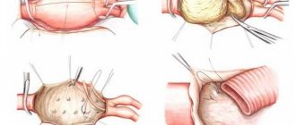
If the rupture occurs in the ascending or thoracic aorta:
- The surgeon opens the patient's chest.
- The vessel defect is removed - the damaged tissue is excised, and a prosthesis for the removed area is individually prepared.
- A synthetic prosthesis is implanted.
During the procedure, there is constant drainage to remove fluid from the cavity, since hemorrhage in this area can lead to instant cardiac arrest.
If a rupture of the main artery in this area has led to a deterioration in the functioning of the heart (defects), further valve replacement is prescribed.
If the rupture occurs in the abdominal part:
- The abdomen is opened (abdominal surgery).
- The intestines are temporarily displaced for better viewing and penetration.
- An incision is made into the peritoneum to gain access to the aorta.
- The blood flow above and below the rupture is blocked (the aorta is compressed).
- The aneurysm is removed along with blood clots.
- The area is undergoing prosthetics.
- The blockage of blood flow stops.
Such an operation can have a number of negative consequences:
- If the aneurysm was close to the renal arteries, renal failure is possible.
- Prolonged disconnection of the aorta during surgery can lead to organ ischemia.
- Failure of the postoperative suture of the vessel leads to acute blood loss and death in the postoperative period.
Cardiac surgeon: how to prevent sudden death from aortic rupture
Gennady Grigorievich, please explain: why does such a disaster happen completely unexpectedly, when a person seemed to feel normal? How high is the risk of such a catastrophe?
Gennady Khubulava
: These are, perhaps, the key words: “I seemed to feel fine,” but in fact, such a catastrophe is in most cases the end of a chronic disease. Aortic ruptures occur as a result of dissection of the aorta changed by a pathological process or an increase in the size of an already existing chronic aneurysm, that is, pathological expansion of the aorta, which a person might not be aware of. The frequency of such ruptures reaches six cases per 100 thousand population per year. It would seem not so much, but look - 6 per 100 thousand, this is already 60 cases per million. For example, in St. Petersburg, where there are five million inhabitants, there are 300 new cases per year - this is a fairly large figure.
How is an aortic aneurysm diagnosed?
Gennady Khubulava:
If we talk about diagnosis, this is most often an accidental finding during examination. That is why doctors insist on regular medical examinations. But it is important to know that if you have any unpleasant sensation in the chest, you need to contact your cardiologist. There are cases of congenital pathology - weakness of connective tissue - the so-called Marfan syndrome. In such patients, aneurysm may occur at a young age, and such patients must be closely monitored.
But, of course, regular examinations are important for all people. Especially if there are risk factors - the person suffers from atherosclerosis, hypertension. Another risk factor is smoking. Well, and of course, old age. The aorta ages just like the rest of the body. This is essentially a tube through which a very large amount of blood passes - about 200 million liters per year - and over time it begins to wear out. Therefore, older patients, especially those suffering from hypertension, atherosclerosis, and coronary heart disease, need more thorough, regular examination and monitoring to prevent acute situations. Medical technologies, and in cardiology, of course, too, are constantly developing and improving. Thus, one of the most important areas in the scientific research of the departments at the S.M. Kirov Military Medical Academy and the I.P. Pavlov First St. Petersburg State Medical University, which I head, we study the strength of the aortic wall, the probabilities of predicting aortic ruptures , we are developing models for organizing the provision of care to patients in acute situations. Modeling these complex processes is impossible without the use of modern information technologies, and in this we work closely with the recently opened Military Innovation Technopolis “Era”. Scientific research put into practice will help prevent many acute vascular accidents.
But you need to understand that maintaining your own health largely depends on the person himself. Patients must take responsibility for their health.
What should you do for this?
Gennady Khubulava:
The simplest thing is that we recommend having a chest x-ray every year. It seems to be the most ordinary, routine study, and, as is often said, not very informative. But, nevertheless, on the fluorogram you can see an increase in the contours of the aorta, and its calcification, and, for example, calcification of the heart valves. And conduct a more in-depth study on such a patient. Echocardiography is usually performed - ultrasound examination of the heart and blood vessels. The third stage, if you need to clarify the diagnosis, is a computed tomography scan. But first of all, we must remember that preventing a disaster is easier than dealing with its consequences.
How is an aortic aneurysm treated?
Gennady Khubulava:
If the increase in the size of the aorta reaches a certain degree, elective surgery is indicated. With a planned operation, the results are much better, since the patient is more thoroughly examined, the operation and everything necessary for it are prepared in advance.
In acute cases - when delamination or rupture occurs unexpectedly - this is a very difficult situation and a big problem. But even here, in many cases, if assistance is provided in a timely manner, there is a chance of saving the patient.
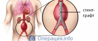
A program for the treatment of acute aortic syndrome has been operating in St. Petersburg for three years now. This is the first program in our country to implement emergency surgical care for patients with acute lesions of the thoracic aorta. During this time, we organized the logistics of providing emergency care to such patients, developed and implemented new organizational and clinical approaches. Both practical doctors and managers and organizers of medical care in our city have worked hard. The results over the three years of work have been improving. But, of course, the most important thing is not to lead to such acute conditions. If the diagnosis is established, surgical treatment is performed as planned. There are many types of operations, but the essence is that the aorta is replaced with various vascular prostheses or stent grafts (special vascular prostheses) are installed endovascularly.
How can this pathology be prevented?
Prevention of aortic rupture is aimed at treating the aneurysm:
- Completion of annual medical examinations (dispensary examinations).
- Timely surgery (if the aneurysm is already more than five centimeters).
- Treatment and prevention of diseases - precursors of diabetes, hypertension, etc.
- Healthy lifestyle, good nutrition, body weight control.
- Control of cholesterol levels, if necessary, a diet that helps reduce its levels.
- To give up smoking.
- Physical training.
- Taking medications that protect the walls of blood vessels (vitamins C and P, dietary supplements with omega-3 fatty acids).
Features of treatment
Resuscitation measures are aimed at stabilizing the patient’s condition, eliminating pain shock, stopping bleeding, and preventing the occurrence of renal failure. Methods of intensive therapy include:
- intravenous administration of glucose-saline solutions;
- diuretics;
- calcium gluconate;
- applying a clamp to the vessels;
- application of a special compress to the aorta area;
- insertion of a balloon catheter into the vessels.
After stopping the bleeding, the aneurysm is removed and blood vessels are replaced.
Life forecast
The chances of survival and further problem-free life are low in any form of the disease other than chronic. However, the latter must be identified early enough to have time to perform surgery or restore damaged vessel walls.
If a complete rupture occurs, the prognosis is disappointing even with a perfectly performed operation. Hemorrhage and circulatory disorders generally lead to shock and damage to internal organs - the heart, kidneys and others in the chosen location.
Even if he survives (less than 10 percent of cases), the person receives a disability, since in the future he will need special living conditions with a prosthesis and the pathologies that arose due to the rupture.
If the gap was incomplete or has not yet occurred, the chances increase. A timely diagnosis and successful prosthetics can guarantee a person’s full return to normal life.
Author of the article: Yulia Dmitrieva (Sych) - In 2014, she graduated with honors from Saratov State Medical University named after V. I. Razumovsky. Currently working as a cardiologist at the 8th City Clinical Hospital in the 1st clinic.
Course of aortic aneurysm
An uncomplicated aortic aneurysm slowly but steadily grows in size and begins to compress the surrounding tissue, causing pain. With the development of thrombosis of the aneurysmal sac and the transfer of thrombus fragments through the bloodstream, signs of circulatory failure of the extremities (trophic ulcers, necrosis of the fingers) or internal organs (renal failure, impaired spinal circulation) may appear. The larger the diameter of the aneurysm, the higher the risk of rupture. If there are symptoms of an abdominal aortic aneurysm and a size greater than 5 cm in diameter, the risk of rupture increases to 20% per year, that is, after 5 years all patients die. An aortic aneurysm, through a protrusion of the wall, can put pressure on surrounding tissues like a tumor, causing destruction of the lumbar vertebrae and even the sternum.