What is atherosclerosis of the aorta
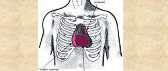
The aorta arises from the left ventricle of the heart, then bends like an arch and leads downward. The pathological condition of damage to large arteries associated with impaired fat metabolism is called atherosclerosis. A decrease in the elasticity of arterial walls and narrowing of the lumens often occurs in elderly people after 60 years of age. Thanks to estrogens, women develop aortic pathology much later than males. Studies have proven that sclerotic changes in the aorta are characterized by a long preclinical course.
Degenerative changes of JSC AK MK: folk remedies, reviews, recipes, in adults
Have you been struggling with HYPERTENSION for many years without success?
Head of the Institute: “You will be amazed at how easy it is to cure hypertension by taking it every day...
Read more "
The aorta is the largest vessel in the human body, through which oxygenated blood is distributed to smaller arteries.
It is directly connected to the left ventricle of the heart, and the flow of arterial blood into it is controlled by the operation of the muscular valve.
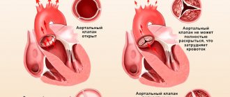
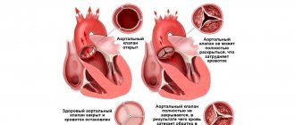
The compaction of the walls of the aorta and the cusps of the aortic valve impedes the flow of blood, which causes rapid wear and tear of the heart and insufficient blood supply to all organs and systems.
Arterial hypertension
Chronic increase in pressure leads to loss of elasticity of the vascular walls. Compensatoryly, the vascular endothelium becomes rigid and thickens due to fibrous growths.
Aortocardiosclerosis
Provoking environmental factors (poor nutrition, obesity, physical inactivity) lead to an increase in cholesterol levels in the blood. It can deposit on the wall of the aorta and heart valves. Over time, portal plaques thicken and significantly narrow the space for blood circulation.
Acute rheumatic fever has many manifestations, one of which is bacterial carditis and valvulitis with subsequent thickening of the mitral or aortic valve.
Endocarditis and myocarditis
Inflammation of the muscular or inner epithelial layer of the heart also leads to scarring at the level of the valves and aortic ring.
Visual inspection
During inspection, pay attention to the following:
OUR READERS RECOMMEND!
Our readers successfully use ReCardio to treat hypertension. Seeing how popular this product is, we decided to bring it to your attention. Read more here...
- characteristic pallor (also called “aortic”): due to a decrease in cardiac output, peripheral capillaries narrow to redistribute blood to the central channel;
- shortness of breath with minimal physical activity – with severe disruption of blood flow;
- acrocyanosis (blueness of the tip of the nose, lips) - not always;
- rarely swelling of the lower extremities.
Physical examination methods
Additionally, the doctor assesses the state of the cardiovascular system using the following diagnostic measures:
- palpation – intensification and displacement of the apex impulse down and to the left (V-Vl intercostal space along the midclavicular line);
- percussion – displacement of the relative dullness of the heart to the left;
- Auscultation – the appearance of a rough noise in the systole phase, weakening of the ll tone over the aorta, moist rales are possible over the surface of the lungs due to left ventricular failure;
- blood pressure measurement - hypotension.
Instrumental diagnostic methods
Additional examination methods are used:
- R-graph – an increase in the size of the heart due to its left parts, expansion of the aortic root;
- electrocardiography – deviation of the electrical axis to the left;
- echocardiography (this examination is also called ultrasound of the heart) - an increase in the thickness of the vascular wall, marginal compaction of the aortic valve leaflets, possible regurgitation (the flow of blood splashed into the aorta back into the heart). EchoCG allows you to assess the degree of destructive changes.
Symptoms
At an early stage of the disease, symptoms of atherosclerosis cannot always be detected. The period of formation of aortic plaque in the heart can last a long time. The manifestation of symptoms in one or another part of the body and the localization of pain will depend on which part of the aorta of the heart is affected. Clinical manifestations of the disease may also be associated with the stage of development of atherosclerosis:
- Ischemic is marked by intermittent claudication, pain in the intestines, and attacks of angina.
- Thrombonecrotic manifests itself as a stroke, gangrene of the feet and myocardial infarction, ischemia, and the formation of thrombosis.
- Fibrous is a chronic type of advanced disease in which the myocardial muscle fibers are replaced with fibrous tissue.
Atherosclerosis of the abdominal aorta
Sclerodegenerative changes in the aorta manifest themselves in the abdominal organs. The initial symptom of aortic pathology is a disruption of the blood supply to the mesenteric vessels that supply the intestinal section. The patient experiences weight loss due to disturbances in the digestive system, pain around the navel, bloating and constipation begin. A patient with atherosclerosis of the abdominal cardiac aorta has the following complaints:
- rapid weight loss;
- stool disorders;
- increased gas formation;
- aching pain after eating;
- increased blood pressure;
- gradual development of renal failure.
Thoracic aorta
Common manifestations of atherosclerosis are deterioration of blood flow through the brain and coronary arteries. Atherosclerosis of the aortic arch and ascending section is manifested by severe aortalgia, which lasts several days or hours and can radiate to the interscapular region, shoulder, upper and lower extremities, and neck. The load on the heart muscle increases due to compaction of the aorta, causing hypertrophy. Atherosclerotic changes in the aorta and its branches cause heart failure and asthma attacks. A patient with atherosclerosis in the thoracic region will experience:
- pain in the heart;
- dizziness;
- increased upper blood pressure;
- the appearance of gray hair;
- manifestation of wen on the face.
Consequences of pathology
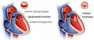
The narrowing of the lumen between the left ventricle and the aorta, as well as the incomplete opening of the valve leaflets, create an obstacle, making it increasingly difficult for the heart to eject blood into the systemic circulation. Excessive work of the heart muscle leads to its thickening (hypertrophy), which in turn is realized in the appearance of so-called diastolic heart failure. The following symptoms occur:
- dizziness and fainting (especially common during physical activity);
- dyspnea;
- rapid heartbeat (as a rule, this is associated with the appearance of atrial fibrillation).
Atherosclerosis of the aorta with damage to the valve and coronary arteries is a fairly common phenomenon. The combination of the increased need for arterial blood of the heart muscle during stenosis and its lack due to atherosclerotic narrowing of the coronary vessels is the cause of chest pain. As a rule, it occurs during emotional or physical stress.
With a pronounced form of narrowing of the aortic mouth, the contractility of the left ventricle weakens, its dilatation (expansion) occurs, which ends in the development of irreversible heart failure. Sudden death is a fairly common occurrence in severe aortic stenosis.
Causes
There are many factors associated with the development of aortic pathology. Atherosclerosis of the heart occurs both for individual reasons and for their combination. The risk group for aortic disease includes people who smoke, are prone to physical inactivity, are obese, have a hereditary predisposition, and patients with diabetes mellitus and infectious diseases. Other causes of aortic disease include:
- a diet that includes the consumption of animal fats;
- decreased physical activity;
- endocrine diseases;
- changes in hormonal levels;
- frequent stressful situations;
- high blood pressure;
- constant intoxication of the body;
- increased cholesterol;
- gender (men are more likely to get sick);
- bad habits;
- genetic predisposition.
Diagnostics
Making an accurate diagnosis of aortic pathology is impossible without adequate medical examinations and correct diagnosis of atherosclerosis. Regular consultations with a therapist will help to detect aortic disease in advance before obvious unpleasant symptoms begin to appear. There are such methods for clarifying atherosclerosis of the heart: angiography, coronography, triplex, duplex scanning, MRI, ECG (with results of ultrasound of the heart and aorta).
Diagnostic methods
Atherosclerosis of the aorta is usually discovered incidentally during examination. After all, the disease is not accompanied by any symptoms for a long time. When listening to the heart and blood vessels, the doctor can detect pathological noises and changes in tones. The pulse in both arms may be asymmetrical.
For a more detailed diagnosis, the patient is referred for instrumental examination:
- Ultrasound of the heart. Allows you to evaluate the size of the ascending aorta, as well as the aortic arch, the thickness of their walls, the presence of defects, and aneurysms.
- Transesophageal echocardiography. A variant of ultrasound of the heart and aorta, in which the sensor is swallowed by the patient. This technique allows you to achieve very high quality images. After all, the vessel is separated from the sensor only by a thin wall of the esophagus. Used to diagnose aortic atherosclerosis when detailed visualization is required.
- Dopplerography. A special type of ultrasound examination that gives the doctor an understanding of the speed of blood flow in different parts of the artery.
- CT, MRI. Both tests allow the doctor to get a picture of the blood vessels. They can be performed with or without contrast. The last examination method involves intravenous injection of a special medical dye, which makes the lumen of the vessel clearer in the image. But since the artery being studied is very large, atherosclerosis of the aorta is well visualized even without the use of contrast.
Treatment
The therapeutic treatment regimen for atherosclerosis is aimed at eliminating pathological processes of the heart, reducing cholesterol, and stimulating its release from the body. Medications must be agreed upon with the attending physician, as there is a high risk of complications. Treatment of atherosclerosis of the aorta of the heart is carried out by taking vitamin complexes, minerals, and products with polyunsaturated fatty acids (Omacor). If life is threatened, emergency surgery may be required. Among the drugs against atherosclerosis, the following names are distinguished:
- Statins: Fluvastatin, Simvastatin, Pravastatin, Atorvastatin, Lovastatin, Rosuvastatin.
- Fibrans: Ciprofibrate, Bezafibrate, Fenofibrate, Gemfibrozil.
- Bile acid sequestrants: Colestipol, Cholestyramine.
- Hypolipid drugs: Ezetimibe, Omega-3-glycerides, Proburcol.
- Nicotinic acid: Enduracin.
Folk remedies
Alternative medicine methods can only be used if atherosclerosis is mild. Among the effective remedies are infusions and decoctions of natural origin from the following plants: viburnum, rose hips, dill, hawthorn, horseradish, parsley, plantain. A special tea made from lemon balm (5 grams of dry raw material per 0.5 liter of water) will help eliminate noise in the head and ears. Below is an effective recipe for a remedy against atherosclerosis. The medicine helps remove salts and cholesterol from the body:
- Take 20 g of wild strawberry leaves and pour a glass of boiling water.
- Boil for 10 minutes.
- Leave for 2 hours, cool.
- Filter and take 3 times daily according to Art. l.
Prevention
In the first place of preventive measures is a diet that restores the metabolic process in the body, which reduces high blood pressure and the blood glucose index.
Proper and balanced nutrition for aortocardiosclerosis:
- daily calorie intake - no more than 2000 kcal, to adjust body weight;
- consume a minimum amount of animal fats - to correct lipoproteins in the blood;
- stop eating simple carbohydrates and sugar - to correct the glucose index;
- minimum salt intake - no more than 5 grams per day. Reducing salt has a beneficial effect on lowering blood pressure in the body;
- consume fiber, which is rich in fresh vegetables, herbs and fruits;
- eat vegetable fats and seafood;
- actively engage in physical activity;
- practice evening walks in the fresh air;
- for pneumosclerosis, a system of breathing exercises is recommended.
Diet for atherosclerosis of the aorta of the heart
Ingestion of excess fat into the body along with food negatively affects blood vessels and leads to the formation of cholesterol plaques. Fat deposits disrupt lipid metabolism, leading to thickening of the walls. An integral part of therapy for atherosclerosis is a balanced diet - a menu adjusted to the consumption of healthy foods and a reduction in carbohydrate and fatty foods. Following a diet leads to the dissolution of plaques and helps cleanse blood vessels. Basic principles of a therapeutic diet:
- easily digestible food;
- reducing salt;
- low-calorie foods;
- fractional meals;
- small portions;
- dairy products;
- cottage cheese;
- fruits;
- high fiber vegetables;
- refusal of sugar, white bread, baked goods.
Sclerotic aorta - what are the dangers?
The aorta is the most important artery of the vascular system in the human body, which supplies blood to human tissues and organs. The main cause of aortic sclerosis is dyslipoproteinemia. Lipoproteins perform cholesterol transport actions in the aorta.
One part of the lipoproteins transfers cholesterol to the walls of the arteries, and the second part removes it from the human body.
When the lipid metabolic process is disrupted, symptoms of atherosclerosis appear. With atherosclerosis, lipids with cholesterol penetrate into the vessel, which thickens the walls of the aorta, and atherosclerotic plaques form.
In the area of these plaques, lesions of fibrous fibers are localized. Over time, these areas grow, which leads to a narrowing of the lumen inside the artery.
Sclerodegenerative changes in the aorta
From about 50 years of age
Sclerotic changes in the heart begin to appear. These changes are more often of the usual nature - “normal”, but they can develop quite quickly and then have a noticeable effect on the function of the heart. Sclerotic changes in the heart develop long before their manifestation. Already in early childhood - from 2-2.5 months of age, cholesterol deposits appear on the anterior leaflet of the mitral valve on the side of the ventricle, which lead to the appearance of a whitish opaque formation, the so-called “white spot”. A “white spot” of the valve occurs in approximately 30% of the corpses of children under 10 years of age and, almost without exception, in all corpses of adults. “At present, most authors agree,” says A.I. Abrikosov, “that the “white spot” of the anterior sail of the bivalve is a particular example of atherosclerosis.”
Sclerotic valve
and fibrous rings of the orifices makes them rigid and inflexible, which leads to insufficient opening and incomplete closure of the valves. Sometimes the valves thicken and curl inward toward the walls of the aorta in such a way that they cannot stretch across its entire opening. As a result, a gap remains between the valve leaflets, through which blood freely returns to the ventricle (O. Kalinsky, 1833).
Hardening of cardiac structures
can occur with the deposition of lime in the “white spot” of the mitral valve, on the aortic valves, in fibrous atrioventricular rings, in thrombotic deposits in endocarditis, in connective tissue, in the tips of the papillary muscles that have become fibrous. At the site of lime deposition, bone tissue sometimes forms, even with bone marrow (A.I. Abrikosov). These deposits can protrude into the lumen of the cavities of the heart and blood vessels, create roughness on the valves and in the valve openings, thereby contributing to the formation of heart murmurs.
On the nature of the blood stream
may be influenced by the special formations that are sometimes found in old people on the arantia nodules of the semilunar valves, most often the aortic and less often on other valves - lamble excretions (Lamble, 1856) - connective tissue outgrowths in the form of ridges, villi or threads up to 4-5 mm in length.
Very often (in 80% of autopsies) observed
the tips of the aortic and pulmonary valves are small through holes. These holes are located above the valve closure line (located on the area of the valve surface that contacts other valves when closed) and therefore do not interfere with their function.
Basic soil
, on which sclerosis of the valvular apparatus of the heart develops, often leading to circulatory disorders, is inflammation of its inner membrane with the involvement of the valves in the inflammatory process. Such sclerosis can, in turn, be complicated by an atherosclerotic process, which usually increases the deformation of the valves.
To the greatest destruction
valve apparatus is caused by ulcerative endocarditis. In contrast to warty endocarditis, alterative changes predominate in ulcerative endocarditis, the whole process proceeds faster, and the phenomena of necrosis and destruction prevail. Destruction processes can lead to rupture, perforation, acute and then chronic valve aneurysms, which, in turn, can be subject to perforation. When the ulcerative process affects the parietal endocardium, the ulcers are sometimes so deep that they can lead to the formation of cardiac aneurysms, perforation of its walls, rupture of the heart, the formation of communications between its cavities, and interruption of the conduction pathways. When the ulcerative process moves from the aortic valves to the inner surface of the valsalvo sinuses, destruction occurs here, including sinus aneurysms and rupture of the aortic wall. Healing of the ulcerative process with the organization of thrombotic masses and sclerosis of the affected areas of the endocardium is rarely observed.
Affected by the inflammatory process
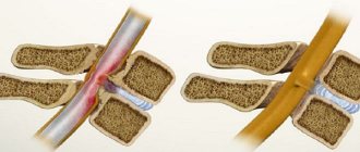
the valves can fuse and lead to narrowing of the valve opening. Fused valves form a kind of funnel, which is more often observed with damage to the mitral valve.
Approximation of valve flaps
, as a condition for the fusion of their edges, can be explained by the action of Bernoulli’s law.
St. Petersburg mathematician Daniel Bernoulli
in the first half of the 18th century found that in a flow of liquid or air, the pressure decreases compared to the pressure outside the flow (or jet).
If a fluid flow passes between two bodies, then these bodies come closer if they are displaceable. The rapprochement is caused by the predominance of pressure on the body from the outside, compared with the pressure from the inside - from the side of the fluid flow. This, by the way, explains the approach of ships by the flow of water passing between them and some other phenomena.
Diagnosis in patients with acquired aortic stenosis
The main Doppler echocardiographic signs of valvular aortic stenosis:
- narrowing of the aortic opening (normally 3–4 cm2);
- acceleration of turbulence of systolic transaortic flow above the level of obstruction (Fig. 8.36-a).
Other characteristic signs of acquired valvular stenosis, determined in M- and B-modes, are:
- thickening and compaction of the valves;
- decrease in systolic opening of the leaflets (if the separation of the right coronary and posterior non-coronary leaflets is less than 1.5 cm) (Fig. 8.36-b).
Additional signs are considered:
- hypertrophy and/or dilatation of the left ventricle;
- decrease in EF tilt of the anterior mitral valve leaflet;
- diastolic flutter of the anterior mitral valve leaflet;
- reduction of aortic excursion;
- post-stenotic dilatation of the aortic root.
Atherosclerosis of the aorta of the heart: causes, symptoms, treatment with folk remedies and medications
Few people know what atherosclerosis of the aorta of the heart is. Treatment with folk remedies and medications should be carried out only after consultation with your doctor.
Atherosclerosis is a fairly common disease, especially among older people . It is a chronic disease of the arteries due to impaired fat and protein metabolism. Accompanied by cholesterol deposition in the aortic lumen.
Cholesterol can be called a bad fat. Entering the body, it sticks to the walls of the aorta, forming growths. They are called cholesterol plaques. Over time, the growths deform the blood vessel, narrow its lumen, and disrupt blood flow. Aortic atherosclerosis leads to coronary heart disease, aortic blockage and death.
Recently, the statistics of atherosclerosis have acquired alarming proportions. It is most often diagnosed in men 45-50 years old, but cases of incidence have also been recorded at a younger age.
Other causes of narrowing of the aortic mouth
Atherosclerosis of the aorta and aortic valve leaflets is the main cause of the accumulation of calcium deposits in older people, which leads to sclerotization of these structures. About 1 in 20 people over the age of 65 have some degree of aortic stenosis. But there are other causes of this pathology that occur at an earlier age. For example:
- Rheumatism of the heart . A disease that 50 years ago was quite common and was the main cause of narrowing of the aortic mouth. Fortunately, with the advent of antibiotics, cases of pathology development have become much less common.
- Bicuspid aortic valve. This congenital pathology is the most common cause of stenosis in young people. Instead of three petals, the human aortic valve consists of two. This makes him more vulnerable to calcium deposits and, accordingly, to the formation of stenosis, the symptoms of which may appear by the age of 40 - 50 years.
Bicuspid aortic valve
- Endocarditis is inflammation, both infectious and autoimmune, of the inner lining of the heart.
- Congenital aortic stenosis is a heart defect (formation of abnormal connective tissue located above or below the valve).
Regardless of the etiology, at a certain stage, severe calcification of the aortic valve occurs, which leads to the appearance of symptoms of narrowing of the aortic ostium. Atherosclerosis of the aorta, aortic and mitral valves is formed mainly in the elderly and senile age, and can also affect other valves of arteries and blood vessels.
Reasons for the development of pathology
The main cause of the development of aortic atherosclerosis is cholesterol deposition. A number of factors lead to this:
The disease affects men more. The thing is that before the onset of menopause, women produce large quantities of the sex hormone estrogen. It is actively involved in cholesterol metabolism. In simple words, it helps break down harmful fats. Men have no such protection. Therefore, up to 50 years of age, men are more likely to suffer from atherosclerosis. When menopause occurs, hormonal imbalances occur in the female body and estrogen production decreases. As a result, the risk of atherosclerosis will increase in women.
With age, all people's bodies age. Blood vessels become less durable and elastic under the influence of multiple factors. At the sites of damage to blood vessels, a loose wall is formed, where it is much better for cholesterol to adhere.
Man, unfortunately, cannot change his genome. If there were relatives in the family with a similar disease, then the person is at risk.
Smoking and excessive alcohol consumption are a direct path to aortic atherosclerosis. Tobacco smoke and ethanol molecules are the strongest poisons for the body. Toxins poison it, disrupting blood circulation, deforming blood vessels, including the aorta.
People suffering from hypertension (also called arterial hypertension) are susceptible to the development of atherosclerosis of the aorta. Hypertension is accompanied by constant surges in pressure. Mostly it is high. At the same time, the blood puts very strong pressure on the walls of the blood vessels, damaging them, making them more vulnerable to narrowing and the formation of cholesterol plaques.
Extra pounds lead to disruption of fat metabolism. At the same time, the balance of “good” and “bad” cholesterol is also disrupted. The result is the formation of plaques in the aorta and atherosclerosis.
This is an endocrine disease accompanied by impaired carbohydrate metabolism. In the body of people with diabetes, such disorders are necessarily accompanied by disruptions in fat metabolism. A person diagnosed with diabetes is much more susceptible to atherosclerosis than healthy people.
- Sedentary lifestyle
Sedentary work and lack of sports lead to stagnation of blood in the vessels. Their deformation and circulatory disturbances occur, which leads to atherosclerosis of the aorta of the heart.
It became known not so long ago that viruses and bacteria can cause atherosclerosis. But still this is a proven fact. People who often suffer from various infectious diseases are at risk for atherosclerosis. Hepatitis and herpes can provoke pathology.
Nervous tension caused by stress is accompanied by the production of large amounts of adrenaline. It has a very dangerous effect on the body: it generates oxygen molecules, which in turn make cholesterol molecules sticky. At high concentrations, fat cells begin to adhere to the wall of the aorta. Therefore, emotional people are more susceptible to aortic atherosclerosis than others.
Fast food, fried, smoked foods contain trans fats. Once in the body, they cause not only atherosclerosis, but also the formation of free radicals. By the way, a fried crust on meat is nothing more than bad cholesterol.
As you can see, multiple causes can become the trigger for the development of aortic atherosclerosis. When exposed to the above factors, damage to the aortic wall occurs. In these places, platelets (red platelets of blood) accumulate and are destroyed. The destruction of platelets is accompanied by the release of active substances, due to the impact of which the muscle cells of the aorta become denser. The result is the formation of atherosclerotic changes in the aorta.
Causes of formation, symptoms and methods of treating aortic stenosis
Aortic stenosis is a reduction in the valve opening. Against the background of this narrowing, the flow of blood from the left ventricle to the heart is significantly limited. This disease is the most common heart defect.
Often the disease progresses in patients who have crossed the sixty-year threshold.
Deformations of the aortic valve leaflets can provoke the appearance of pathology. This happens eighty-five percent of the time. And only in ten percent of cases the root cause of the disease is the process of rheumatization.
general information
Aortic stenosis that has not been acquired is considered separately by doctors. The presence of a congenital anomaly leads to an increase in the load on the left ventricle.
Provoking factors
There are a considerable number of reasons that provoke the development of aortic stenosis. The most common reasons for which the disease develops include:
- Congenital cardiac anomaly.
- Deposition of calcium salts on the aortic valve.
- Rheumatic process.
Features of congenital anomaly
Congenital aortic stenosis should be considered a pathology when a baby is born with a pre-existing abnormal process in the actual valve. This is a fairly rare pathology. A bicuspid valve is no less rare. When a person is in childhood, the anomaly has no consequences. A completely different picture is observed in adult patients. Due to the narrowing of the valve, a person often needs to have it replaced.
When is calcium deposited?
Over time, calcium salts are deposited on the walls of the heart valves. This element is found in human blood, being dissolved. As a person ages, salts deposit on the valves.
In this case, the manifestations of a serious disease do not make themselves felt until the person reaches the age of seventy.
Signs of atherosclerosis
The aorta is divided into two parts: abdominal and thoracic. Symptoms of atherosclerosis of the thoracic aorta:
- attacks of dizziness that end in fainting;
- impaired swallowing reflex, sensation of a lump in the larynx;
- voice disorder – it becomes hoarse, hoarse;
- cramps in the neck;
- fast fatiguability;
- shortness of breath even with little physical activity;
- increased blood pressure;
- pain in the heart area can radiate under the shoulder blades, in the neck, left and right arms, and spine.
The pain does not go away, even if the person has taken medications with nitroglycerin. Due to the compacted aorta, the load on the heart greatly increases. Hypertrophy (thickening) of the heart muscle develops. This condition is characterized by regular attacks of suffocation.
Atherosclerosis of the abdominal aorta is much more common. Manifests:
- digestive disorders (a person suffers from constipation, flatulence, diarrhea, lack of appetite);
- paroxysmal aching pain in the abdomen, developing 3-4 hours after eating.
Stenosis of the aortic mouth (aortic stenosis) and all its features
general information
Have you been struggling with HYPERTENSION for many years without success?
Head of the Institute: “You will be amazed at how easy it is to cure hypertension by taking it every day...
Read more "
Aortic stenosis (aortic stenosis) is one of the most common heart defects in modern society. It is diagnosed in every fifth patient over the age of 55, and 80% of patients are men.
In patients with this diagnosis, there is a narrowing of the aortic valve opening, which leads to disruption of blood flow into the aorta from the left ventricle. As a result, the heart has to make significant efforts to pump blood into the aorta through the reduced opening, which causes serious problems in its functioning.
Causes and risk factors
Aortic stenosis can be congenital (occurs as a result of intrauterine developmental anomalies), but more often it develops during a person’s life. The causes of the disease include:
- heart disease of a rheumatoid nature, which usually occurs as a consequence of acute rheumatic fever due to infections caused by a certain group of viruses (group A hemolytic streptococci);
- atherosclerosis of the aorta and valve - a disorder that is associated with lipid metabolism disorders and cholesterol deposition in the vessels and valve leaflets;
- degenerative changes in heart valves;
- infective endocarditis.
Classification and stages
Aortic stenosis has several forms, which are distinguished according to different criteria (localization, degree of blood flow compensation, degree of narrowing of the aortic opening).
- according to the location of the narrowing, aortic stenosis can be valvular, supravalvular or subvalvular;
- by the degree of blood flow compensation (by how much the heart can cope with the increased load) - compensated and decompensated;
- According to the degree of narrowing of the aorta, moderate, severe and critical forms are distinguished.
The course of aortic stenosis is characterized by five stages:
- Stage I (full compensation). There are no complaints or manifestations; the defect can only be determined through special studies.
- Stage II (hidden insufficiency of blood flow). The patient is concerned about mild malaise and increased fatigue, and signs of left ventricular hypertrophy are determined by x-ray and ECG.
- Stage III (relative coronary insufficiency). Chest pain, fainting and other clinical manifestations appear, the heart increases in size due to the left ventricle, and the ECG shows its hypertrophy, accompanied by signs of coronary insufficiency.
- Stage IV (severe left ventricular failure). Complaints of severe malaise, congestion in the lungs and a significant enlargement of the left heart.
- Stage V, or terminal. Patients experience progressive failure of both the left and right ventricles.
See more about the disease in this animation:
Is this scary? Danger and complications
The quality and life expectancy of a patient with aortic stenosis depends on the stage of the disease and the severity of clinical signs. People with a compensated form without severe symptoms do not have a direct threat to life, but the symptoms of left ventricular hypertrophy are considered prognostically unfavorable.
In patients with the “classic triad” (angina, syncope, heart failure), life expectancy rarely exceeds five years. In addition, in the later stages of the disease there is a high risk of sudden death - approximately 25% of patients diagnosed with aortic stenosis die suddenly from fatal ventricular arrhythmias (usually these include people with severe symptoms).
The most common complications of the disease include:
- chronic and acute left ventricular failure;
- myocardial infarction;
- atrioventcular block (comparatively rare, but can also lead to sudden death);
- swelling and congestion in the lungs;
- systemic emboli caused by calcium particles from the valve can cause strokes and visual impairment.
Symptoms
Often, signs of aortic stenosis do not appear for a long time. Among the symptoms that are characteristic of this disease are:
- Shortness of breath. Initially, it appears only after physical exertion and is completely absent at rest. Over time, shortness of breath occurs in a calm state and intensifies in stressful situations.
- Chest pain. Often they do not have an exact localization and appear mainly in the heart area. The sensations can be pressing or stabbing in nature, last no more than 5 minutes and intensify with physical activity and stress. Angina pain (acute, radiating to the arm, shoulder, under the shoulder blade) can be observed even before the appearance of pronounced symptoms and is the first signal of the development of the disease.
- Fainting. Usually observed during physical activity, less often - in a calm state.
- Rapid heartbeat and dizziness.
- Severe fatigue, decreased performance, weakness.
- A feeling of suffocation, which may become worse when lying down.
When should you see a doctor?
Often the disease is diagnosed accidentally (during preventive examinations) or in the later stages due to the fact that patients attribute symptoms to overwork, stress or adolescence.
Diagnostics
Diagnosis of stenosis of the defect is complex and includes the following methods:
- Anamnesis collection. Analysis of patient complaints, past illnesses and family history (cases of heart disease or sudden death in close relatives).
- External inspection. Patients have pallor and cyanosis of the skin, heart murmurs and wheezing in the lungs, and the peripheral pulse in the radial arteries is weak and rare.
- Auscultation of aortic stenosis. The method involves listening to the sounds and rhythms of the heart - with aortic stenosis, the second sound is usually weakened or completely absent, and systolic and diastolic murmurs are also noted.
- General blood analysis. It is carried out to determine the level of red blood cells, platelets, leukocytes, as well as the level of hemoglobin.
- General urine analysis. Makes it possible to identify disorders that may affect the course of the disease.
- Electrocardiography. A method for assessing the electrical activity of the heart, allowing to identify disturbances in its functioning.
- Echocardiography. An ultrasound examination that determines the degree of narrowing of the aorta and the most significant indicators of heart function.
- Coronary angiography with aortography. An invasive procedure that involves penetrating the vessels of the arms and legs to examine the vessels of the heart and aorta.
- Exercise tests. Exercise tests include walking, exercise bike and treadmill tests.
Why do you feed pharmacies if hypertension is afraid of the usual like fire...
Tabakov has revealed a unique remedy against hypertension! To reduce blood pressure while preserving blood vessels, add to…
Treatment methods
There is no specific therapy for aortic stenosis, so treatment tactics are selected based on the stage of the disease and the severity of symptoms. In any case, the patient must register with a cardiologist and be under strict supervision. It is recommended to undergo an ECG every six months, give up bad habits, diet and a strict daily routine.
Patients with stages I and II of the disease are prescribed drug therapy aimed at normalizing blood pressure, eliminating arrhythmia and slowing the progression of stenosis. It usually includes taking diuretics, cardiac glycosides, and drugs that lower blood pressure and heart rate.
Radical methods for the initial stages of aortic stenosis include cardiac surgery. Balloon valvuloplasty (a special balloon is inserted into the aortic opening, after which it is inflated mechanically) is considered a temporary and ineffective procedure, after which in most cases a relapse occurs.
In childhood, doctors usually resort to valvuloplasty (surgical valve repair) or Ross operation (transplantation of the pulmonary valve to the position of the aorta).
At stages III and IV of aortic stenosis, conservative drug treatment does not give the desired effect, so patients undergo aortic valve replacement. After surgery, the patient must take blood thinners for the rest of his life, which prevent the formation of blood clots.
Prevention
There are no methods to prevent congenital aortic stenosis or to diagnose it in utero.
Preventive measures for acquired defects include a healthy lifestyle, moderate physical activity and timely treatment of diseases that can provoke narrowing of the aorta (rheumatic heart disease, acute rheumatic fever).
Any heart disease, including aortic stenosis, is potentially life-threatening. To prevent the development of heart pathologies and defects, it is very important to take a responsible attitude towards your health and lifestyle, as well as regularly undergo preventive examinations, which can detect diseases at the initial stages of their development.
Complications
If the pathology is not treated, the consequences may be as follows:
- Hypertensive crisis.
- Myocardial infarction.
- Stroke (hemorrhagic/ischemic).
- Heart failure.
- Dissecting aortic aneurysm.
The prognosis directly depends on the damage to the aorta. The fact that people are ready to fight the disease also plays a role. If a person notices its symptoms in time and undergoes comprehensive treatment, the prognosis is favorable. People live with the disease, but they need to be diagnosed regularly.
In forensic medicine there is the concept of sudden death, which in 89% is caused by atherosclerosis of the aorta of the heart.
Clinical picture
Symptoms of aortic atherosclerosis are quite varied and specific. In most cases, their severity and nature directly depend on the period of development of the disease itself and the location of the affected area of the aorta.
There are two main periods of development of the disease in question, namely: preclinical (initial) and clinical (progressive).
In the first case, there are no general symptoms of the disease, and the only evidence of its development is only pathological changes in blood vessels recorded during laboratory tests. In the second case, the disease begins to progress and manifest itself with the appearance of a fairly pronounced clinical picture. This period is divided into three successive stages of development, called ischemic, necrotic and sclerotic stages.
As soon as atherosclerosis reaches the clinical period in its development, the patient begins to appear the main pathological signs, differentiated depending on the location of the aortic lesion.
If the abdominal part of the vessel is affected, atherosclerosis of the abdominal aorta is diagnosed. As a rule, it does not appear immediately, but is asymptomatic over a fairly long period of time. Symptoms of atherosclerosis of the abdominal aorta include: constipation, digestive disorders, aching pain in the abdomen.
In the case of damage to the thoracic aorta, the disease manifests itself in a more varied clinical picture, characterized by increased blood pressure, dizziness, convulsions, prolonged burning and pressing pain behind the sternum or in the upper abdomen.
Atherosclerosis of the aorta of the coronary arteries usually gradually weakens health, provoking the slow development of pathological disorders in the functioning of various organs and systems (kidneys, nervous system, gastrointestinal tract). Most often, the course of this type of atherosclerosis is accompanied by the appearance of the main signs of angina pectoris; in more rare cases, coronary atherosclerosis can manifest itself as myocardial infarction, as well as sudden death of a person.
If the atherosclerotic process affects, in addition to the aorta, also the leaflets of the aortic valve, this pathology is defined as atherosclerosis of the aorta and aortic valve and indicates the development of insufficiency of this valve. In such cases, the main clinical signs of pathology include: angina pectoris, general weakness, dizziness, rapid heartbeat, pronounced pain in the chest, as well as increased pulsation in the head and neck.
If any signs of atherosclerotic damage to the aorta appear, you should immediately consult a doctor, since this disease, if not treated in a timely manner, can provoke life-threatening consequences for the patient.
Diagnosis, treatment
The following methods are used for diagnosis:
- blood test for cholesterol;
- ultrasound examination of blood vessels;
- heart doppler;
- electrocardiography during physical activity;
- contrast X-ray examination of blood vessels;
- electrocardiogram;
- comprehensive study of lipid metabolism;
- computed and magnetic resonance imaging.
Based on the results of these studies, a diagnosis is made and a course of therapy is prescribed.
Pathology needs to be treated for a long time and comprehensively. It cannot be said that there is one single treatment regimen. All drugs and techniques are prescribed by the attending physician in each individual case. The age of the person, the general condition of the body, the degree of damage to the aorta, and the presence of secondary pathologies are taken into account.
Drug treatment
One of the important conditions for treatment is physical therapy and adherence to a cholesterol-free diet. The following drugs are used for treatment:
- Statins. Slow down the synthesis of cholesterol molecules and help reduce the concentration of fat in the blood.
- Fibrates. Prescribed to reduce the concentration of fats.
- A nicotinic acid. Helps speed up the breakdown of fats.
- Sequesters of bile acids. Prevents the absorption of bile acids from the gastrointestinal tract. This reduces the amount of bad cholesterol.
At the same time, medications are prescribed for the treatment of concomitant pathologies, for example, diabetes mellitus, hypertension, angina pectoris, and obesity.
An equally important condition during therapy is the absolute rejection of bad habits and alcohol consumption.
Surgery
In severe cases, treatment of atherosclerosis of the aorta of the heart is carried out surgically. During the operation, a section of the damaged part of the aorta is removed and a prosthesis (synthetic explant) is placed.
Treatment of aortic stenosis
If there are no symptoms, then the patient does not need treatment. When symptoms occur, valve replacement is usually recommended. Today, replacing it is considered the most effective way to get rid of the disease. But this is not always possible, so the patient may be offered drug therapy. Drugs are mainly prescribed to reduce the manifestations of heart failure. The most commonly used drugs are:
- ACE inhibitors (angiotensin-converting enzyme inhibitors);
- diuretics.
Since the operation helps to remove a serious obstruction to blood flow, in most cases the pumping function of the heart is restored in a short time. Therefore, even if the patient is elderly, his postoperative period proceeds quite favorably.
Depending on each specific situation, the following types of surgical correction of stenosis may be offered:
- Valvotomy is a dissection of the valve leaflets; the operation is performed on an open heart.
- Valve replacement . Also requires open heart surgery. Prostheses are divided into mechanical and biological. The first are made from materials of non-biological origin. The latter partly consist of specially processed animal tissues (for example, pigs).
- Valvuloplasty is stretching of a narrowed lumen using a special balloon. This is a minimally invasive option that does not require open heart surgery. A catheter is brought through the femoral artery to the mouth of the aorta, at the end of which there is a balloon; when expanded, the aortic valve leaflets “fused with each other” are stretched.
- Transcatheter aortic valve implantation . This is another minimally invasive approach to stenosis surgery, where an artificial valve is implanted through a very complex catheterization procedure. Nevertheless, the technique is quite new and not yet well developed. Therefore, it is used in cases where it is impossible to replace the valve in an open heart.
Transcatheter aortic valve implantation
Balloon valvuloplasty, compared with valvotomy and valve replacement, has worse long-term results. Its therapeutic effect is often not so long-lasting. As a rule, relapse of stenosis occurs over time.
To learn how transcatheter aortic valve implantation is performed, watch this video:
Treatment with folk remedies
For atherosclerosis of the aorta of the heart, treatment with folk remedies can also be carried out. But they can only be a method of secondary therapy, and not the main one. The most effective herbal teas:
- Dandelion root; stems, leaves, roots of strawberries; nettle; lemon balm are taken in equal proportions, pour 1 liter of boiling water. Leave the composition for 2-3 hours. Take 0.5 cups every day 3 times.
- Burdock root (50 g), dill (30 g), horsetail (60 g), drop cap (50 g). 1 tbsp. l. collection, pour 200 ml of boiling water, leave until cool, strain. Drink during the day.
- Take hawthorn, yarrow, and birch leaves in equal quantities. Pour 100 g of collection into 1 liter of water and boil in a water bath for 5 minutes. After cooling, take 2 tbsp. l. 2 times during the day.
- Rose hips, strawberry leaves, mint, oats. Take 50 g of each ingredient. Pour in 500 ml of water. Boil for 10 minutes in a water bath. Take half a glass 2 times a day.
Many people live with aortic atherosclerosis for a long time, not paying attention to pain in the abdomen and chest. We must not forget that cholesterol plaque can grow along the aorta, but it can also grow into the lumen. If the aorta is purchased, death will be instantaneous. If a person is at risk or has bad habits, he must undergo diagnostics at least 3 times a year in order to prevent the development of complications and irreversible consequences.
Complicated form of the disease
The development of thrombosis leads to blockage of some vessels, and if the thrombus is large, it can block the coronary vessel and cardiac arrest will follow.
Destruction of the vessels of the peritoneum threatens the disease peritonitis and non-healing trophic ulcers in the lower extremities.
Oxygen starvation when the blood supply system does not function properly can lead to infarction of the brain and heart muscle, strokes in the brain and kidneys.
Failure of the brain and heart muscle (myocardium), and internal vital organs, also develops. The most terrible complication for life is coronary insufficiency, which can be instantly fatal.