What does it represent?
Ventricular extrasystole is a type of arrhythmia, as a result of which the heart muscle contracts under the influence of a pathological focus of excitation concentrated in one of the ventricles.
The ICD-10 code is 149.3.
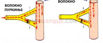
Attention! If any of the components of the conduction system of the ventricular myocardium can self-reproduce an excitation wave, then an ectopic focus of depolarization is formed. During an ECG, a similar condition will be manifested by extrasystoles.
Features of bradyarrhythmia in children
Children and teenagers have a faster heart rate, which is required to support their growing bodies.
Heart rate norms in children differ from adults:
| Age | Normal heart rate (beats per minute) |
| Infants up to one year old | 130-150 |
| Children from two to 7 years old | 100-110 |
| Children from 8 to 10 years old | 80-90 |
| Teenagers from 10 to 14 years old | 70-90 |
It should be noted that girls of the same age may have a pulse 10-15 units less, which is explained by the characteristics of the body.
Bradyarrhythmia in children is a less common problem than respiratory tachycardia. Correction is required only if there are pathologies of the heart muscle or a non-standard pacemaker. In the absence of symptoms, the disease is left under observation without treatment.
Causes
All reasons for the development of pathology can be divided into 3 groups:
- organic;
- functional;
- idiopathic.
Organic
Formed with more serious heart problems. Premature heart beats can develop as a result of the following diseases:
- insufficient heart function;
- ischemia;
- myocardial infarction;
- rheumatic damage to the valve apparatus;
- cardiosclerosis;
- hypertension.
Functional
This category includes:
- stress;
- smoking;
- drinking alcoholic drinks, coffee and energy drinks in large quantities.
Important! If pathology develops for these reasons, treatment is not carried out. It is important to eliminate the underlying factor and undergo diagnostics again after 2 months.
Idiopathic
In this condition, a completely healthy person will experience ventricular extrasystoles, the cause of which is not clear. The patient has no clinical manifestations of the disease, so therapy is not prescribed.
Classification
Before prescribing a treatment regimen to a specialist, it is important to find out the type of pathology.
According to the source of occurrence, ventricular extrasystoles are of the following types:
- monotopic - have one ectopic focus of the impulse;
- polytopic - several secondary sources of impulse.
Based on the duration of the compensatory pause, they are divided into:
- full - the total duration of the pre- and post-ectopic interval is equivalent in duration to two main cardiac cycles;
- incomplete - the total duration of the intervals is less than the duration of the two main cardiac cycles.
According to Lown-Wolf-Ryan, ventricular extrasystoles are divided into 5 degrees:
- monomorphic (less than 30 per hour);
- monotopic, more than 30 per hour;
- polymorphic extraordinary contractions;
- is divided into subcategories “a” – twin, “b” – salvo;
- salvo polymorphic PVCs (3-5 in half a minute. In this case, it is important for the patient to provide emergency assistance.
There is also a zero degree, which implies the complete absence of extrasystoles.
The pathology is also classified according to the time of development:
- early – formed during the passage of an impulse through the atria;
- interpolated - the right and left ventricles contract simultaneously with the atria;
- late – formed during the “rest” of the upper chambers of the heart.
Circadian types of extrasystole are:
- mixed;
- daytime;
- nightly.
Diagnostics
The diagnosis can be suspected by the patient independently when the slightest signs of bradyarrhythmia appear and when counting the pulse. Only a doctor can make an accurate diagnosis after interpreting the electrocardiogram. Therefore, if one of the types of bradyarrhythmia is suspected, especially with an extremely low pulse, an ambulance team should be immediately called.
The criteria for ECG diagnosis of bradyarrhythmia are as follows:
- Rare complexes reflecting ventricular contractions
- Prolongation of intervals between ventricular complexes,
- Irregular heart rhythm.
The doctor will begin therapy at the prehospital stage, in particular, the administration of atropine. After this, the patient is usually taken to the hospital emergency department and examined by a cardiologist or therapist, where the type of bradyarrhythmia is determined.
In the hospital, additional research methods may be prescribed - ultrasound of the heart, 24-hour monitoring of blood pressure and ECG, etc.
Diagnostic methods
The following research methods are used:
- ECG - this diagnostic option allows you to determine what happens to the organ during contraction and relaxation.
- ECG monitoring according to Holter - the essence of the method is that the patient is attached to a device with sensors, and he must do this all day. Thanks to such daily monitoring, it is possible to track the periods and frequency of occurrence of extraordinary ventricular contractions. This diagnostic method is the most basic.
- Bicycle ergometry is used to confirm or refute the relationship with physical activity.
Why does bradyarrhythmia occur in athletes?
Physical activity causes the heart rate to increase as a response to a lack of oxygen for muscle cells to function.
With constant movement and stress, the body adapts to hypoxia by increasing hemoglobin and red blood cells. At rest, the heart reduces the heart rate, causing bradyarrhythmia without symptoms, since there is enough oxygen even with rare contractions of the heart. Most often, this condition develops in runners and swimmers, that is, those athletes who need increased endurance.
Treatment
A treatment regimen is prescribed by a doctor only after a diagnosis has been made and the cause of the pathology has been discovered.
Medication
Treatment with medications is prescribed privately and is determined taking into account morphological information, frequency of arrhythmia and other associated cardiac signs. In practice, antiarrhythmic drugs are used, which are divided into the following subtypes:
- sodium channel blockers - Novocainamide, Gilurythmal, Lidocaine;
- beta-blockers - Cordinorm, Carvedilol, Atenolol, Anaprilin;
- potassium channel blocker medications – Sotalol, Amiodarone;
- calcium channel blockers – Verapamil, Amlodipine, Cinnarizine;
- antihypertensive medications (indicated for patients with extrasystole accompanied by high blood pressure) - Captopril, Enaprilin, Ramipril;
- prevention of thrombosis - Clopidogrel, Aspirin.
After 2 months of therapy, the patient must perform a control ECG. If extrasystoles become rare or absent altogether, then treatment is canceled. If the results of the studies are poor during therapy, then the treatment course continues for several months.
Reference! In case of malignant pathology, medications are taken throughout life.
Surgical methods
Surgical treatment is indicated only in cases where drug methods are ineffective. This mainly applies to patients with diagnosed organic ventricular extrasystole.
The following types of surgical treatment exist:
- Introduction of a pacemaker - This device is equipped with electronics and a battery that lasts 10 years. Electrodes extend from the pacemaker, and during surgery they are fixed to the ventricle and atrium. They produce electronic impulses, due to which the myocardium begins to contract. The pacemaker is a substitute for the sinus node, which is responsible for rhythm. An electronic device relieves a person from extrasystole and allows him to return to a full life.
- Radiofrequency ablation - the essence of the method is that a small catheter is inserted through a large vessel into the cavity of the heart. Using radio waves, pathological areas are cauterized. Electrophysiological monitoring is used to find the “operated” area. The effectiveness of the method is 75-90%.
For reference! Cardiologists recommend installing a pacemaker for patients who need to regulate their heart rhythm with medications throughout their lives. Most often these are elderly people, because sometimes taking a pill on time is a difficult task for them.
Folk remedies
Non-traditional methods can only be used as an addition to the main therapy. Popular recipes:
- Add grated lemon zest (4 tbsp) to 50 ml of honey, 10-120 pcs. apricot kernels, ground in a coffee grinder. Mix everything well and drink 1 tbsp. 2 times a day.
- Pour 10 g of mistletoe herb into 250 ml of boiling water. Leave for 10 hours, filter and take 250 ml 3 times a day. The tea must be heated before drinking.
- Mix the following herbs: lemon balm, motherwort, heather, hawthorn, hops in the following proportion: 5:5:4:3:2. Pour 30 g of the mixture with a glass of boiling water. Leave for 30 minutes and take 20 ml 3-4 times a day. The prepared tea has cardiotonic, antiarrhythmic, sedative and sedative effects.
Attention! Before using traditional methods of treatment, you should consult a doctor, as some herbs can lead to an allergic reaction.
Symptoms (signs)
Clinical manifestations • Dizziness, short-term loss or confusion, darkening of the eyes, fainting (in 50–70% of cases), sudden falls with injuries and fractures • Feeling of palpitations or frequent interruptions in heart rhythm • The first manifestations of SSPS may be paroxysmal tachycardia, atrial fibrillation arrhythmia or Morgagni–Adams–Stokes attack.
Clinical and electrocardiographic signs. SSSPU can manifest itself in seven variants • Constant or episodic sinus bradycardia • Rigid sinus rhythm • SUS failure (see Sino-atrial node failure) • Sinauricular block of the second degree • Brady - tachyarrhythmia - alternation of sinus bradycardia with flutter, atrial fibrillation or atrial tachycardia • Post-tachycardia depression of SPU - slow restoration of sinus rhythm after pacemaker, cardioversion or cessation of supraventricular tachycardia (recovery time of sinus node function exceeds 1400 ms) • Chronotropic insufficiency - discrepancy between the frequency of sinus rhythm and the needs of the body.
Consequences, what is the danger
To give a prognosis, it is necessary to take into account the severity of the impulse disturbance and the degree of dysfunction of the ventricles. If pathological changes in the myocardium are pronounced, this leads to atrial and ventricular fibrillation, persistent tachycardia, which can subsequently lead to the death of the patient.
When the shock during relaxation of the ventricles, which occurs out of turn, coincides with the contraction of the atria, the blood, without having time to empty the upper compartments, is sent back to the lower chambers of the circulatory organ. This phenomenon is fraught with the development of thrombosis.
The danger is that a blood clot, when entering the bloodstream, leads to thromboembolism . If the lumen of the vessels is blocked, this can lead to the development of the following pathologies:
- stroke;
- heart attack;
- ischemia.
Timely consultation with a doctor will help prevent complications. Timely prescribed treatment and compliance with all recommendations are the key to a speedy recovery and transition to a full life.
Is it possible to play sports and serve in the army with bradyarrhythmia?
Sinus bradyarrhythmia has its own ICD code (International Classification of Diseases) - R00.1 and refers to pathologies that are divided into physiological and organic.
If the disease does not have pronounced symptoms and is the norm for a particular person (with good physical preparation), then he will be called up to serve in the army. If during a medical examination it was proven that the bradyarrhythmia is organic (the result of serious disorders in the body), then the conscript is exempt from military duty.
With this disease, activities that involve moderate cardio exercise (for example, running) are not prohibited, but strength training should be avoided.