Types of extrasystole
1. By localization:
- Sinus.
- Atrial.
- Atrioventricular.
- Ventricular
2. Time of appearance in diastole:
- Rare (up to 5/min).
- Medium (6-15/min).
- Frequent (more than 15/min).
5. By frequency:
- Sporadic (random).
- Allorhythmic – systematic – bigeminy, trigeminy, etc.
6. On carrying out:
- Repeated impulse entry using the re-entry mechanism.
- Blockade of conduction.
- Supernormal conduction.
8. By number of sources:
Sometimes the so-called interpolated ventricular extrasystole - it is characterized by the absence of a compensatory pause, that is, the period after the extrasystole when the heart restores its electrophysiological state.
Lown and its modification according to Ryan has gained great importance .
Classification of extrasystole according to Lown
The creation of the Lown classification of ventricular extrasystole is an important step in the history of arrhythmology. Using the classification in clinical practice, the doctor can adequately assess the severity of the disease in each patient. The fact is that PVC is a common pathology and occurs in more than 50% of people. In some of them, the disease has a benign course and does not threaten their health, but others suffer from a malignant form, and this requires treatment and constant monitoring of the patient. The main function of ventricular extrasystole, Lown classification, is to distinguish malignant from benign pathology.
Ventricular extrasystole gradation according to Laun includes five classes:
1. Monomorphic ventricular extrasystole with a frequency of less than 30 per hour.
2. Monomorphic PVC with a frequency of more than 30 per hour.
3. Polytopic ventricular extrasystole.
4. The fourth class is divided into two subclasses:
- Paired VES.
- 3 or more VES in a row – ventricular tachycardia.
5. VES type R on T. ES is assigned the fifth class when the R wave falls on the first 4/5 of the T wave.
Lown's classification of PVCs has been used by cardiologists, cardiac surgeons and doctors of other specialties for many years. Appeared in 1971 thanks to the work of B. Lown and M. Wolf, the classification, as it seemed then, would become a reliable support for doctors in the diagnosis and treatment of PVCs. And so it happened: until now, several decades later, doctors focus primarily on this classification and its modified version from M. Ryan. Since that time, researchers have not been able to create a more practical and informative gradation of VES.
However, attempts to introduce something new have been made repeatedly. For example, the already mentioned modification from M. Ryan , as well as the classification of extrasystoles by frequency and form from RJ Myerburg .
Classification of extrasystole according to Ryan
The modification made changes to grades 4A, 4B and 5 of ventricular extrasystole according to Lown. The complete classification looks like this.
1. Ventricular extrasystole grade 1 according to Ryan – monotopic, rare – with a frequency of less than 30 per hour.
2. Ventricular extrasystole grade 2 according to Ryan – monotopic, frequent – with a frequency of more than 30 per hour.
3. Ventricular extrasystole grade 3 according to Ryan – polytopic VES.
4. The fourth class is divided into two subclasses:
- Ventricular extrasystole grade 4a according to Ryan – monomorphic paired VVCs.
- Ventricular extrasystole grade 4b according to Ryan is a paired polytopic extrasystole.
5. Ventricular extrasystole grade 5 according to Ryan – ventricular tachycardia – three or more VVCs in a row.
Ventricular extrasystole - classification according to RJ Myerburg
The Myerburg classification divides ventricular arrhythmias depending on the shape and frequency of PVCs.
Frequency division:
- Rare - less than one ES per hour.
- Infrequent - from one to nine ES per hour.
- Moderate frequency - from 10 to 30 per hour.
- Frequent ES - from 31 to 60 per hour.
- Very frequent - more than 60 per hour.
Division by shape:
- Single, monotopic.
- Single, polytopic.
- Double.
- Ventricular tachycardia lasting less than 30 seconds.
- Ventricular tachycardia lasting more than 30 seconds.
- RJ Meyerburg published his classification in 1984, 13 years later than B. Lown. It is also actively used, but significantly less than those described above.
Treatment of ventricular extrasystole of the heart
Depending on the type of ventricular extrasystole, the prescribed treatment may vary. It depends on what signs of ventricular extrasystole are present, and what is the degree of complexity of the disease. The following treatments may be used to restore heart rhythm.
Therapeutic
There is no need to treat extrasystole if it is not clearly detected. Patients are advised to follow a specific diet and increase physical activity if their life activities involve inactivity.
For prevention, it is advised to get rid of bad habits (alcohol, smoking, strong coffee and tea).
Drug
If the pathology occurs at a serious stage with objective symptoms, then the patient is prescribed drug therapy.
Among the medications prescribed by the doctor, there may be tranquilizers and adrenergic blockers. They allow you to reduce the frequency of extraordinary heart contractions, which improves their general condition.
With the help of anticholinergic drugs, the patient’s heart rhythm is restored in a short time and the condition is improved if there are manifestations of bradycardia. If previously used drugs did not give a positive result, then additional antiarrhythmic drugs may be prescribed.
Attention! The medications used must be prescribed by the attending physician. Self-medication will not lead to a positive effect, but can only worsen the patient’s situation.
Surgical intervention
If the patient has a severe degree of the disease, and drug treatment of ventricular arrhythmia is unsuccessful, then they may resort to radiofrequency ablation (RFA) using a catheter.
Treatment with folk remedies
This method may not always give a positive result, therefore it is used mainly in combination with a therapeutic method. In folk medicine, a couple of well-known relaxing and sedatives are often used: valerian and motherwort.
Classification of extrasystole according to JT Bigger
The diagnosis of VES in itself does not say anything about the patient’s condition. Much more important is information about concomitant pathologies and organic changes in the heart. To assess the likelihood of complications, JT Bigger proposed his own version of the classification, on the basis of which one can draw a conclusion about the malignancy of the course.
Read also: Transluminal angioplasty
In the JT Bigger classification, PVCs are assessed according to a number of criteria:
- clinical manifestations;
- VES frequency;
- the presence of a scar or signs of hypertrophy;
- the presence of persistent (lasting more than 30 seconds) or unstable (less than 30 seconds) tachycardia;
- left ventricular ejection fraction;
- structural changes in the heart;
- influence on hemodynamics.
A PVC with pronounced clinical manifestations (palpitations, fainting), the presence of scars, hypertrophy or other structural lesions, a significantly reduced left ventricular ejection fraction (less than 30%), a high frequency of PVC, the presence of persistent or non-sustained ventricular tachycardia, a minor or pronounced effect is considered malignant. on hemodynamics.
Potentially malignant PVC : mildly symptomatic, occurs against the background of scars, hypertrophy or other structural changes, accompanied by a slightly reduced left ventricular ejection fraction (30-55%). The frequency of PVCs can be high or moderate, ventricular tachycardia is either unstable or absent, hemodynamics suffer slightly.
Benign PVC : not clinically manifested, there are no structural pathologies in the heart, the ejection fraction is preserved (more than 55%), the frequency of ES is low, ventricular tachycardia is not recorded, hemodynamics do not suffer.
The extrasystole criteria of the JT Bigger classification give an idea of the risk of developing sudden death - the most dangerous complication of ventricular tachycardia. Thus, with a benign course, the risk of sudden death is considered very low, with a potentially malignant course - low or moderate, and the malignant course of VES is accompanied by a high risk of developing sudden death .
Sudden death refers to the transition of PVCs to ventricular tachycardia and then to atrial fibrillation. With the development of atrial fibrillation, a person goes into a state of clinical death. If resuscitation measures are not started within a few minutes (best defibrillation with an automatic defibrillator), clinical death will be replaced by biological death and it will become impossible to bring the person back to life.
Classification of housing and communal services options
In some cases, ectopic rhythms occur with regular intervals. This is a persistent disorder called allorhythmia. The ratio of extrasystoles and the number of subsequent contractions is classified:
- bigeminy - the appearance of extrasystole after each regular contraction (ratio 1:1);
- trigeminy - every two normal contractions (1:2);
- quadrigeminy - after every three normal contractions (1:3);
- verse - two paired extrasystoles;
- unsustained ventricular tachycardia (or short run of ventricular tachycardia) - three or more consecutive extrasystoles.
Allorhythmias characterize the post-infarction state and occur with heart valve defects and cicatricial changes in the heart muscle. In the absence of organic myocardial defects, a possible cause may be vegetative-vascular dystonia.
Classification of PVCs according to prognosis is important for assessing the risk of developing other heart diseases and their outcome:
| Criteria for evaluation | Benign PVC | Potentially malignant PVC | Malignant PVC |
| Sudden cardiac death | Low risk | Low to moderate risk | High risk |
| Organic heart damage | No | Yes | Yes |
| Left ventricular ejection volume | Normal | Moderately reduced | Significantly reduced |
| VES frequency | Minor | Moderate | Significant |
| Paired VES | No | Yes | Yes |
| Sustained ventricular tachycardia | No | No | Yes |
| Hemodynamic disorders | No | Moderate | Expressed |
| Clinical signs | Palpitation detected during routine examination | Palpitation detected during routine examination | Palpitations, history of brief loss of consciousness or cardiac arrest |
Benign ventricular extrasystole in the absence of organic changes in the myocardium does not threaten the patient’s life. Treatment of potentially malignant and malignant forms of PVCs is aimed at preventing the development of severe arrhythmias that cause sudden coronary death. According to medical statistics, life-threatening forms of ventricular extrasystole are observed in people over 50 years of age, and the risk of complications increases with age.
General information
The factors that caused the development of the disease can be of physiological and pathological origin. An increase in the tone of the sympathetic-adrenal system leads to an increase in the occurrence of extrasystoles. Physiological factors influencing this tone include the consumption of coffee, tea, alcohol, stress and nicotine addiction. There are a number of diseases that lead to the formation of extrasystole:
- cardiac ischemia;
- myocarditis;
- cardiomyopathy;
- heart failure;
- pericarditis;
- hypertonic disease;
- osteochondrosis of the cervical spine;
- prolapse of the mitral valve leaflets;
- cardiopsychoneurosis.
There is a certain connection between the patient’s age, time of day and the frequency of extrasystoles. Thus, more often the ventricular type is present in people over 45 years of age. Dependence on circadian biorhythms is manifested in the registration of extraordinary heart contractions, more in the morning hours.
Ventricular extrasystole threatens the patient’s life. Its formation increases the risk of sudden cardiac arrest or ventricular fibrillation.
Causes
Several causes of disorders in the heart can be identified.
The main reason for the development of left and right ventricular extrasystole is heart disease, but heavy physical activity and stressful situations can also cause this disease.
Cardiac causes include the following types of factors:
- Heart failure. Pathologies in the heart tissues lead to insufficient blood circulation. Because of this, oxygen starvation of organs and tissues and other abnormalities in metabolism are detected.
- Coronary heart disease (CHD). This disease is a consequence of impaired coronary circulation. It may manifest itself as an attack of myocardial infarction or periodic attacks of angina.
- Cardiomyopathy. Pathologies of the myocardium, which lead to a lack of blood flow, extraordinary contractions and enlargement of the heart.
- Heart disease. It may be congenital or acquired. The essence lies in physiological deviations in the abduction.
- Myocarditis. The occurrence of inflammatory processes can cause arrhythmia, excite and contract the myocardium.
Pathology can also develop while taking medications, an increased dosage of which leads to abnormalities in cardiac activity:
- Diuretics. Diuretics can reduce the amount of potassium in the body, which is necessary for the formation of heart impulses.
- Glycosides. Used to increase myocardial strength and reduce rhythm, but may have side effects such as arrhythmia or ventricular fibrillation.
There are also non-cardiac causes of the development of ventricular extrasystole.
- Diabetes. During the course of a disease associated with a violation of the regulation of the balance of carbohydrates in the body, damaged nerve fibers arise, which also affect the functioning of the heart, causing rhythm disturbances.
- Thyrotoxicosis.
- Diseases of the adrenal glands.
If ventricular extrasystole is caused by non-cardiac diseases, then it is not organic, but functional. At the same time, by eliminating the negative factor, you can bring the heart rhythm back to normal.
Here are possible functional factors:
- Disturbed electrolyte balance. As already noted, the amount of potassium in the body may decrease when urination changes, and the balance of other electrolytes - calcium and sodium - may also be disrupted after surgery on the liver or small intestine.
- Bad habits. Excessive levels of alcohol or drugs in the body lead to tachycardia. This is caused by a metabolic disorder, which results in poor oxygen supply to the myocardium.
- Stress. Due to panic and stressful situations, a disorder of the autonomic nervous system can develop, and this also affects cardiac activity, causing surges in blood pressure.
Attention! Ventricular extrasystole affects the entire body very negatively. Heart rhythm disturbances such as ventricular extrasystole can cause serious disturbances in the myocardium.
Classifications
There are many classifications of ventricular extrasystoles. Each of them is based on some criterion. Having determined whether the pathology belongs to one type or another, the doctor will determine the level of its danger and the method of treatment.
What subgroups are ventricular arrhythmias with extraordinary systoles usually divided into:
- according to the form of rhythm disturbance (mono-, polymorphic, group);
- by the number of sources (mono-, polytopic);
- depending on the frequency of occurrence (rare, infrequent, moderately rare, frequent, very frequent);
- by stability (stable, unstable);
- from the time of appearance (early, late, interpolated);
- according to the pattern of abbreviations (disordered, ordered);
- classification of ventricular extrasystoles according to Lown and Bigger.
Ordered ventricular extrasystoles form a special pattern of development, which determines their name. Bigemeny is an extraordinary contraction of the ventricles, recorded every second normal cardiac cycle, trigemeny - every third, quadrigymeny - every fourth.
Ventricular extrasystole - how dangerous is it?
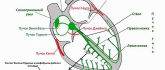
Ventricular extrasystole (VES) is an extraordinary contraction of the heart that occurs under the influence of premature impulses emanating from the wall of the left or right ventricle, the fibers of the conduction system.
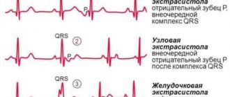
Usually, extrasystoles that occur during PVCs affect only the ventricular rhythm, i.e. without affecting the upper parts of the heart. At the same time, extraordinary contractions that “originate” higher up – in the atria and atrioventicular septum (supraventricular extrasystole) can also provoke ventricular premature contractions.
In the group of arrhythmias of the extrasystolic type, PVCs are detected in 40-75% of cases among the population over 50 years of age.
Premature ventricular contractions on ECG
Gradation of ventricular extrasystoles according to Laun-Wolf
In the medical community, the most common classification of ventricular extrasystole according to Lown.
Its last modification was in 1975, but it still has not lost its relevance and contains the following classes:
- 0 (no arrhythmia);
- 1 (extrasystoles less than 30/hour, from one source and one form);
- 2 (one source and form, 30 or more extrasystoles per hour);
- 3 (multifocal extrasystoles);
- 4a (paired extrasystoles from one focus);
- 4b (polymorphic extrasystoles accompanied by other arrhythmias - ventricular fibrillation/flutter, tachycardia paroxysm);
- 5 (early extrasystoles “type R on T”).
Read also: Is pulmonary edema treatable?
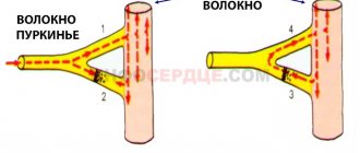
The mechanism of development of extrasystoles may differ. There are two main ones - reciprocal and automatic. Reciprocal arrhythmias arise when a vicious circle of intraventricular excitation is formed, the so-called “re-entry” mechanism. Its essence lies in the disruption of the passage of a normal signal, which is associated with the presence of at least two paths for the impulse. In this case, the signal for one of them is delayed, which causes the formation of an extraordinary contraction. This mechanism plays a role in the formation of such arrhythmias as paroxysm of ventricular tachycardia and extrasystoles, Wolff-Parkinson-White syndrome, atrial/ventricular fibrillation. An ectopic focus of excitation can occur with increased automatism of the pacemaker cells of the heart. Arrhythmias with such a development mechanism are called automatic.
Classification of ventricular extrasystoles
Under certain circumstances, ventricular extrasystole causes a severe form of arrhythmia - ventricular tachycardia, which turns into fibrillation. And this condition is the most common cause of sudden coronary death.
Lown classification
The classification of PVCs has changed several times following diagnostic and prognostic needs. Extrasystoles in them were distributed according to quantitative values, location and frequency of occurrence. For about 15 years in cardiology they used the classification of ventricular extrasystoles according to Lown and Wolf (B. Lown and M. Wolf). They proposed it for the gradation of gastric extrasystoles in post-infarction patients. A few years later, it was adapted for patients without a history of heart attack.
This classification reflects the quantitative and morphological signs of PVCs (based on the results of a 24-hour ECG):
| Class | Characteristics of ventricular extrasystole |
| Extraordinary layoffs are not recorded | |
| 1 | Less than 30 single extrasystoles in any 60 minutes during the day |
| 2 | More than 30 single extrasystoles during any hour of monitoring |
| 3 | There are (polymorphic) extraordinary contractions occurring in different foci of excitation |
| 4A | There are two extrasystoles in a row emanating from one focus of excitation (monomorphic) |
| 4B | There are paired polymorphic extrasystoles |
| 5 | There are polymorphic PVCs passing in one gulp of 3–5, registration of paroxysmal ventricular tachycardia is possible |
According to Lown's classification, class 1 is a benign condition, rather considered as a functional disorder without circulatory impairment and clinical signs. Starting from class 2, ventricular extrasystole has a poor prognosis, being associated with an increased risk of developing fibrillation and cardiac arrest.
Gradation of extrasystoles by location, time and frequency of occurrence
There is a classification of ventricular extrasystole according to the place of origin of impulses and the number of foci of excitation:
- extrasystoles emanating from one focus are called monotopic;
- PVCs that originate in several foci are polytopic;
- right ventricular extrasystole;
- left ventricular extrasystole.
According to the time of occurrence, ventricular extrasystoles are divided into early (recorded at the beginning of diastole), interpolated, appearing in the middle between two beats, and late, occurring at the very end of diastole. Early extraordinary contractions according to the ECG sign are called “R on T” (layering of two teeth on top of each other). They are caused by organic changes in the myocardium. According to Lown, this type of ventricular extrasystole is classified as class 5.
In cardiology, there is a concept of a statistical daily “norm” of VES if they are well tolerated. There is no such norm for early extrasystole - it should not exist at all. Average or interpolated is the majority of the VES (up to 80%). Its average normal indicator is considered to be up to 200 extraordinary contractions per day. Late extrasystoles almost overlap the subsequent normal contraction. Their permissible daily quantity is up to 700. Today, the medical community uses the RJ Myerburg classification, which reflects the shape and frequency of ventricular extrasystoles:
| By frequency | According to morphology | ||
| 1 | less than 1 in 60 minutes - rare; | A | Single from one source |
| 2 | 1–9 per hour - infrequent; | B | Single from different foci |
| 3 | 10–30 per hour – moderately frequent; | C | Doubles |
| 4 | 31–60 per hour - frequent; | D | Unsustained ventricular tachycardia |
| 5 | more than 60 per hour - very frequent. | E | Sustained ventricular tachycardia |
Classification of extrasystoles according to Bigger
Bigger's classification provides for the formation of groups of patients according to the degree of increase in the risk of complications.
It includes the following course of extrasystole:
- malignant;
- potentially malignant;
- benign.
With benign extrasystoles, the risk of complications is extremely low. Moreover, such patients have no signs of pathology of the cardiovascular system in the anamnesis and during examination (normal left ventricular ejection fraction, no hypertrophy or cicatricial changes in the myocardium). The frequency of ventricular extrasystoles does not exceed 10 per hour and there is no clinical picture of paroxysmal ventricular tachycardia.
A potentially malignant disease is characterized by a moderate or low risk of sudden death. The examination reveals structural changes in the heart in the compensation stage. Ultrasound of the heart reveals a decrease in LV ejection fraction (30-55%) and the presence of scar or myocardial hypertrophy. Patients complain of a feeling of interruptions in the work of the heart, accompanied by short-term episodes of ventricular tachycardia (up to 30 seconds).
Malignant extrasystoles are those whose manifestation causes a disturbance in the general well-being of the patient (palpitations, fainting, signs of cardiac arrest). Patients exhibit a critical decrease in ejection fraction - less than 30%. Persistent ventricular tachycardia is also noted.
The most dangerous ventricular ecstasystoles include 3 gradations in the Lown classification - 4a, 4b and 5 classes.
Types and classification of cardiac arrhythmia
Arrhythmia is a pathology in which there is a disturbance in the rhythm of the heart. It manifests itself in different variations, which differ from each other in the cause of the failure, symptoms, localization and other characteristics. What types of cardiac arrhythmia exist?
The principle of dividing arrhythmia into types
There are several types of arrhythmia. The division into types is important because the patient’s further treatment, life prognosis, the likelihood of developing complications and much more depend on it. In addition, a person often exhibits several types of heart rhythm disturbances, which must be differentiated.
Arrhythmia is divided into types according to the principle of localization of the pathological focus, according to the frequency of heart contractions, and according to the nature of the failure. The Lown classification is also used.
One of the classifications of arrhythmia
Types of arrhythmia according to the location of the source
This group includes the following types of arrhythmia, which differ from each other not only in location, but also in clinical picture.
Atrial arrhythmias
This type of arrhythmia is localized in the atria, and more specifically in their inner part or in the septum between them. Pathology has several more subtypes:
- Atrial extrasystole.
- Atrial tachycardia.
- Atrial flutter.
Atrial extrasystole is a condition when the heart contracts prematurely due to an impulse from the atria. A common cause of this disease is an increase in diastolic pressure in the left ventricle, as well as stretching of the left atrium.
The disorder can develop against the background of other heart diseases, for example, myocardial infarction, left ventricular failure. Complications of the pathology include atrial fibrillation and impaired blood circulation.
Patients with atrial extrasystole experience symptoms such as:
- Feeling your own heartbeat.
- Pale skin.
- Suffocation.
- Darkening in the eyes.
- Panic.
- General weakness.
Darkening in the eyes is one of the symptoms of atrial extrasystole
The second subtype is atrial tachycardia. It differs in that the heart rate increases. This occurs in short-term attacks, which can last for different times depending on the severity of the disease. Pathology often occurs due to factors such as:
- High blood pressure.
- Heart defect.
- Failure of metabolic processes in the body.
- Obesity.
The clinical picture is as follows:
- Attacks of dizziness.
- Dyspnea.
- Pain syndrome in the chest area.
- Spots before the eyes.
- Increased anxiety.
- Panic attacks.
Tachycardia is not considered life-threatening, but it significantly worsens the quality of life in advanced cases, when arrhythmic attacks last up to several days. As a result, not only does the patient’s well-being worsen, but the heart muscle is also depleted.
The third subtype is atrial flutter. With it, there is a strong increase in the number of heart contractions - over 200 beats per minute. The cause of the development of pathology may be problems with blood pressure and heart disease. Symptoms include severe shortness of breath, poor exercise tolerance, anxiety, and chest pain.
Ventricular arrhythmias
This type of cardiac arrhythmia is also divided into several subtypes:
- Extrasystole.
- Tachycardia.
- Fibrillation.
Ventricular extrasystole is characterized by non-premature contraction of the ventricles of the heart. In this case, the patient is concerned about such manifestations as:
- A pronounced sensation of interruptions in the functioning of the heart muscle.
- General weakness.
- Dyspnea.
- Painful sensations in the heart area.
- Suffocation.
- Dizziness.
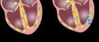
Ventricular tachycardia is a rapid contraction of the ventricles. Manifests itself in the form of short-lived attacks. The danger of the pathology is that it can lead to impaired blood flow and cardiac arrest. In severe forms, the pulse rate reaches 300 beats per minute.
Ventricular fibrillation is a condition where the ventricles do not contract fully. This occurs due to a malfunction of individual muscle tissues of the heart. It also manifests itself in attacks that do not last long. With this pathology, serious complications are also possible, including sudden cardiac arrest.
Tachycardia and fibrillation have a clinical picture similar to extrasystole.
Ventricular (left) and supraventricular (right) extrasystole
Sinus arrhythmias
Sinus arrhythmia differs in that the time period between contractions of the heart muscle changes. Occurs in the form of attacks, which are most often provoked by such factors as:
- Physical activity.
- Emotional stress.
- Severe diseases of the heart and blood vessels, for example, coronary artery disease.
- Excess body weight.
- High blood pressure.
Also, this type of arrhythmia is often diagnosed in women who are pregnant and in teenage children.
There are two subtypes of the disease. The first is sinus bradycardia, which is characterized by a slow heart rate. The main manifestations of this disorder:
- General weakness.
- Attacks of dizziness.
- Paleness of the skin.
- Darkening in the eyes.
The second subtype is sinus tachycardia. It is distinguished, on the contrary, by an increased heart rate. When an attack occurs, patients complain of chest pain, shortness of breath, increased anxiety, palpitations, and suffocation.
Atrioventricular arrhythmias
This type of arrhythmia is characterized by impaired cardiac conduction, failure of the passage of impulses between the atria and ventricles. The cause of the development of the disease is atrioventricular block, which leads to disruption of not only rhythm, but also blood circulation.
There are several degrees of blockade:
- First. With it, slow conduction of impulses from the atria to the ventricles is observed. Patients do not notice the failure because it is asymptomatic. Therefore, the disease is detected by chance when performing electrocardiography. Also, grade 1 does not require treatment, only medical supervision.
- Second. Some signals are lost, as a result of which they do not fully reach the ventricles. In this case, symptoms already appear - discomfort in the heart area, weakness, darkening of the eyes, loss of consciousness. Such a blockade already needs to be treated.
- Third. The passage of impulses is completely blocked. Patients experience a slow pulse, not exceeding 45 beats per minute. This is fraught with the development of oxygen starvation of the brain, fainting and even sudden death. Treatment is required.
As you can see, the clinical signs of different types of cardiac arrhythmia are the same, so it is possible to accurately determine the type of pathology only after an instrumental examination of the patient.
Correct interpretation of the ECG will allow you to determine the type of arrhythmia
Division into types according to heart rate
Based on the number of beats per minute, arrhythmia is divided into two main groups - tachycardia and bradycardia. In the first case, an increased heart rate is observed when the pulse exceeds 90 beats. As a result of this disease, the activity of the heart deteriorates, since less nutrition is supplied to it.
The ventricles do not have time to fully fill with blood, which causes a drop in blood pressure and a decrease in the performance of many internal systems.
Bradycardia, on the contrary, is characterized by a decrease in heart rate when the number of beats per minute does not exceed 50. This condition is no less dangerous than tachycardia. It can lead to cardiac arrest, especially if the person also has AV block.
Types of arrhythmia according to electrophysiological criteria
This classification of cardiac arrhythmia takes into account the type of disorder that occurs during the pathology. It looks like this:
- Impulse creation failed.
- Violation of signal conduction.
- A combination of these pathological changes.
The first type refers to tachycardia, bradycardia, the second – to heart block, the third combines both options.
Types of arrhythmia according to Lown
Lown classification involves dividing pathology into classes as the disease progresses. The higher the class, the more dangerous the disease and the higher the risk of developing serious complications.
Progressive disease has a higher Lown class
The following classes are distinguished:
- 0 – no arrhythmia.
- 1 – assigned when there are less than 30 extrasystoles per hour, and there is only one source.
- 2 – extrasystoles increase by more than 30 per hour, the source remains the same.
- 3 – several foci of pathological impulses are already observed.
- 4a – paired extrasystoles arise from one source.
- 4b – polymorphic extrasystoles appear, which are combined with other arrhythmias.
- 5 – extrasystoles “type R to T”.
This classification was created many years ago, but is still used in medical practice.
Rare variants of arrhythmia on ECG
When conducting electrocardiography, doctors are able to identify any type of cardiac arrhythmia. Some disorders are more common, others less common. Most rarely, doctors encounter such a manifestation on the electrocardiogram as sinus node dysfunction syndrome.
This disease is characterized by a disruption of the heart rhythm resulting from a deterioration in automaticity or a sudden cessation of the functioning of the atrial node. The following factors can provoke the development of the disease:
- Violation of impulse formation.
- Slow heart rate.
- Ectopic pathologies.
This syndrome poses a danger to human life, as it can cause sudden cardiac arrest. It can also be combined with diseases such as pulmonary edema, coronary insufficiency, and heart attack.
Thus, there are several classifications of arrhythmia, which differ from each other by different criteria. It is impossible to determine the type of pathology by symptoms alone; this requires a heart examination. This is important, since further treatment tactics depend on the type of disorder.
Clinical manifestations
In most patients, in the absence of damage to the cardiovascular and nervous systems, extrasystole occurs hidden. There are no specific complaints inherent to the disease. Its pronounced clinical picture is usually represented by the following symptoms:
- weakness;
- irritability
- dizziness/headaches;
- feeling of discomfort in the chest (pain, tingling, heaviness);
- heart sinking feeling
- a push in the chest with frequent extrasystoles;
- arrhythmic pulse;
- feeling of pulsation in the veins of the neck;
- dyspnea.
The presence of concomitant cardiac pathology aggravates the course of the disease.
Diagnostics
Making a diagnosis is based on the results of collecting complaints, the history of the patient’s development and life, data from a comprehensive examination and additional studies. Assessing the patient’s condition, the doctor pays attention to increased pulsation of the neck veins, changes in the pulse wave and auscultatory pattern of heart sounds. A standard set of laboratory tests is prescribed (general blood and urine analysis, blood glucose and biochemical blood test), as well as an analysis of thyroid and pituitary hormones.
To obtain an accurate diagnosis, the mandatory criterion is the result of an ECG and daily Holter monitoring. Using these methods, it is possible to accurately determine the source of the pathological focus, the frequency of extrasystoles, the number and relationship with the load. Echo-CG is performed to identify the left ventricular ejection fraction and the presence/absence of structural changes in the heart. If it is difficult to diagnose the disease, MRI, CT, and angiography may be prescribed.
If there are no patient complaints, with a benign course of extrasystole, only monitoring the state of the cardiovascular system is indicated. Such patients are recommended to undergo examination 2 times a year with mandatory ECG registration. The tactics of patient management depend on the number of extrasystoles per day, the course of the disease, and the presence of concomitant pathology. The dosage of drugs is selected individually by the attending physician.
Antiarrhythmic drugs are divided into 5 classes:
- 1a – Na + channel blockers (“Procainamide”, “Disopyramide”);
- 1c – activators of K + channels (“Difenin”, “Lidocaine”);
- 1c – Na + channel blockers (“Flecainide”, “Propafenone”);
- 2 – beta-blockers (“Metaprolol”, “Propranolol”);
- 3 – K + channel blockers (“Amiodarone”, “Ibutilide”);
- 4 – Ca 2+ channel blockers (“Diltiazem”, “Verapamil”);
- 5 – Other drugs with antiarrhythmic effects (cardiac glycosides, calcium, magnesium preparations).
For ventricular extrasystole, class 2 drugs are widely used. They help reduce symptoms of arrhythmia and also have a positive effect on the quality of life of patients.
Scientific studies have proven that beta-adrenergic blockers improve the prognosis regarding the risk of cardiac death in patients with cardiovascular pathology.
Read also: Balloon angioplasty cost
Persistent ventricular extrasystole according to Lown, which is not amenable to drug treatment, requires surgical intervention. For the success of the operation, it is necessary to accurately know the source of pathological activity. When it is determined, patients undergo implantation of cardioverter-defibrillators or radiofrequency catheter ablation.
Principle of classification
There are many factors that characterize a particular disease. As for extrasystoles, the following signs are distinguished:
- number of ectopic areas (mono-, polytopic);
- form of arrhythmia (mono-, polymorphic);
- frequency of occurrence (rare, moderately frequent, frequent);
- localization (right, left ventricular);
- pattern of abbreviations (ordered, unordered);
- frequency (spontaneous, regular).
In accordance with these parameters, many options were proposed: according to Bigger, Mayerburg. However, the Laun-Wolff classification turned out to be the most practical and popular. Ventricular extrasystole according to Laun is determined using so-called gradations, each of which is assigned one number :
- 0 – no arrhythmias during the last 24 hours of observation;
- I – no more than 30 arrhythmias are observed during an hour of monitoring, monotopic and monomorphic;
- II – more than 30 per hour of the same type;
- III – polymorphic extrasystoles appear;
- IVa – paired monomorphic;
- IVb – paired polymorphic;
- V – characterized by the presence of ventricular tachycardia (extrasystoles that occur more than 3 times in a row).
The use of gradations for the treatment of extrasystole
The presence of the degree of arrhythmia in the formulation of the diagnosis is very important. The treatment tactics chosen by the doctor will depend on this.
Thus, the presence of extrasystoles of the first gradation in a patient indicates the functional nature of the resulting abnormal contractions. About 60-70% of people have a similar phenomenon, and this is considered the absolute norm. The only thing required is to periodically check the ECG. However, if you have any symptoms of cardiovascular pathologies, you should undergo additional examination, as this may be one of the debuts of the disease.
In the presence of the second gradation without signs of hemodynamic impairment, non-drug treatment is indicated - auto-training, psychotherapy, avoidance of risk factors. If there are accompanying symptoms or the appearance of polymorphic foci is noticed (third gradation), an appropriate course of antiarrhythmic drugs is required.
Finally, grades four and five, as well as grade three refractory to conservative therapy, especially in the presence of hemodynamic disturbances, require surgical treatment. In this case, surgical interventions such as catheter radiofrequency ablation or pacemaker implantation may be indicated.
This classification is also used to make a forecast. Ventricular extrasystole grade 3-5 according to Lown is considered threatening. These are the so-called malignant arrhythmias. They are characterized by a high risk of sudden death. In this case, the patient should be transferred to the intensive care unit.
The location of the lesions also matters. The prognosis is less favorable in the presence of left ventricular arrhythmias
Symptoms
Among young people, about half suffer from single premature ventricular extrasystoles. Daily Holter monitoring allows you to observe this. At the same time, they do not notice extraordinary contractions of the heart. Symptoms begin only after the normal heart rhythm is seriously disrupted due to premature contractions.
For ventricular extrasystole that occurs without other heart diseases, the following symptoms characteristic of the patient can be noted:
- “Suspension” of the heart with a series of strong contractions after this.
- Periodic strong contractions of the heart.
- Extrasystole occurs after eating.
- Heart rhythm disturbances can also occur during rest.
- Physical activity practically does not cause rhythm disturbances.
Against the background of other organic heart diseases, this disease usually occurs without symptoms. Arrhythmia develops under the influence of physical activity and goes away after calming down. As a rule, such arrhythmia develops against the background of tachycardia.