5 / 5 ( 1 voice )
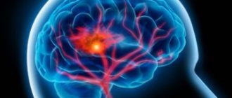
Ventricular tachycardia is a heart rhythm disorder that is caused by damage to the muscles of this organ. If left untreated, the pathology can cause death. In this article we will answer the question of what are the symptoms and treatments for ventricular tachycardia.
Ventricular tachycardia is often the cause of death
The mechanism of pathology development
The pathogenic process forms relatively quickly. The impetus for the beginning of changes is injury, violation of the integrity of the heart or other organic damage as a result of a heart attack, cardiosclerosis, or infectious moments.
To understand how the problem arises, we need to look at anatomy and normal physiology.
An adequate state of affairs is characterized by the full functioning of the heart. Contractions of the muscular organ occur autonomously under the control of the brain stem.
External factors can only slightly affect the functioning of the heart, but not change it radically.
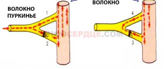
Rhythmic beats are possible due to the generation of an electrical impulse by a special bundle of cardiac cells - the so-called sinus node. From the driver (an alternative name for this formation), the signal is transmitted along the bundles of His to other tissues of the organ. This leads to muscle contraction. This happens all the time.
Ventricular tachycardia (VT) is characterized by pathological localization of the generation of an electrical impulse. The remaining cardiac structures should not participate in creating the signal; they are conductors and a kind of “pump” that redirects the blood in the right direction.
In this case, the ventricles join in generating the signal.
This ends with catastrophic consequences:
- The intensity of blood ejection decreases. Organs and systems do not receive enough nutrients and oxygen. The deficit is significant. Hence hypoxia, which often ends fatally.
- Even if this does not lead to the death of the patient, the ventricles contract chaotically. Hence the likelihood of cardiac arrest at one moment. Resumption of organ activity is possible after resuscitation measures, but the main process continues to flow. There is a high probability of relapse. It is not known when this will happen: this very minute, in an hour or a month. Such patients are sent to a cardiology hospital for urgent diagnosis and emergency treatment.
The myocardium of the left ventricle is most often affected due to its anatomical structure.
A problem of this kind is never natural. This is a direct indication of a pathological process. When a pronounced clinical picture develops, there is almost no time left for diagnosis and treatment.
Description of ventricular tachycardia
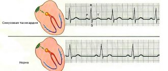
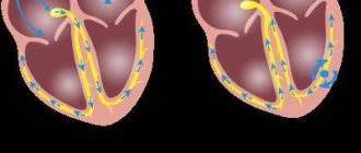
The heart muscle in its normal state conducts electrical impulses regularly and orderly, with a frequency of 60-90 times per minute. In this case, the atria contract first, and then the impulse travels through the atrioventricular node into the ventricles, which also contract a few milliseconds later. This process takes place so quickly that it is practically not felt by a person, and in medicine it is defined as sinus rhythm.
In ventricular tachycardia, the sinus node is not the main pacemaker because it is not able to control the contractility of the heart.
Ventricular tachycardia (VT) is a violation of the normal (sinus) rhythm of the heart, characterized by an increase in the number of ventricular contractions. This occurs due to the disturbed structure of the myocardium, as a result of which the electrical impulse cannot be transmitted normally through the fibers. If it passes through the atria and through the AV node normally, then in the ventricles it begins to be interrupted and circulate in a vicious circle. Or ectopic foci are formed in the ventricles themselves, which become additional and extraordinary generators of the excitation signal. As a result of their activity, chaotic contraction of the ventricular myocardium begins at a crazy pace.
With VT, hemodynamics are disrupted. This is due to the influence of two factors:
- with an increase in the frequency of ventricular contractions, the release of blood into the pulmonary and systemic circulation decreases, which negatively affects the general condition of the patient.
- Discoordination of the heart reduces its functionality, which also affects hemodynamics.
Classification
The pathological process can be classified on various grounds.
Depending on the intensity of the onset and time of occurrence of the phenomenon:
| Paroxysmal ventricular tachycardia | Non-paroxysmal form |
| It starts abruptly, for no apparent reason. But there is always a trigger factor, although it is not obvious to the patient. This could be taking a drug, coffee, stress, etc. It flows spasmodically. In the early stages, the duration ranges from a few minutes to a couple of hours. Then the pathology “fixes”, becomes a constant companion of the patient, and the rapid pulse persists for weeks. | Occurs more often. Characterized by the appearance of multiple extrasystoles. Requires quick treatment. Preferably in a hospital setting. Less dangerous than the previous variety. |
Based on the severity of the current, we can name:
- Paroxysmal form. Observed in 55% of clinical cases. Corresponds to the early stage of the pathological process. Each paroxysm lasts from 2 minutes to several hours.
- Permanent variety. The same type of pathogenic process, but the duration of the attacks is longer. 2-3 days is not the limit. Prevalence of the phenomenon - 20%
- Chronic type. It is characterized by persistent heart rate disturbances. However, the patient gets used to the condition and almost does not feel the problem, which complicates late diagnosis.
Finally, depending on the form, there are:
- Monoform variety. One area of pathological rhythm generation appears. This is usually the left ventricle. A direct indication of organic heart damage.
- Polymorphic type. There are 2 or more places where an electrical impulse is produced (not counting the sinus node). This is a consequence of noncardiac factors, overdose of psychoactive substances, including medications.
The ventricular type of tachycardia is multifaceted. Since patients seek help relatively late, especially in the absence of a normal screening program, there is not much time to thoroughly identify the underlying cause and form of the process.
Everything has to be done in emergency mode. Hence the high mortality rate: the drugs themselves, if used incorrectly, can aggravate the course of the disease. Conclusion: at the first suspicion of problems with the heart, you should contact a cardiologist.
Types of ventricular tachycardia
Due to various factors, VT can occur in several forms: unstable and stable. There are also types of ventricular tachycardia that are potentially dangerous due to the high risk of developing ventricular fibrillation.
In a small amount, about 2%, ventricular tachycardia develops in young people. At the same time, there are no special violations in their health. In such cases they speak of idiopathic VT.
Sustained and unsustained ventricular tachycardia
The unstable type of VT is characterized by an unstable course. The ECG records paroxysms with a frequency of half a minute. Their number is more than three for a certain period. Hemodynamic disturbances occur, but the prognosis for death is insignificant. Unsustained ventricular tachycardia is a common complication of ventricular extrasystole, therefore, when they are combined, a diagnosis is made in the form of “extrasystole with jogging ventricular tachycardia.”
The persistent type of VT is no longer prognostically favorable. The resulting paroxysm lasts at least 30 seconds, determined by ECG. The ventricular complexes in this case are greatly changed. Due to the increased risk of sudden cardiac death against the background of developed fibrillation, tachycardia of this type is considered life-threatening.
Classification of ventricular tachycardia
According to this division, the types of VT that are potentially dangerous due to the possible development of fibrillation are determined.
- Monomorphic VT, which often occurs due to organic heart damage.
- Polymorphic, or multiform, VT are ventricular complexes of different amplitudes and directions, formed as a result of the action of two or more ectopic foci. They occur mainly without structural changes in the heart, although in some cases organic changes are detected. There are bidirectional-fusiform polymorphic VTs and polytopic or multifocal ones.
Sometimes tachycardia of the “pirouette” type occurs, when the QRS complexes progressively change and repeat against the background of a prolonged QT interval.
Characteristic changes on the ECG
Electrocardiography is the main method for diagnosing ventricular tachycardia at any stage.
Typical signs:
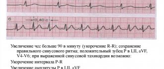
- Increasing heart rate to 100-300 beats per minute. Even in a state of complete rest. After exercise tests, your heart rate may drop sharply.
- The width of the QRS complex is over 0.12-0.14 seconds.
- The axial value of the same complex in degrees is more than 30.
- Ventricular capture and AB dissociation are present.
- The R wave is high, wide, not split as in the supraventricular type.
- S jagged.
The ventricular form of tachycardia on the ECG is characterized by an increase in heart rate and deviation of the peaks. It is relatively simple to delimit this form of the pathological process using a cardiogram if you have the qualifications.
Independent decryption is impossible. If the results are questionable, it is recommended to obtain the opinion of another specialist.
Diagnosis of fusiform tachycardia
Diagnosis of the disease is quite complex, so a diverse study is required. First of all, an electrocardiogram is prescribed, which shows the following signs:
- irregular sinus rhythm;
- ventricular extrasystoles (rare, early);
- deviation of the electrical axis of the heart;
- increase in heart rate 160–240 beats/min.
Additionally, an analysis of the QT interval is carried out, with the help of which its increase or normal state is determined. An ultrasound of the heart, chest x-ray, Holter monitoring, and general laboratory tests are also required.
Cardiac causes of failure
As was said, always pathological. Mainly of cardiac origin. Among the probable phenomena causing ventricular tachycardia:
- Dysplastic process in the ventricles of the heart. Simply put, the replacement of functional active tissue, cardiomyocyte cells, with adipocytes.
- Myocardial infarction. It consists of an acute malnutrition of the muscular structures of the organ. The result is necrosis of the muscles on scar tissue, which is not capable of automatism or even contraction and acts as a kind of “filler”. The healing process ends with the formation of persistent cardiosclerosis. Ventricular tachycardia in this case acts as a strange adaptive mechanism that has nothing to do with the normal functioning of the system.
- Heart defects, both congenital and acquired. They go unnoticed in most cases. There are no symptoms; they are discovered by chance during a more in-depth examination of patients. However, this is not a guarantee either. There are many cases of diagnosis after the fact, when the patient died. This is due to screening errors and insufficient attention of people to their own health.
- Cardiac ischemia. Chronic disturbance of myocardial nutrition in a certain area. This is a precursor to a heart attack. In the next couple of years, an emergency will arise. Qualified assistance is needed at an early stage.
- Rheumatism. Autoimmune damage to a muscle organ. There is no cure, it can only be corrected to a small extent. The goal of therapy lies in the need to prevent the generation of pathological impulses.
- Inflammatory heart diseases. Usually of infectious origin. Myocarditis and others. Without competent and urgent help, they lead to total destruction of cardiac structures. In such a situation, tachycardia is not the biggest problem. There is a risk of cardiac arrest. In case of successful treatment, cardiosclerosis is still likely with understandable consequences.
- Aneurysm of a vessel localized in the ventricle. Bulging of the wall of a large artery leads to malnutrition. The result may be rupture and massive bleeding, fatal in 100% of cases.
Treatment and rehabilitation
Treatment of gastrointestinal tract is carried out on an individual basis, depending on the patient’s condition and the cause of the pathology.
Electrical pulse treatment (restoration of heart rhythm using electric current pulses) is mainly used as therapeutic measures
Conservative (drug) therapy for gastrointestinal tract includes the use of the following drugs:
- Antiarrhythmic drugs that restore and maintain heart rhythm;
- Beta-adrenergic blockers - reduce heart rate and lower blood pressure;
- Calcium channel blockers - restore normal heart rhythm, dilate blood vessels, reduce blood pressure;
- Omega 3 fatty acids lower cholesterol levels in the blood, prevent the formation of blood clots and have an anti-inflammatory effect.
Surgical treatment is performed if the following indications exist:
- History of ventricular fibrillation;
- Serious changes in hemodynamics in patients with post-infarction GVT;
- Persistent extrasystolic allorhythmia;
- Frequent, recurrent attacks of tachycardia in patients who have had myocardial infarction;
- Disorders, pathologies and diseases resistant to drug therapy, as well as the inability to use other treatment methods.
Surgical treatment methods include implantation of electrical defibrillators and pacemakers , as well as destruction of the source of arrhythmia using a radiofrequency pulse.
During the rehabilitation period, patients who have suffered an attack of gastrointestinal tract disease are advised to adhere to a diet, eliminate physical and psycho-emotional stress, regularly spend time in the fresh air and follow all doctor’s instructions.
This video highlights new research and treatment options for this disease:
Noncardiac causes
Occur less frequently. Among them:
- Endocrine disorders associated with deficiency of hormones of the adrenal cortex, thyroid gland or pituitary gland.
- Dehydration or excess water in the body.
- Genetic syndromes and deviations. Many congenital pathologies affect the functioning of the heart. In some cases, children do not live to be diagnosed; the disease is determined after the fact.
- Lack of potassium and magnesium. Caused by nutritional factors. Poor nutrition is another risk point.
- Excessive consumption of drugs for the treatment of arterial hypertension, problems with the cardiovascular system.
- Intoxication with alcohol, caffeine, nicotine, psychoactive substances (cocaine and heroin), salts of heavy metals, compounds of toxic chemical elements.
In the absence of evidence for organic changes in the heart or other systems, they speak of an idiopathic form. Her treatment comes down to stopping the picture throughout her life.
Causes
A single extrasystolic complex from the left or right ventricle can be recorded in a healthy person and is not considered a pathology. Problems with the appearance of frequent abnormal contractions of the myocardium with hemodynamic disturbances most often arise as a result of severe diseases of the heart and blood vessels. In this case, the reason may be:
- ischemia and its consequences (angina pectoris, cardiosclerosis, heart attack, aneurysm);
- cardiomyopathy;
- inflammation of the myocardium and pericardium;
- heart failure (both chronic and acute);
- hypertonic disease;
- valve disorders;
- surgical intervention.
Non-cardiac causes can also provoke an attack:
- severe lung diseases;
- electric shock;
- intoxication as a result of severe poisoning, liver or kidney failure;
- the use of a number of medications (bronchodilators, cardiac glycosides, diuretics);
- a decrease in potassium levels in the blood and an increase in calcium.
Predisposing factors to the development of ventricular arrhythmia are bad habits (smoking, drugs, alcohol, large doses of coffee), constant physical and psycho-emotional overload.
Symptoms
Manifestations of ventricular tachycardia require qualified assessment. You need to pay attention to the following signs of a possible problem:
- Increased heart rate. It feels subjective, but not for long.
- Panic attack. Characteristic of the paroxysmal course of the disease.
- Loss of orientation in space. Dizziness.
- Nausea.
- Vomiting (rare).
- Fainting state.
- Dyspnea. Caused by tissue hypoxia. This is an alarming manifestation, indicating rapid progression of the process.
- Cyanosis of the nasolabial triangle. Blue area.
- Paleness of the skin.
- Increased sweating.
- Weakness.
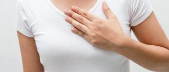
- Chest pain.
These are typical manifestations of paroxysm or a long-term, but not yet chronic process. As we move forward, the clinical picture smoothes out and the patient stops feeling anything. Life seems to be returning to normal, but this is only an appearance. The problem continues to exist and progress. In the early stages, there are also no symptoms, or they are so weak that the person does not pay attention.
Complications of ventricular tachycardia occur in almost 70% of cases.
The following signs indicate an emergency:
- A sharp headache for no apparent reason, in the back of the head or crown. Beats to the beat of the heart.
- Pressing discomfort behind the sternum. Doesn't let me breathe.
- Repeated loss of consciousness.
- Focal neurological disorders of vision, hearing or touch.
- Paralysis, paresis, feeling of numbness in the limbs.
- Facial distortion.
- Inability to speak normally.
All these are moments inherent in two terrible conditions: heart attack and stroke. The second one is easier to recognize; there are simple tests available (raise your hands, smile, say a phrase, if at least one action is impossible - call an ambulance). The first one is diagnosed only in the hospital.
Symptoms of ventricular tachycardia from the brain, nervous, and cardiovascular systems are explained by impaired hemodynamics at the general level and a decrease in oxygen concentration in the blood.
Signs and symptoms of arrhythmia
Any type of VT, especially polymorphic ventricular tachycardia, is usually poorly tolerated by patients. Main symptoms:
- dizziness, possible fainting;
- cardiopalmus;
- feeling of lack of air;
- sometimes nausea;
- confusion and loss of consciousness;
- heart failure.
Heart rate ranges from 140 to 250 per minute. The duration of the attack usually ranges from several seconds to several hours. Sometimes VT lasts several days.
Arrhythmia is accompanied by signs of oxygen starvation:
- burning, squeezing pain appears behind the sternum, which is not relieved by nitroglycerin;
- Arrhythmic shock may occur with a sharp decrease in pressure and loss of consciousness;
- urine output decreases;
- There is abdominal pain and bloating (with prolonged or frequent attacks).
Some people do not feel attacks of tachycardia, although the danger to life remains.
Emergency assistance during a seizure
You need to act quickly.
- Blood pressure and contraction frequency of the organ are measured. If blood pressure is low (less than 90 over 60), call an ambulance immediately; you cannot help yourself. There is a high probability of arrhythmic collapse.
- Next, you should take a tablet of Anaprilin or Carvedilol.
- Drink motherwort, valerian (1-2 tablets).
- Provide a flow of fresh air into the room.
- Do not make sudden movements, get to bed as carefully as possible, and lie down.
- Loosen tight jewelry and wardrobe items. The neck should be free.
- After 20 minutes, check your blood pressure and heart rate again.
If there is no effect, call an ambulance. Before arrival, maintain a stable physical position.
Attention:
Under no circumstances should you resort to contrast showers, hot baths, or the use of herbal remedies. This is dangerous and often fatal. Large dosages of drugs are also contraindicated. They can aggravate the course of the attack. No alcohol, glycosides, or other drugs.
Disease prognosis
If the left ventricle is functioning normally, this indicates that the risk of relapse is minimal and also excludes the occurrence of ventricular fibrillation. The prognosis of the disease is favorable.
The risk of recurrence of arrhythmic attacks is characterized by a low cardiac ejection fraction, namely low efficiency of the heart and small volumes of blood ejected into the vessels. This prognosis is disappointing and requires urgent action, otherwise there is a risk of death.
Bidirectional ventricular tachycardia is also dangerous; the risk of sudden death in this case is very high.
Unsustained ventricular tachycardia may resolve quickly and not recur. As a rule, it is asymptomatic. The cause of this form of illness is usually a previous myocardial infarction.
Diagnostics
It is carried out under the supervision of a cardiologist. Third party specialists are involved as necessary. However, the main “burden” falls on the specialized doctor.
Among the examination methods:
- Patient interview. Health complaints are identified. A person must tell everything, even if it seems that the moment is not relevant to the issue. The doctor himself will sort the information.
- Anamnesis collection. Bad habits, lifestyle, heredity and family history of diseases. Here are just some important facts.
- Measurement of blood pressure and heart rate. Several times, with an interval of 5-15 minutes or more.
- 24-hour Holter monitoring. Better in an outpatient setting. This way the data will be more accurate and the patient will be in a natural, familiar environment. Stress and phobias are excluded. Physical activity at an adequate level. The dynamics of the process are revealed accurately.
- Electrocardiography. It is applied first. Specific data allows you to quickly make a diagnosis. It is important to correctly decipher the information.
- Echocardiography. Ultrasound examination. Imaging is indicated to detect organic changes.
- To obtain a more detailed picture, MRI or CT is used.
A complete diagnosis is carried out within 2-4 days. In stationary conditions it is faster. There's no point in wasting time. In the early stages there is no urgency; in the next few years it will worsen.
Types of paroxysmal tachycardia
Paroxysmal tachycardia can occur in different parts of the heart. In this regard, atrial and ventricular tachycardia are diagnosed. Atrial tachycardia occurs as a result of:
- oxygen starvation of the heart muscle;
- disturbances in the content of calcium, chlorine, potassium in the blood;
- endocrine disorders.
The patient becomes dizzy and weak, and may experience pain in the heart area. The disease is diagnosed mainly by ECG indicators. Short-term attacks cannot be examined using this method.
Ventricular tachycardia can progress to ventricular fibrillation. The complication leads to poor circulation and pulmonary edema.
Therapeutic course
Treatment of ventricular tachycardia is medicinal, surgical or mixed. Lifestyle changes, folk recipes, diet correction, all these methods have no effect, since the cause of the process is organic damage to the heart.
Preparations:
- Cardiac glycosides. They normalize work by weakening the generation of pathological signals. Digoxin and lily of the valley tincture. In strictly controlled quantities.
- Calcium antagonists. Verapamil or Diltiazem.
- Beta blockers. Carvedilol, Metroprolol, Anaprilin.
- Antiarrhythmic drugs. To restore normal heart rate. Amiodarone, Hindin and others.
Surgery is indicated in extreme cases. It consists of cauterization of a pathological bundle of cardiac cells (endovascular ablation), implantation of a cardioverter or artificial pacemaker. When tissue is destroyed, prosthetics are indicated.
The ventricular form of tachycardia is treated mainly with medications using several groups of drugs. Lifelong prescription of symptomatic medications often occurs.
Secondary prevention of ventricular tachycardia
When attacks occur for the first time, it is necessary to diagnose and treat the disease that caused VT as soon as possible. Afterwards, therapy is selected individually, which is essentially secondary prevention of ventricular tachycardia.
With the development of frequent paroxysms, which are difficult to stop and significantly affect the patient’s quality of life, the doctor may give a referral for implantation of a defibrillator.
In order to prevent relapses of VT, it is useful to adhere to general recommendations for adjusting your usual lifestyle:
- Regularly monitor blood pressure, blood glucose, and body weight.
- Eat right, including heart-healthy foods.
- Bad habits must be eliminated.
- Engage in physical therapy and perform acceptable physical activities.
Video: New treatment options for ventricular tachycardia
4.75 avg. rating ( 93 % score) - 4 votes - ratings
First aid and treatment
Treatment for any abnormalities in the functioning of the heart of a pathological nature should be prescribed by a doctor. It would be useful not only for sick people, but also for healthy people to visit a cardiologist from time to time. The danger of tachycardia lies in the need to provide first aid in the first seconds of the attack. It is called the leading cause of sudden death in humans. Voluntary myocardial contractions can cause ventricular fibrillation and death.
Therefore, it is important to get ready and competently help the victim before the ambulance arrives.
- A precordial blow is a sharp blow to the lower third of the chest from a distance of approximately 20 cm with a fist.
- Taking a deep breath can have an effect. At this moment, the victim strains, thereby affecting the vagus nerve
- Carotid sinus massage. It is performed with the patient lying on his back, squeezing the right carotid artery.
The difficulty of providing first aid for tachycardia is the need to constantly monitor the heart rhythm . It is extremely dangerous to perform mechanical effects on the heart during myocardial infarction.
The attack can be controlled medically with the following drugs:
- "Verapamil";
- "Lidocaine" - it has the least toxicity, the traditional dose is 6 - 8 mg. intravenous 2% solution;
- "Procainamide."
Then, as a rule, the administration of Amiodarone begins. Droppers with adenosine triphosphate ATP show greater effectiveness. They are placed in a hospital setting and constant monitoring of heart rhythm on an ECG. It is important to eliminate the underlying cause of the attack, ischemia or electrolyte disturbances.
It is very dangerous to take any medications on your own. The type of tachycardia is very important when prescribing medications.
For tachycardias associated with intoxication, glycosides are not used.
In atrial tachycardia, mechanical stimulation of the myocardium and ATP may be ineffective.
In paroxysmal tachycardia with premature ventricular excitation syndrome, drugs that provoke increased heart rate are dangerous.
Causes of polymorphic tachycardia
The disease develops against the background of rare genetic mutations that contribute to the development of dysfunction of ion channels located in heart cells.
Ion channels are molecular structures that are included in the lipid layer of the cell membrane. The dimensions of the channels are very small and their main task is the exchange of substances with the environment. With their help, excitation and inhibition of muscle fibers and nerve cells are carried out.
Today, research has confirmed that the development of a typical clinical picture of polymorphic tachycardia is associated with mutations determined in at least three genes. At the same time, it is not completely clear whether the localization of a defect in one or another component of the gene can influence the clinical picture of the disease. For example, the slowing of the rhythm detected between attacks does not depend on the location of the mutation. Therefore, research continues to be done to determine such details.
Risk factors associated with polymorphic tachycardia:
- previously diagnosed ventricular fibrillation;
- death of relatives from sudden cardiac arrest;
- identification of signs of illness in childhood;
- excessive physical activity.
Complications and prognosis
In the absence of timely treatment of tachyarrhythmias, complications may develop. The most common causes are pulmonary embolism, arrhythmogenic shock, ischemic stroke, and acute myocardial infarction.
PE (thromboembolism) is manifested by sudden shortness of breath, suffocation, and bluish discoloration of the skin of the face and neck. With arrhythmogenic shock, the patient’s extremely serious condition immediately occurs, accompanied by loss of consciousness (collapse), a drop in pressure and pale skin. Stroke and heart attack have their own symptoms with paralysis of the limbs, chest pain and other symptoms.
Ventricular tachyarrhythmia can lead to sudden cardiac death.
To ensure that the patient does not develop complications, he should be immediately examined by a doctor at the first appearance of signs of tachyarrhythmia.
The prognosis for sinus tachyarrhythmia, as well as for paroxysmal and permanent forms of atrial fibrillation, is favorable in the absence of complications.
In patients with ventricular tachyarrhythmia, especially in those who have experienced clinical death, the prognosis is unfavorable, since fatal rhythm disturbances develop in the first year after this in more than 50% of cases.