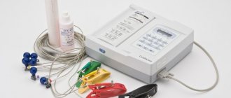
Electrocardiography. ECG. Leads and electrode application points for ECG readings
The category was prepared based on materials from the publication “Cardiology.
National leadership”, ed. Yu.N. Belenkova (GEOTAR-Media, 2007) Navigation menu.
– ECG leads – Electrocardiographic topography (ECG topography). – Location of electrodes. Points for applying electrodes for ECG readings. – Electrocardiogram components and their normal values .
ECG - leads.
Electrocardiographically recorded:
3 standard leads:
I – left hand (+) and right hand (-), II – left leg (+) and right hand (-), III – left leg (+) and left hand (-);
3 reinforced unipolar limb leads:
aVR – enhanced abduction from the right arm, aVL – enhanced abduction from the left arm, aVF – enhanced abduction from the left leg;
6 chest leads:
V1, V2, V3, V4, V5, V6; It is also possible to remove additional leads:
3 additional chest leads (targeted diagnosis of focal myocardial changes in the posterobasal regions of the LV):
V7, V8, V9;
3 bipolar leads along the Palate (additional diagnosis of focal changes in the myocardium of the posterior, anterolateral and upper sections of the anterior wall of the LV):
D – Dorsalis, I – Inferior, A – Anterior. There are also extremely rare types of leads:
Lead S5 - used for poorly defined atrial ECG complex, helps in the differential diagnosis of ventricular and supraventricular cardiac arrhythmias.
Orthogonal leads according to Frank - as orthogonal leads, an ECG is taken in three chest leads. The simplest are leads X, Y, Z. The axes of these leads are located perpendicular to each other and perpendicular to the horizontal, vertical and sagittal plane of the person.
Esophageal leads - used to identify the atrial ECG complex. To record them, an electrode connected to a cardiograph is inserted into the esophagus using a probe. In the esophageal leads, the wave caused by the excitation of the atria is clearly visible, which helps in the diagnosis of various arrhythmias.
Intracardiac leads - used to record the EMF of the heart in the cavity of the atrium or ventricle. To do this, a special electrode probe is inserted into the heart cavity during probing.
Leads according to Arrighi. The axes of the Arrighi leads are located in the sagittal plane and form a triangle, in the center of which the heart is located. For any type of location of the heart in the chest (asthenic, hypersthenic), one of the axes remains parallel to the posterior wall of the left ventricle and captures signs of myocardial infarction somewhat better than standard III and lead aVF.
An ECG is taken in Arrighi leads in the following switch positions: in the first position, lead A1 is recorded, in the second - lead A2, in the third - A3.
Up
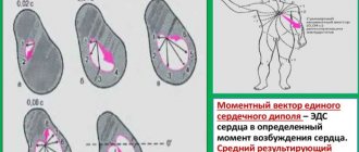
Electrocardiographic topography (ECG topography). Designations: - RCA-Right Coronary Artery (right coronary artery); - SVC-Superior Vena Cava (superior vena cava); - IVC-Inferior Vena Cava (inferior vena cava); - RA-Right Atrium (right atrium); - RV -Right Ventricle (right ventricle); - LAD-Left Anterior Descending artery (anterior descending artery); - LV-Left Ventricle (left ventricle); - LCX-Left CircumfleX artery (circumflex artery). If we recall the skeletotopy of the heart in a healthy person, then 2/3 of the right parts of the heart (right atrium and right ventricle) and 1/3 of the left ventricle are projected onto the anterior surface of the chest. Since the left ventricle is “electrically” more active and stronger, the ECG topography is perceived somewhat differently: 2/3 of the anterior wall is occupied by the left ventricle, and 1/3 of the right border is occupied by the right ventricle. Accordingly, the lower and left side walls are represented by the left ventricle. CONDITIONALLY! Conventionally, it is accepted that the first two chest electrodes (V1, V2) are located on the border of the right and left ventricles, that is, on the septum. Therefore, it is they who demonstrate both the electrophysiological characteristics of the left ventricle (septal and posterior high infarctions) and the activity of the right one (hypertrophy and block of the right bundle branch). Leads from the limbs “look” at the heart in a vertical plane, respectively, showing only the lower and lateral walls. Looking at the picture, and visually, if you imagine, the lateral wall is “showed”: l and aVL leads. The lower wall: lll, aVF and ll. The chest leads “show” the heart in a horizontal plane, in a kind of semicircle. The first four leads show the anterior wall, and the last two show the lateral wall. -V1-V2-septum; -V3-V4-actually, the front wall; -V4 is usually called the apex. -V5-V6-side wall. Additional chest leads: V7-V9 show the posterior wall, and additional RIGHT chest leads: V3R and V4R show the right ventricle.
Up
Location of electrodes. Points for applying electrodes for ECG readings.
In standard leads and 3 enhanced leads from the limbs, the electrodes are located: Red - right hand, Yellow - left hand, Green - left leg, Black - right leg.
In the chest leads, the electrodes are located:
V1 (red) – in the fourth intercostal space along the right edge of the sternum, V2 (yellow) – in the fourth intercostal space along the left edge of the sternum, V3 (green) – approximately at the level of the fifth rib along the left parasternal line, between the fourth and second electrodes, V4 (brown ) - in the fifth intercostal space along the left midclavicular line, V5 (black) - on the horizontal line V4 along the left anterior axillary line, V6 (blue) - on the horizontal line V4-V5 along the left midaxillary line.
In additional chest leads the electrodes are located:
V7 – at the level of V4-V6 along the left posterior axillary line, V8 – at the level of V4-V6 along the left scapular line, V9 – at the level of V4-V6 along the left paravertebral line.
In the leads along the Sky, the electrodes are located:
Red standard - in the second intercostal space along the right edge of the sternum, Green standard - in the fifth intercostal space along the left midclavicular line, Yellow standard - on a horizontal line with a green electrode along the posterior axillary line.
In lead S5, the electrodes are located:
The red electrode is installed on the manubrium of the sternum, the Yellow electrode is installed in the fifth intercostal space on the left, directly next to the sternum.
In orthogonal leads according to Frank, the electrodes are located:
Chest electrodes are placed at the level of the fifth intercostal space when the patient is sitting and at the level of the fourth when lying down.
The locations for applying the electrodes are as follows: point E is located along the midsternal line; point M - on the spine, symmetrical to point E; point A - center of the left mid-axillary line; point C - between electrodes E and A; point I - along the right midaxillary line; point H - on the back of the neck or on the head and point F - on the left leg. The polarity proposed by Frank is as follows: lead X (horizontal spatial component) is obtained by commutating electrodes E, C and A (positive pole) and I (negative pole); lead Z (sagittal spatial component) - electrodes A and M (positive pole) and 1, E, C (negative) and lead V (vertical spatial component) - electrodes F and M (positive pole), and electrode H - (negative) .
In the Arrighi leads, the electrodes are located:
The yellow (active, positive) electrode is strengthened with the help of a flat plate at the angle of the left shoulder blade, the Red (negative) electrode on the suction cup is above the middle of the left collarbone, and the Green electrode is on the left shin.
Up
Components of the electrocardiogram and their normal values.
Up
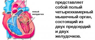
Electrocardiography is one of the basic research methods that does not lose its relevance, allowing a doctor of any specialty to determine the functional state of the heart and the presence of possible pathology recorded on an electrocardiogram (ECG).
The ECG technique is quite simple, but there are a large number of specific leads and electrodes used for this.
In this category you can find basic and rarely used ECG leads, rules for applying electrodes for taking an electrocardiogram when recording various ECG leads.
ECG with mirror arrangement of internal organs
In order to determine whether the position of the patient’s heart is normal, it is necessary to listen to it with a phonendoscope. If this is not possible, then just put your palm to your chest and use it to feel the heartbeat; use your fingertips to determine the maximum impulse of the heart - this is its apex. Its location will correspond to the location of the heart in the chest cavity.
With dextracardia, the application of electrodes corresponds to the usual one, with the difference that the electrodes are applied on the right side of the chest:
- red – along the left edge of the sternum in the 2nd intercostal space;
- yellow - along the right edge of the sternum in the 2nd intercostal space;
- green – between the yellow and brown electrodes in the middle;
- brown – in the 5th intercostal space along the midclavicular line;
- black - in the 5th intercostal space along the anterior axillary line;
- purple – in the 5th intercostal space along the posterior axillary line.
Correct application of electrodes plays a very important role in deciphering the cardiogram and making a diagnosis. Therefore, circuits designed to make it easier to remember electrode sequences save time and allow you to quickly and efficiently establish or disprove a diagnosis. Sometimes a person's life depends on it.
ECG in the sky - features of the procedure, differences, level of information content
Cardiovascular problems are a common concern for most people. Some people are worried about going to the doctor, because a lack of understanding of many terms and concepts puts them in an awkward position.
An electrocardiogram is the main method for determining the quality of heart function. It is recommended to all patients immediately upon admission to the hospital.
How is an ECG performed across the Sky? What is the difference between the method? When is such a study recommended?
The essence of the ECG
An electrocardiogram is a procedure that allows you to reliably assess the work of the heart, heart rate and characteristics of the transmission of electrical impulses. To obtain true data, it is important to strictly follow the rules for conducting an ECG and correctly apply the electrodes.
ECG according to the Sky is a special technique for applying electrodes. The Neb leads are slightly different from the standard ones, which allows for a more detailed assessment of the work of the heart. When the procedure is performed by an experienced doctor, he can obtain a lot of additional data about the functioning of the heart muscle. An ECG records heartbeats.
Correct placement of electrodes plays a key role in obtaining reliable data when examining a patient. An electrocardiogram is recorded only after making sure that everything is installed correctly and the patient has not experienced physical exertion before the procedure.
How does the procedure work?
An electrocardiogram of the heart is performed in an outpatient or hospital setting. However, this technique is also used as an emergency method for diagnosing pathologies, which is why ambulance crews have an electrocardiograph.
The procedure is sometimes carried out directly in the car or at the patient’s home.
Important! When conducting an ECG, regardless of where it is performed, the distance from the patient to the nearest power supply must be at least 2 meters.
This will avoid unnecessary electrical interference and record accurate information.
ECG on the Sky is carried out as follows:
- First, the attending physician examines the patient, checks all devices for functionality,
- a special electrically conductive gel is applied to the body at the sites where the electrodes are supposed to be installed,
- treats the skin with an antiseptic,
- correctly applies electrodes,
- performs ECG recording.
When performing electrocardiography, the patient should be in a calm, relaxed state, preferably lying down.
Features of applying electrodes
Depending on the chosen research technique, there are differences in the number of electrodes and the nature of their application. When conducting an ECG according to Slopak, 9 leads are used. There are only 3 of them to take a cardiogram across the Sky.
Important! The patient's skin must be treated with an antiseptic.
Almost all types of electrodes are considered reusable, so disinfecting them after each use will prevent the spread of infection from person to person.
An ECG according to the Sky is performed in only three leads. The device involves the use of electrodes of three colors:
- red,
- yellow,
- green.
The red electrode is installed on the right side of the chest at the level of the 5th intercostal space. The yellow lead is installed on the posterior axillary line at the same level. The green electrode is placed on the left at the midclavicular line.
Why this type of ECG
Many patients cannot understand: why perform additional electrocardiograms using other methods? A standard ECG in some cases does not provide a complete picture. Conducting a Sky survey is recommended in the following situations:
- for monitoring the condition of athletes,
- if a posterobasal myocardial infarction is suspected,
- with coronary heart disease.
The value of this research method is that the results of cardiography show the state of those areas of the myocardium that cannot be seen when performing a classical ECG.
Lead D shows in detail what changes the wall of the left ventricle itself has undergone, lead A shows what pathologies are on the anterior wall of the heart, and lead I helps to see the stage of ischemic disease in the lower zone of the anterolateral wall of the left ventricle.
It is obvious that in cardiology there are no unnecessary research methods. The use of various techniques and equipment helps to give people reliable diagnoses and provide high-quality and effective treatment.
In addition to the classic three-lead cardiogram across the Sky, a stress ECG is practiced. In this case, an electrocardiogram is recorded after the patient performs physical activity. Sometimes drugs are introduced into the patient’s body that provoke spasm of the coronary artery. After this, an ECG is performed, which helps to identify hidden atherosclerotic changes.
Is it possible to replace one method with another?
Although the results of the Neb electrocardiogram are very valuable for determining some pathologies, there are diseases that cannot be diagnosed only by taking readings from three leads. Therefore, in order for the doctor to obtain complete information about the state of the heart muscle and the quality of its work, a classic ECG in 12 leads and the Sky is required.
Only a specialist interprets the results of the cardiogram. Correct interpretation of the results obtained and diagnosis becomes the main condition for successful treatment.
If you carefully monitor your well-being and do not ignore the appearance of alarming symptoms, you can recognize the problem in time and conduct an examination. Any pathology is more successfully treated in the early stages of development than in an advanced stage. By following the recommendations of doctors, you can maintain your health at the proper level and avoid serious complications.
Loading…
Source: https://dlja-pohudenija.ru/serdcze/diagnostika/mozhet-li-ekg-po-metodu-neba-polnostyu-zamenit-klassicheskuyu-kardiogrammu
How does the procedure work?
An electrocardiogram of the heart is performed in an outpatient or hospital setting. However, this technique is also used as an emergency method for diagnosing pathologies, which is why ambulance crews have an electrocardiograph. The procedure is sometimes carried out directly in the car or at the patient’s home.
ECG on the Sky is carried out as follows:
- first, the attending physician examines the patient, checks all devices for functionality;
- a special electrically conductive gel is applied to the body at the sites where the electrodes are supposed to be installed;
- the skin is treated with an antiseptic;
- correctly applies electrodes;
- performs ECG recording.
When performing electrocardiography, the patient should be in a calm, relaxed state, preferably lying down.
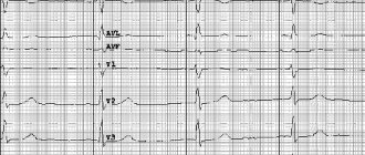
ECG sky leads
The following arrangement of teeth in the leads is considered normal ECG indicators:
- equal distance between R-teeth;
- the P wave is always positive (it may be absent in leads III, V1, aVL);
- the horizontal interval between the P-wave and the Q-wave is no more than 0.2 seconds;
- S and R waves are present in all leads;
- The Q wave is exclusively negative;
- The T wave is positive, always shown after the QRS.
ECG is taken on an outpatient basis, in a hospital setting, and at home. The results are decoded by a cardiologist or therapist. If the obtained indicators do not correspond to the established norm, the patient is hospitalized or prescribed medication.
Sections of the heart and the leads responsible for them
Each of the six main leads displays one or another part of the heart muscle:
- I and II standard leads are the anterior and posterior cardiac walls, respectively. Their totality reflects standard lead III.
- aVR – lateral cardiac wall on the right;
- aVL – lateral cardiac wall anterior to the left;
- aVF – posterior inferior wall of the heart;
- V1 and V2 – right ventricle;
- VZ – septum between the two ventricles;
- V4 – upper cardiac section;
- V5 – lateral wall of the left ventricle in front;
- V6 – left ventricle.
Thus, deciphering the electrocardiogram is simplified. Failures in each individual branch characterize the pathology of a specific area of the heart.
More about leads
For accurate diagnosis, the difference in indicators of the electrodes (electrical potential of the lead) attached to the patient’s body is recorded. In modern cardiological practice, 12 leads are accepted:
- standard – three leads;
- reinforced - three;
- chest – six.
Diagnostics are carried out only by specialists who have received the appropriate qualifications
Blockade on ECG
- left hand – electrode “ ”, right – minus (first lead – I);
- left leg – sensor “ ”, right arm – minus (second lead – II);
- left leg – plus, left arm – minus (third lead – III).
Electrodes for standard leads are secured with clips in the lower part of the limbs. The conductor between the skin and the sensors is wipes treated with saline solution or medical gel. A separate auxiliary electrode installed on the right leg performs the grounding function. Reinforced or unipolar leads, according to the method of fixation on the body, are identical to standard ones.
The electrode, which registers changes in the potential difference between the limbs and the electrical zero, has a “V” designation in the diagram.
The left and right arms are designated “L” and “R” (from English “left”, “right”), the leg corresponds to the letter “F” (leg).
Thus, the place of attachment of the electrode to the body in the graphic image is determined as aVL, aVR, aVF. They record the potential of the limbs on which they are attached.
Reinforced electrodes are necessary for convenient decoding of the cardiogram, since without them the waves on the graph will be weakly expressed.
Bipolar standard and unipolar reinforced leads determine the formation of a coordinate system of 6 axes.
The angle between the standard leads is 60 degrees, and between the standard and the adjacent enhanced lead is 30 degrees. The cardiac electrical center splits the axes in half.
The negative axis is directed towards the negative electrode, the positive axis, respectively, is directed towards the positive electrode.
ECG chest leads are recorded by single-pole sensors attached to the skin of the chest using six suction cups connected by tape. They record impulses from the circumference of the cardiac field, which is equally potential to the electrodes on the limbs. On a paper chart, the chest leads are designated “V” with a serial number.
Cardiac examination is performed according to a specific algorithm, therefore the standard system for installing electrodes in the chest area cannot be changed:
- in the area of the fourth anatomical space between the ribs on the right side of the sternum - V1. In the same segment, only on the left side - V2;
- connection of the line coming from the middle of the clavicle and the fifth intercostal space - V4;
- lead V3 is located at the same distance from V2 and V4;
- connection of the anterior axillary line on the left and the fifth intercostal space - V5;
- intersection of the left middle part of the axillary line and the sixth space between the ribs - V6.
Additional electrodes are used when it is difficult to make a diagnosis, when decoding the six main indicators does not provide an objective picture of the disease
Each lead on the chest is connected with an axis to the electrical center of the heart. In this case, the position angle V1–V5 and the angle V2–V6 are equal to 90 degrees. The clinical picture of the heart can be recorded by a cardiograph using 9 branches. Three unipolar leads are added to the usual six:
- V7 – at the junction of the 5th intercostal space and the posterior line of the armpit;
- V8 – the same intercostal area, but in the midline of the armpit;
- V9 is the paravertebral zone, parallel to V7 and V8 horizontally.
ECG across the sky
These are bipolar leads that record the potential difference between two points located on the chest wall. There are three leads in the Sky:
- Lead D (Dorsalis) – used to diagnose focal changes in the posterior wall of the left ventricle.
- Lead A (Anterior) – used to diagnose infarctions of the anterior wall of the left ventricle.
- Lead I (Inferior) – used to diagnose infarctions of the lower parts of the anterolateral wall.
Cardiograph lead wires are marked in different colors: red is connected to the right arm; yellow - to the left hand; green - to the left leg. For registration, electrodes used to record ECG in standard leads are used.
The red electrode is installed on the second intercostal space of the right side of the sternum; the yellow electrode is placed at the level of lead V7; green electrode – at level V4.
Lead D records the potential difference between the red and yellow electrodes. Lead A records the potential difference between the red and green electrodes. Lead I records the potential difference between the yellow and green electrodes.
The main advantage of Neb leads is that to record ECG readings it is not necessary to apply electrodes to the limbs.
There are a number of leads that are not often found in clinical practice:
- lead S5 - used for poorly defined atrial ECG complex, helps in the differential diagnosis of ventricular and supraventricular cardiac arrhythmias. The red electrode is installed on the manubrium of the sternum, and the yellow electrode is installed in the fifth intercostal space on the left, directly next to the sternum.
- orthogonal leads - found application in ECG analysis using a computer. An ECG is taken in three chest leads as orthogonal ones. The axes of these leads are located perpendicular to each other and perpendicular to the horizontal, vertical and sagittal plane of the person.
- esophageal leads - used to identify the atrial ECG complex. To record them, an electrode connected to a cardiograph is inserted into the esophagus using a probe. In the esophageal leads, the wave caused by the excitation of the atria is clearly visible, which helps in the diagnosis of various arrhythmias.
- intracardiac leads - used to record the EMF of the heart in the cavity of the atrium or ventricle. To do this, a special electrode probe is inserted into the heart cavity during probing.
In the palate ECG technique, it is common to use only three electrodes. Red and yellow sensors are fixed in the fifth intercostal space. Red on the right side of the chest, yellow on the back of the axillary line.
The green electrode is located on the line of the middle of the collarbone.
Most often, an electrocardiogram according to the Sky is used to diagnose necrosis of the posterior heart wall (posterior basal myocardial infarction), and to monitor the condition of the heart muscles in professional athletes.
Schematic arrangement of the ventricles and atria, based on the location of which the electrodes are placed
Source: https://moeserdce.net.ru/otvedeniya-po-nebu-ekg/
General rules for applying electrodes
When recording an electrocardiogram, electrodes are installed on several areas of the body. This ensures the conduction of electrical impulses through the heart, and the results are more accurate. The correct location of the terminals is the key to reliable recording of heart function.
General rules for installing electrodes:
- The skin at the site where the electrode is applied is degreased with alcohol;
- Pronounced hair when using reusable electrodes is treated with a soap solution (otherwise the hair is shaved off);
- The electrodes are coated with a special gel that improves electrical conductivity (it can be replaced with an isotonic solution, but this is not recommended, as the contact will worsen);
- The use of gauze pads instead of a special gel is also not an alternative to the gel, since they dry out quickly (such pads are absolutely prohibited for long-term studies, for example, Holter monitoring);
- It is important to follow safety rules when working with electrical devices, in particular grounding (not required when recording ECGs using portable battery-powered electrocardiographs).
All electrodes are divided into reusable and disposable. Each type has advantages and disadvantages and, as a rule, the option for recording is selected by medical personnel.
Features of disposable electrodes
You can purchase disposable electrodes in the Avicena-med online store, where only high-quality elements made in Italy are sold. They are suitable for daily monitoring or stress tests, which involve the patient’s physical activity.
Advantages of disposable electrodes:
- No risk of transmission of infectious diseases;
- Easy to install (doctors consider them more practical);
- High degree of adhesion (does not fall off after prolonged use);
- Good conductivity and high-quality contact;
- Suitable for patients with excessive sweating.
Unlike disposable electrodes, reusable designs are often used in government agencies, as they are more economical and durable.
ECG in the sky - algorithm, indications for the procedure
An electrocardiogram is a procedure that allows you to reliably assess the work of the heart, heart rate and characteristics of the transmission of electrical impulses. To obtain true data, it is important to strictly follow the rules for conducting an ECG and correctly apply the electrodes.
Conducting standard electrocardiography by a diagnostician
ECG according to the Sky is a special technique for applying electrodes. The Neb leads are slightly different from the standard ones, which allows for a more detailed assessment of the work of the heart. When the procedure is performed by an experienced doctor, he can obtain a lot of additional data about the functioning of the heart muscle. An ECG records heartbeats.
Application of electrodes when performing a cardiogram on the patient according to the Sky
Correct placement of electrodes plays a key role in obtaining reliable data when examining a patient. An electrocardiogram is recorded only after making sure that everything is installed correctly and the patient has not experienced physical exertion before the procedure.
Normal ECG pattern
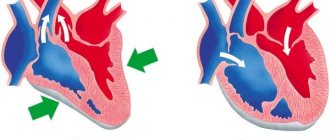
An electrocardiogram is a graph. The horizontal axis marks time, the vertical axis represents the amount of electrical charge generated when the heart contracts. One cycle of cardiac contraction is reflected in the form of teeth and intervals between them.
Main teeth:
- P-reflects contraction of the right and left atria.
- QRS is a complex consisting of three teeth. Characterizes electrical activity at the time of ventricular contraction.
- T-wave, which occurs at the end of the cardiac cycle. Plays an important role in characterizing ischemic phenomena. A high or negative potential indicates a possible ischemic process.
Installation of electrodes across the Sky
The chest area is moistened with saline solution or gel. Each sensor, using a suction cup, is installed in place for leads.
Important! It is necessary to remove electronic watches, metal chains and bracelets.
- It is installed along the second intercostal space to the right of the sternum.
- Along the midaxillary line into the space between the fourth and fifth ribs.
- Midclavicular line, just above the fifth rib.
Depending on the chosen research technique, there are differences in the number of electrodes and the nature of their application. When conducting an ECG according to Slopak, 9 leads are used. There are only 3 of them to take a cardiogram across the Sky.
An ECG according to the Sky is performed in only three leads. The device involves the use of electrodes of three colors:
- red;
- yellow;
- green.
The red electrode is installed on the right side of the chest at the level of the 5th intercostal space. The yellow lead is installed on the posterior axillary line at the same level. The green electrode is placed on the left at the midclavicular line.
Sign up for a study
There is no special preparation procedure provided.
Important! Before the procedure, it is recommended to get enough sleep and avoid physical activity.
To sign up for a study at one of the NEARMEDIC network clinics without queues, call a convenient clinic or a general multi-line phone, or fill out a form on the website.
You will find the names of cardiologists with biographies and photographs in the “Doctors” section.
You can also make an appointment with a therapist or cardiologist for a preliminary consultation and development of recommendations for the procedure.
Indications
- Diagnosis of hidden rhythm disorders.
- Detection of hypoxic phenomena in the heart that are not determined by the standard method.
- Determining the possibility of playing sports for patients with pathology of the cardiovascular system.
Detection of heart disease at an early stage of development allows timely treatment to begin, preventing irreversible consequences from developing in the tissues.
Latent myocardial ischemia may not affect a person’s life, but if it is present in a person undergoing severe training, then the impact of extreme loads can be fatal. That is why an ECG is carried out once a year for everyone who has devoted their life to sports. Monitoring the condition of the heart and blood vessels is especially important in athletes in the older age group.
Source: https://remson58.ru/nalozhenie-elektrodov-nebu/
Normal ECG pattern
An electrocardiogram is a graph. The horizontal axis marks time, the vertical axis represents the amount of electrical charge generated when the heart contracts. One cycle of cardiac contraction is reflected in the form of teeth and intervals between them.
Main teeth:
- P-reflects contraction of the right and left atria.
- QRS is a complex consisting of three teeth. Characterizes electrical activity at the time of ventricular contraction.
- T-wave, which occurs at the end of the cardiac cycle. Plays an important role in characterizing ischemic phenomena. A high or negative potential indicates a possible ischemic process.
How to do an ECG on the Sky
To obtain accurate results on the condition and functioning of the heart in electrocardiography, the use of additional leads is often practiced.
As a rule, this additional measure allows for timely diagnosis of the development of myocardial infarction and other pathologies.
Symptoms of this problem are not always noticed during routine diagnostics using standard parameters, which is explained by the varying strength of the manifestation of changes. The most popular is the Sky ECG, less used are the orthogonal lead and Arrighi.
A person who specializes in this area knows exactly how the axes of the leads along the Sky are connected and the probable possibilities of diagnosing them. This knowledge helps not to repeat standard techniques.
Carrying out an ECG using Neb
To carry out Neb diagnostics, it is first necessary to correctly position the electrodes. There are three of them in total, painted in different colors.
To improve the contact properties between the electrode and the patient’s skin, a special paste with a slightly thick consistency, made from baby soap and plain water, is used.
To avoid staining the sheet that is placed under the body of the subject, you need to use oilcloth.
The connection occurs as follows:
- The first to attach is the electrode, which is active and yellow in color. Using a special flat electrode plate, it is installed under the left shoulder blade in its lower corner. Installation in position V7 is allowed - it was noticed that in this position the result is more accurate. In addition, this method greatly facilitates the diagnosis of bedridden patients.
- The next one is a red electrode, which, unlike yellow, is passive. It is attached using a pear-shaped suction cup. The location of this electrode is the second intercostal space on the right side of the chest.
- The last electrode is painted green and is attached to the body, like the red one, using a suction cup. Unfortunately, it is often the green conductor that is not fixed where it is needed. Its correct location is at the apex of the heart. The correct place can be determined by palpation - apical impulses are felt in the right place. Often the location is determined incorrectly and the electrode is installed in the fifth intercostal space, on the left side of the clavicular line.
After connecting all the electrodes, you can determine the triangle with the heart inside. The ECG procedure occurs by switching a toggle switch to connect various axes in series:
- the first axis is formed from the red and yellow electrodes. This lead is parallel to the back of the heart. This standard position is written with the letter “D” and is considered the most accurate indicator for determining the presence of cardiac pathologies, and more precisely, for myocardial infarction associated with the left ventricle;
- the second position is indicated by the letter "A". The axis, which is formed when the toggle switch is switched for the second time, is located on the side of the front wall of the organ. The data that needs to be taken at this stage does not greatly affect the originally obtained values;
- the third switch remains similar to axes V2 and V3, the only difference is the location of the heart organ in the chest. The results of the third switch are written with the letter “I”, but they also do not greatly affect the data obtained during the first switch.
So, we can draw the following conclusion - the most accurate and important data in diagnostics is obtained when the toggle switch is first switched and the axis with the letter “D” is formed.
The ECG showed distinctive signs, which consisted of the ST line. It can be raised and have a hump-shaped shape, which directly indicates myocardial infarction, or it can be lowered and imply hypertrophy of the left ventricle of the heart. When analyzing the results, it is also important to pay attention to the shape of the tooth - it can be negative symmetrical and negative asymmetrical.
Other additional methods of performing an ECG
There are several types of additional leads that increase the efficiency of the study: ECG according to the Sky: orthogonal and the Arrini method.
Diagnosis by ECG can be carried out in several ways
Orthogonal leads for ECG
Orthogonal leads are measured exclusively in three planes. Moreover, they are further divided into two types – corrected and uncorrected.
Orthogonal leads are measured along three types of axes. For each measurement, the most accurate and important are the indicators recorded when the toggle switch was first switched. The difference lies in the different locations of the electrodes during formation:
We recommend you read:Additional ECG leads
- in the first method, the red electrode is fixed on the right side of the axillary line, focusing on the apex of the heart, and the yellow electrode is attached in accordance with the standard position of V5 on the left side. Thus, the specialist receives X-axis indicators;
- with the second method, a positive yellow electrode is attached to the left leg, and a red electrode is attached slightly above the middle of the left collarbone. According to this scheme, the Y axis is formed;
- the third axis, labeled Z, is measured with the red electrode directly at the angle of the left shoulder blade, and the yellow electrode in the standard V3 position.
With this arrangement of the axes, three planes are formed, which are perpendicular to each other. If we display these planes schematically, they will intersect in the region of the heart.
It is worth noting that experts do not highlight any major advantages in this method of conducting an ECG over the standard system. But in this way, it is possible to obtain additional information about the patient’s heart.
The orthogonal lead method is most often used in sports medicine and in computer-monitor studies, when the goal is to obtain as much information as possible about the organ with the least amount of time spent on diagnosis.
Sign up for a study
To sign up for a study at one of the NEARMEDIC network clinics without queues, call a convenient clinic or a general multi-line phone, or fill out a form on the website. You will find the names of cardiologists with biographies and photographs in the “Doctors” section. You can also make an appointment with a therapist or cardiologist for a preliminary consultation and development of recommendations for the procedure.
There is no special preparation procedure provided.
Important! Before the procedure, it is recommended to get enough sleep and avoid physical activity.
How is an ECG interpreted?
Cardiogram analysis is deciphered exclusively by a specialist. Indicators include P, Q, R, S, T waves and ST and PQ segments. In turn, teeth directed upward are called positive, and teeth directed downwards are called negative.
Key ECG indicators:
- the source of excitation under normal conditions is accompanied by sinus rhythm;
- rhythm frequency – the gap between the R waves is no more than 10%;
- normal heart rate is 60-80 beats/min;
- rotation of the electrical axis of the heart muscle - from semi-horizontal to semi-vertical;
- The R wave is accompanied by a positive character;
- T wave – must be positive;
- PQ section – from 0.02 to 0.09 sec;
- section ST – runs along the isoline; normally there may be deviations of no more than 0.5 mm.
Electrocardiography is a method often used in medical practice that allows one to obtain detailed information about the condition of the heart and some other organs in a short period of time. The data obtained during diagnosis is used to identify many diseases, help to start treatment in a timely manner, and prevent serious complications.
Preparing for an ECG
Rules for preparing for an ECG
- The patient must remain calm during the ECG recording. You should not worry, be nervous, or experience excessively strong emotions. Breathing should be smooth and not rapid. If the patient experiences excitement or anxiety, the doctor must reassure the patient and explain the safety and painlessness of the manipulation. 10-15 minutes before taking the cardiogram, it is advisable to sit, adapt to the functional diagnostics room and medical staff, and restore breathing.
- Preparation for an ECG excludes smoking, drinking alcoholic and caffeinated drinks, strong tea, and coffee before the procedure. Smoking and caffeine stimulate the heart, which may cause the ECG reading to be unreliable.
- It is not recommended to eat 1.5-2 hours before the procedure, and it is better to conduct an ECG on an empty stomach.
- After taking a morning shower on the day the cardiogram is taken, it is not advisable for the patient to apply oil-based creams and lotions to the body. This may create some obstacle to good contact between the electrodes and the skin.
- The patient's clothing should be comfortable and loose, so that it is possible to freely expose the hands and ankles, and quickly remove or unbutton clothes to the waist.
- There should be no metal jewelry, chains, or bracelets on the chest and limbs.
ECG recording algorithm
For correct diagnosis, making an accurate diagnosis and prescribing optimal treatment, it is important to adhere to the following algorithm of actions, namely:
- preparing the patient for the procedure;
- checking the operation of the device;
- application of electrical transmission gels and antiseptic liquids;
- adherence to clear rules on where to place electrodes depending on color;
- selection of lead registration scheme;
- directly recording.
In most cases, the electrocardiogram is recorded in a separate room in a medical facility, but sometimes it can even occur at home, in a ward, or in an ambulance. The room in which the procedure will be performed must be at a sufficient distance from possible electrical interference. As for the couch itself, on which the patient lies, it is located approximately two meters from the power supply.
An ECG is an absolutely painless procedure that does not cause absolutely any negative emotions
The procedure is carried out in a lying position, with free access to the upper torso, legs and arms. If there are contraindications, then an ECG can be performed in a sitting position. Before an electrocardiogram, it is better not to engage in intense physical activity, and also not to take food or drinks that could activate the heart.