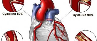
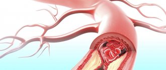
Angina pectoris is a form of coronary heart disease (CHD). Characterized by narrowing of the coronary arteries that supply the heart.
The attack is caused by intense physical or emotional activity (that is, “stress”). Each episode leads to the death of part of the muscle layer.
However, over time, the disease transforms into another form that is not associated with the determining factor (an attack occurs even at rest). This significantly aggravates the treatment and the overall course of the pathology. The forecasts are much worse.
Effective elimination of the deviation is possible only at the first stage (FC 1), when the disease begins. The duration of exposure is 2-4 months.
Then you have to fight the investigation, the issue is not resolved fundamentally. Specialist - cardiologist.
How the disease develops
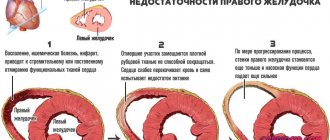
Normally, the heart is supplied with blood through the coronary arteries. Adequate trophism is considered a guarantee of the functional activity of muscle tissue.
Angina pectoris develops due to dual influence.
- The nervous system provokes a narrowing of the blood supply structures. The amount of incoming oxygen and nutritional compounds drops to critical levels at one point. This is fraught with an attack of ischemia. In advanced situations, the death of cardiac structures and scarring of areas (heart attack and subsequent sclerosis) are likely.
- Another pathogenetic mechanism concerns the release of large amounts of cortisol and adrenaline (hormones of the adrenal cortex). They also cause coronary artery stenosis and disruption of local blood flow.
Accordingly, as part of the treatment, a solution to two problems is indicated: reducing the concentration of corticosteroids and correcting the functioning of the central nervous system.
The directions are implemented by different specialists. Cardiologist as primary.
Causes of angina pectoris
The study of angina pectoris received a good impetus in the middle of the last century, when the problem of atherosclerotic damage to the arteries and lipid metabolism became especially urgent. With the advent of various fast food chains and a significant acceleration in the pace of life in large cities, people began to pay less attention to their diet in order to maximize time savings for work.
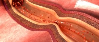
Cardiologists say that angina pectoris develops as a result of decreased elasticity of the coronary arteries; this phenomenon occurs due to progressive atherosclerosis. Atherosclerosis itself has many causes, including endocrine diseases, disturbances in the functioning of the heart muscle itself or the kidney filter. Hypertension is now recognized as the most common factor in damage to the vascular wall.
It is with an increase in blood pressure that microtears occur in the inner lining of the arteries (intima), which are subsequently filled with connective tissue and lead to many problems. According to WHO, approximately two thirds of patients suffering from angina pectoris have hypertension and lipid metabolism disorders.
Other, less frequent and more specific causes of angina include the following factors::
- presence of diagnosed hypertrophic cardiomyopathy;
- pulmonary hypertension;
- predisposition of heart vessels to spasms due to impaired mineral metabolism;
Angina pectoris 2 FC
- aortic valve insufficiency;
- inflammatory processes localized in the vessels supplying the heart muscle.
In addition to the serious pathologies described above, other factors not related to cardiac activity can lead to the development of angina.:
- age over sixty years;
- the presence of nutritional obesity of the second and higher stages;
- menopause period;
- hypodynamic lifestyle;
- predisposition due to a family history;
- presence of diabetes mellitus type 1 or 2;
- abuse of alcohol and tobacco products.
Classification by shape
Typing of the pathological process is carried out according to a group of criteria. The basis is the division into forms.
Stable
Characterized by a slow, sluggish course. The development period is long. More than one year passes from the onset of the pathological process to the appearance of pronounced symptoms.
The main feature of the condition in question is the minimal rate of progression or its complete absence.
Therefore, a stable form is considered less dangerous and easier to correct. There are also good prospects for a cure.
Unstable variety
Has a tendency to progress. How development will occur, abruptly or smoothly, depends on a lot of factors: age, weight, accompanying physiological data, especially cardiac changes.
Patients with a group of negative aspects are at greater risk. Their final state occurs over a period of several years; treatment brings minimal effect.
Stable exertional angina is less common (30% versus 70%). The main patient population is middle-aged men (45 and over). Young people rarely get sick.
Classes of variant angina
It is a form of resting angina and can cause heart attack and sudden death. The pathology has other names: unstable vasospastic, spontaneous, Prinzmetal's angina.
The main feature of the pathology is a prolonged and severe pain attack, which can manifest itself even without physical or emotional stress, in a state of complete rest. Most often this happens in the morning.
Variant angina can occur without pain. In this case, there are two main types (classes) of this form of pathology:
- The first type is different in that there is no pain during an attack. There is only a feeling of squeezing in the chest. This type of resting angina occurs in people with a high pain threshold, as well as in those who suffer from diabetes, since they have decreased sensitivity as a result of polyneuropathy.
- The second type of Prinzmetal's angina is alternating attacks without pain and episodes of angina with severe pain. This type is much more common than the first.
Functional classes (FC)
Can be considered as stages of development of a pathological process. In total, the classification distinguishes 4 functional classes:
- FC1. This is the initial degree of angina. Provoking an attack is relatively difficult. An episode can only occur during intense physical activity. The type of mechanical activity sufficient to cause symptoms varies. It all depends on the individual preparation of a particular patient. The moments themselves are minimal in duration, the approximate time is 2-5 minutes. The main manifestation is intense chest pain. The frequency is also not high. About 1-2 times a month.
- FC2. Most often, angina is diagnosed at this stage. Limitation of life activity occurs. Moderate, habitual physical activity leads to prolonged attacks. The main symptom is pain, supplemented by shortness of breath, disturbances of consciousness, duration – 5-10 minutes or a little less. Recovery using drastic methods is impossible; lifelong maintenance therapy under the supervision of a cardiologist is required. There are chances to put the condition into remission, especially if the form is stable.
- FC3. The third functional class makes a person disabled. The risk of death and the development of acute circulatory disorders in the myocardium increases significantly. To provoke an attack, a small amount of physical activity is enough. Climbing to the 2-3 floor, jogging, swimming and others of a similar nature.
- FC4. Terminal phase. A walk is enough to start the episode. Physical activity is basically impossible and is given to the patient with great difficulty. The probability of death is maximum, there is no way to help. There is no chance of cure. Development also occurs at rest.
It is important to keep in mind:
A standard angina attack, even in the most severe cases, lasts no more than 20 minutes. Anything above that is most likely a heart attack. Therefore, it makes sense to call the doctors.
Canadian classification
To determine the severity of symptoms of angina pectoris, the Canadian Society of Cardiology has developed a special classification in the form of a table, which includes the following functional classes of angina pectoris:
Functional class 1
When performing normal physical activity for a person, he feels good. Pain only appears during intense and prolonged work, such as weightlifting or long-distance running.
Functional class 2
Pain occurs even during normal walking, when a person walks more than 200 meters. Also, angina pectoris develops if the patient climbs stairs above the 2nd floor, goes outside in very cold weather, or overeats.
Functional class 3
The attack begins when walking from 100 to 200 meters, or when climbing to the 2nd floor.
Functional class 4
Doing any physical work causes pain. An attack can develop even in a completely calm state.
Causes
As the name of the disease suggests, the main factor in the development of the process is tension. Physical and psychological.
An approximate list of provocateurs is as follows:
- Anger. Expressed negative emotion. It provokes an episode of cardiac pain especially often.
- Stress of any kind. Usually accompanied by excitement and fear. May lead to rhythm disturbances and other conditions.
- Excessive mechanical load. Running, fast walking, climbing stairs, carrying heavy objects and others.
- Hypothermia. It is considered an exception from this list.
Strictly speaking, the above reasons provoke the angina attack itself, but they do not affect the origin of the process.
What are the main pathogenetic points?
Vasculitis
Inflammatory damage to the walls of blood vessels. Usually viral or autoimmune in nature. May be part of other conditions, for example, lupus erythematosus and others.
Treatment is carried out urgently. In the active phase, disruption of normal blood flow occurs due to stenosis of the coronary arteries.
Further, if therapy is inadequate or absent altogether, the pathology goes into spontaneous remission. Possible formation of adhesions, fusion of blood vessels and stable ischemia of cardiac structures.
These are dangerous anatomical complications. Recovery is usually surgical. It consists of excision of strands or artificial expansion of the lumen (stenting).
Anomalies in the structure of the coronary arteries
They have an innate character. The reasons are spontaneous genetic mutation or intrauterine development disorders.
Surgical treatment is aimed at restoring the anatomical shape of the vessels or prosthetics, creating bypasses.
Change in heart size
Develops as a result of exposure to one of a group of factors. Cardiomegaly or spontaneous tissue proliferation, chamber hypertrophy (thickening of the myocardium), scarring after necrosis (cardiosclerosis) and other conditions as an option.
In all cases, compression of the coronary arteries occurs by pathologically altered tissues. This leads to impaired blood flow.
Endocrine diseases
Hyperthyroidism (excess of thyroid hormones), excessive synthesis of cortisol and others. This includes diabetes mellitus. The main factor in the development of angina pectoris is vasospasm.
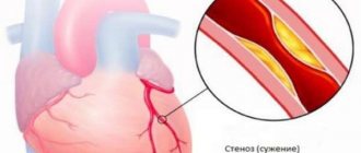
Atherosclerosis of the coronary arteries
The main reason for the formation of the condition. Mass fraction - 90% or more, according to some estimates 95-96%.
The essence is the deposition of cholesterol on their walls.
Lipid formations can be eliminated by conservative means only in the early stages.
As it progresses, surgical treatment is required. Because plaques calcify and harden.
Angina pectoris FC-1 is the most favorable stage for treatment. Everything starts from this stage, regardless of the determining moment.
Unstable angina
Risk factors
Risk assessment for unstable angina, taking into account clinical symptoms and ECG changes:
| High risk | Medium risk | Low risk |
| At least one of the following signs | Not meeting high risk criteria + at least one of the following | Failure to meet high and medium risk criteria |
| Prolonged attack of angina (> 20 min), continuing to the present day | Prolonged (> 20 min), but currently resolved angina attack | Increased or worsening angina |
| Pulmonary edema, most likely caused by myocardial ischemia | Angina at rest (> 20 minutes or resolved with rest or sublingual nitroglycerin) | Reducing the level of stress that causes angina pectoris |
| Angina at rest with ST segment elevation or depression > 1 mm | Nocturnal angina attacks | New-onset angina (from 2 weeks to 2 months) |
| Angina pectoris with the appearance or intensification of moist rales, III tone or murmur of mitral regurgitation | Angina with transient T wave changes | No new ECG changes or normal ECG |
| Angina with arterial hypotension | Severe angina for the first time in the last 2 weeks | |
| Increased levels of markers of myocardial necrosis | Pathological Q waves or ST segment depression of several leads at rest | |
| Age over 65 |
Read also: Differential diagnosis of hemorrhagic diathesis
A high risk for unstable angina is indicated by attacks of angina without a provoking load (longer than 20 minutes), heart failure (III heart sound, congestion in the pulmonary circulation, moist rales in the lungs), systolic dysfunction of the left ventricle, arterial hypotension, newly detected or intensified murmur mitral insufficiency, elevation or depression of the ST segment by 0.5-1 mm or more in several leads + increased levels of markers of myocardial necrosis. Low and moderate risk is indicated by short anginal attacks, absence of ischemic changes in the ST segment, normal levels of myocardial necrosis markers and stable hemodynamics.
Unstable angina is a transient clinical condition in which painful attacks are marked by greater intensity, duration, and less effect from taking nitrates. There are two possible ways of developing this condition: regression of symptoms with a possible increase in functional class or the development of acute myocardial infarction.
There are first-time, progressive, post-infarction and vasospathic forms (Prinzmetal) of angina:
- For the first time, it occurs 1-2 months after the first painful attack.
- Progressive – worsening pain syndrome in patients with stable angina pectoris, often with an increase in FC.
- Early post-infarction angina is the appearance of anginal (painful) attacks within 24 hours to two weeks after myocardial infarction (NYHA criteria). In domestic cardiology, a longer time interval is traditionally used - from 3 days (after 72 hours) to 4 weeks (up to the 28th day inclusive) from the onset of a heart attack.
- Vasospastic (Prinzmetal, variant) – pain occurs unrelated to exercise, often during sleep, without any visible provoking factors. Unlike other forms, Prinzmetal's angina develops when the heart vessels are intact, not affected by atherosclerosis, and is associated with a sudden spasm of the coronary (supplying the heart muscle) vessels.
Braunwald classification
The Braunwald classification of unstable angina assesses the risk of acute myocardial infarction based on the characteristics of the anginal attack and the causes of its occurrence.
Read also Atherosclerosis - causes, symptoms, treatment, prevention
| Class | Characteristics |
| I | Angina pectoris |
| New, severe or progressive angina pectoris within the last 2 months | |
| Increased frequency of angina attacks | |
| Reducing the level of stress at which angina occurs | |
| No angina at rest during the last 2 months | |
| II | Angina at rest, subacute |
| Angina at rest within the last month but not within the last 48 hours | |
| III | Angina at rest, acute |
| Angina at rest within the last 48 hours | |
| Circumstances of occurrence | |
| A | Secondary - provoked by non-coronary pathology, for example anemia, infection, thyrotoxicosis, hypoxia |
| IN | Primary |
| WITH | Post-infarction - within 2 weeks after myocardial infarction |
Rizik classification
The Rizik classification of unstable angina assesses risk based on the characteristics of the angina attack and ECG changes.
| Class | Characteristics |
| I.A. | Increased angina without ECG changes |
| I.B. | Worsening angina with ECG changes |
| II | New-onset angina pectoris |
| III | New-onset angina at rest |
| IV | Prolonged angina at rest with ECG changes |
In a study of 1,387 patients admitted to the emergency department with unstable angina, the risk of in-hospital complications (myocardial infarction, severe angina, death) increased proportionally with the class of angina: Rizik class IA was 2.7%, and class IV was 42. 8%.
TIMI Risk Assessment Scale
The TIMI scale is based on the TIMI IIB and ESSENCE studies. It takes into account age, clinical picture, ECG changes and increased levels of markers of myocardial necrosis.
| Points (each risk factor adds one point, maximum 7 points) | |
| Age > 65 years | |
| The presence of three or more risk factors for atherosclerosis | |
| Previously diagnosed coronary artery stenosis of more than 50% diameter | |
| Elevation or depression of the ST segment on ECG upon admission | |
| Two or more attacks of angina in the last 24 hours | |
| Taking aspirin within the last 7 days | |
| Increased markers of myocardial necrosis | |
| Number of points | Risk of death or myocardial infarction in the next 2 weeks, % |
| 0-1 | 4,7 |
| 2 | 8,3 |
| 3 | 13,2 |
| 4 | 19,9 |
| 5 | 26,2 |
| 6-7 | 40,9 |
A high TIMI score indicates a high risk of death, myocardial infarction, and recurrent ischemia requiring revascularization.
GUSTO Risk Score
Additional risk factors were identified in the PURSUIT and GUSTO IV–ACS studies. All these classifications make it possible to assess the risk and, on this basis, choose treatment tactics.
| Age | Points |
| 50-59 | 2 |
| 60-69 | 4 |
| 70-79 | 6 |
| 80 and older | 8 |
| Anamnesis | Points |
| Heart failure | 2 |
| Strokes, transient cerebral ischemia | 2 |
| Myocardial infarction, revascularization, stable angina | 1 |
| Symptoms and laboratory parameters | Points |
| Heart rate more than 90 min-1 | 3 |
| Increased levels of troponin or CPK MB fraction | 3 |
| Creatinine > 1.4 mg% | 2 |
| C-reactive protein > 20 mg/l | 2 |
| C-reactive protein 10-20 mg/l | 1 |
| Anemia | 1 |
| Sum of points | 30-day mortality |
| 0-5 | 0,4 |
| 6-10 | 2,8 |
| 11-15 | 8,7 |
| 16-19 | 25,0 |
| 20-22 | 41,7 |
Risk factors
In addition to these, there are also so-called risk factors.
- Consumption of coffee in large quantities. Systematically. Also other tonic drinks, such as tea and specialized energy drinks.
- Smoking. There is a direct connection between the amount of tobacco consumed per day, length of service and the likelihood of coronary disease in the form of angina pectoris.
- Physical inactivity. Forced, as a result of prolonged immobilization or due to the nature of professional activity.
- Ventricular or atrial hypertrophy. Usually a symptomatic manifestation and not an independent disease.
- Persons prone to negative emotions. Hot-tempered.
- Patients with lipid metabolism disorders. Outwardly manifested by obesity.
- Diabetics, hypertensives, people suffering from pathologies of the thyroid gland and adrenal glands.
- Male representatives. The ratio between them and women is defined as 3-4:1 according to various estimates.
- Age from 45 years and over.
Many of these factors can be adjusted on your own, without the help of a doctor. Lifestyle changes are an item on the list of therapeutic measures and at the same time a preventive measure.
Diagnosis of angina pectoris
Angina pectoris is a syndromic diagnosis. The patient needs to ask questions regarding attacks of chest pain, the time of their occurrence, duration, circumstances and how these pains are relieved.
Photo: https://pixabay.com/photos/heartache-chest-pain-hurt-pain-1846050/
But often patients suspect heart disease in vain, confusing pain caused by heart problems with non-anginal pain.
Signs of non-cardiac pain may include:
- Changes in the location of the pain - it occurs either on the right or on the left of the sternum.
- Point and stabbing nature of pain - patients indicate the specific point of pain. This can also include “shooting” pain.
- The duration of the pain syndrome is more than 15-30 minutes - the pain can last for days or weeks.
- Pain occurs when palpating the sternum, adjacent ribs or intercostal spaces. Or the reason for their occurrence is a turn of the body, bending, changing body position, deep sharp breathing, but not physical activity or stress.
- The nature and duration of pain is not affected by the patient's intake of nitrates.
Correctly collected complaints and anamnesis are 75% of the diagnosis. Correct questioning of a patient with suspected angina will help to make the correct diagnosis, exclude other causes of pain, and determine the functional class of angina. But the standard for diagnosing angina pectoris includes a number of studies that can confirm or refute the diagnosis.
Clinical diagnostic methods
General tests
A general blood test and a general urinalysis are included in the standard for diagnosing all diseases. A decrease in hemoglobin in the blood can increase the number and intensity of angina attacks, and changes in urine analysis indicate other, non-cardiac pathologies with chest pain syndrome
Lipidogram
Lipidogram, or lipid profile, or biochemical blood test with lipid testing. As we remember, in the case of angina pectoris, cholesterol is our enemy, but it is worth distinguishing between the types of cholesterol and knowing what works to harm and what is beneficial to health.
The parameters that the doctor is interested in in the analysis are low-density lipoproteins (LDL), high-density lipoproteins (HDL), triglycerides (TG) and very low-density lipoproteins (VLDL).
Cholesterol as a macromolecule enters the human body with food or is synthesized in the liver. Then he moves to the department of carrier proteins (lipoproteins).
LDL cholesterol and LDL cholesterol are “atherogenic” - atherosclerosis-causing lipoproteins. They are responsible for transporting cholesterol from the liver to tissues and accumulating it there. A large amount of cholesterol in tissues increases the size of plaques in blood vessels and accelerates the processes of atherosclerosis.
HDL cholesterol is considered anti-atherogenic, “good” cholesterol.
In case of exertional angina, the main cause of myocardial ischemia is atherosclerotic plaques, so doctors are trying their best to reduce the amount of LDL and VLDL in order to reduce the risk of deterioration of the patient’s condition. And increase the content of HDL in the body to enhance the anti-atherogenic effect.
Blood glucose test
A study of blood glucose levels, since diabetes mellitus or impaired glucose tolerance is an important provoking factor in the development of angina pectoris.
Photo: https://pixabay.com/photos/laboratory-medical-medicine-hand-3827745/
Instrumental diagnostics
ECG
An electrocardiogram at rest does not provide accurate data about the existing pathology; more often it reveals nonspecific changes in the myocardium, which occur in various diseases.
Echocardiography
Indicated for all patients to clarify areas of impaired myocardial contractility, left ventricular hypertrophy, and differentiate cardiac causes of pain from non-cardiac ones.
Stress electrocardiography with physical activity
With increased physical activity, the myocardium needs oxygen more, and the ECG reveals changes characteristic of myocardial ischemia. Types of physical activity can be a treadmill - treadmill test, exercise bike - bicycle ergometry. If the patient's condition is severe, medication may be required.
ECG monitoring
It is carried out in patients to search for concomitant arrhythmia.
Coronary angiography
The gold standard for clarifying the diagnosis and stratifying the risk of angina pectoris. This is an invasive research method in which a guidewire is inserted into the patient through the radial artery through the vessels to the coronary arteries. Water-soluble contrast is then injected into these arteries and spreads through the bloodstream.
Next, a series of x-rays are taken, which show the distribution of contrast in the coronary arteries, the degree and location of narrowing of the arteries.
There are many indications for coronary angiography:
- carried out in patients with severe angina of functional class 3-4, with progression of pain or when symptoms do not respond to standard therapy;
- in patients whose results from other research methods are questionable;
- in patients who need to reassess the degree of stenosis after multislice cardiac computed tomography.
Stress echocardiography
A rarely used method based on the same principles as stress ECG, but during stress an ultrasound of the heart is performed. It has greater specificity and reliability than an ECG, allows you to diagnose atypical forms of angina, but is not readily available in regular hospitals.
Myocardial perfusion scintigraphy
Study of blood filling of the coronary arteries by introducing a radioisotope and conducting a test with physical activity or loading with pharmaceuticals.
Multislice computed tomography of the coronary arteries
Used as an alternative to all imaging techniques.
Symptoms
Manifestations in full occur only in the acute period, when an attack develops. Then everything fades away until the next moment.
Episodes are accompanied by the following symptoms:
- Chest pain. They differ significantly from other types of discomfort in other diseases. Lasts up to 20-30 minutes. It develops sharply, spontaneously, the intensity is average, pronounced, and is more typical for a heart attack. It squeezes, presses, burns. It radiates to the left shoulder blade and arm. The strength of the manifestation does not deviate when inhaling or changing body position, which excludes a connection with intercostal neuralgia or pulmonary conditions. Treated with Nitroglycerin. These features are pathognomonic.
- Dyspnea. In an acute moment it is impossible to take a single step. The sign intensifies significantly.
- Cyanosis of the nasolabial triangle. Blue discoloration of the area around the mouth.
- Impaired consciousness up to severe, deep fainting.
- Increased heart rate.
Symptoms of angina pectoris cease to exist after the attack ends. The clinical picture in the early stages is incomplete.
The main specific symptom is chest pain.
Attention:
You should not rely on your own instincts. If a manifestation occurs, it is better to call an ambulance to rule out a heart attack. It often disguises itself as angina pectoris.
Symptoms of angina
With stable angina, pain occurs during physical activity, under the influence of provoking factors (cold wind, stress, large meals, leaving a warm room in the cold, smoking). Pain or burning is localized behind the sternum, less often in the left half of the chest. They can radiate to the arms, neck, lower jaw, interscapular space, and upper abdomen. The pain does not change with pressure, turning the body or raising the arms, or taking a deep breath. The duration of the attack is no more than 15 minutes. Goes away with rest or after taking nitrates within 1-3 minutes.
Read also DIC syndrome - causes, symptoms, treatment, stages
Symptoms of angina pectoris may also include shortness of breath, sudden weakness, fatigue, arrhythmias, neurological disorders, which may accompany a painful attack, or be the only manifestations.
Unstable angina is characterized by chest pain at rest or with minimal physical activity, another option is new or progressive exertional angina (progression can be expressed in increased or more frequent attacks of pain and their occurrence with less physical activity than before). Compared to exertional angina, chest pain with unstable angina is usually more severe and prolonged, often resolving only after several doses of sublingual nitroglycerin or prolonged periods of rest.
Painful attacks with vasospastic angina develop in a younger age group, more often at night, without connection with physical or emotional stress. There is no or minimal effect from nitrates.
First aid for an attack
You need to stabilize the situation on your own until the brigade arrives. Therefore, the first step is to call the emergency room and explain the typical symptoms. The following is the algorithm:
- Measurement of blood pressure, heart rate. Provides an opportunity to evaluate objective indicators. During an attack, both are elevated.
- Take a Nitroglycerin tablet (1). One. To relieve intense pain.
- In case of severe tachycardia, it is permissible to take Anaprilin (1 tablet). It is better not to touch blood pressure unless it is at critically high levels. Otherwise, Captopril (1/4 tablet) is recommended.
- Open a vent or window to provide fresh air into the room.
- Remove pressure jewelry from the neck, loosen the collar to normalize blood flow to the brain and prevent reflex heart rhythm disturbances.
- Lie down, calm down. Move as little as possible.
It is possible that the episode of angina will go away on its own. Upon arrival, doctors will tell you about your condition. Resolve the issue of hospitalization.
If you are prone to fainting, it is better to have someone nearby.
What not to do
It is strictly prohibited:
- Violate the rules for taking medications listed above. An excess of antiarrhythmics, beta blockers, and antihypertensives will most likely lead to an emergency such as a heart attack, stroke, or cardiac arrest.
- You cannot take baths, wash your face, or use the shower. Water, both hot and cold, will lead to further vasospasm. Sudden death is likely.
- Move actively. You're supposed to lie down. Otherwise, there is a risk of heart attack or stroke.
- It is prohibited to use folk remedies. Not all of them are harmless. Thus, the widely recommended black rowan provokes a sharp drop in blood pressure. Alcohol tinctures aggravate the spasm. Therefore, it is better to put “grandmother’s” recipes in the far corner.
- Eat, drink. Possible loss of consciousness. If there were no syncope before, it is naive to believe that everything will continue like this. If you faint, vomiting is likely. In a helpless state, the patient is unable to help himself.
- Do not panic. Stress increases heart rate and blood pressure.
By adhering to the recommendations, the patient has a good chance of saving health and life.
Diagnostics
It is carried out under the supervision of a cardiologist. Clarifying the origin of the pathological process requires the “connection” of third-party specialists, at the discretion of the attending physician.
- Oral questioning of the patient, collection of anamnestic data. The first step to identifying the problem.
- Measurement of blood pressure and heart rate. As a routine method and assessment of vital signs.
- 24-hour Holter monitoring. Records the same levels for 24 hours. A special programmable tonometer is used. Perhaps no seizures will occur in a day. Then a repeat study is indicated.
- Electrocardiography. Coronary insufficiency in the form of angina pectoris does not give typical manifestations. But it is possible to identify functional disorders (arrhythmias).
- Echocardiography. Visual technique. It also does not guarantee early diagnosis.
Stress tests are appropriate only at the first stage, when the clinical picture is not clear enough and there are no organic defects on ECHO.
As part of an extended diagnosis, after establishing the fact, it is recommended to undergo blood tests, a sugar curve, an assessment of the neurological status, and an EEG.
MRI as indicated if there is a suspicion of malformations of the coronary arteries or advanced atherosclerosis.
What are the symptoms of exertional angina?
The main symptom of angina is pain.
There are several mandatory characteristics of pain of coronary origin for a better differential diagnosis of coronary pathology.
- The pain is pressing, burning, squeezing from the inside.
- It is localized behind the sternum; patients, when describing it, put their fist to their chest.
- Pain is preceded by physical activity or stress.
- Irradiates to the left hand, fingertips of the left hand, left scapula, interscapular region, lower jaw, epigastric region.
- When the load stops, the pain stops.
Slightly less common, but angina pectoris is manifested by shortness of breath or a feeling of burning or heaviness in the chest.
Treatment methods
Conservative or operational, depends on the origin of the process and the stage of development.
Drug treatment is carried out according to the ABCDE scheme (step by step).
- Restoring the rheological properties of blood, preventing the formation of blood clots. Aspirin-Cardio.
- Elimination of coronary artery stenosis using beta blockers. Carvedilol. Additionally, Nitroglycerin is used.
- Fighting cholesterol deposits with statins. Atoris as the main drug.
- Proper nutrition. Minimum fats and fast carbohydrates.
- Normalization of myocardial condition. Potassium and magnesium drugs are prescribed.
Normally, treatment should lead to at least partial improvement from stage B. This is approximately the second week of therapy.
The basis for the operation is the ineffectiveness of the presented scheme.
Surgical methods:
- Prosthetics of arterial sections.
- Stenting, ballooning to restore the lumen of blood vessels.
- Shunting. The extreme option, the most traumatic and radical.
Treatment of angina pectoris is carried out in a hospital, then outpatient observation is indicated.
Treatment
The main drug for relieving an attack of angina is nitroglycerin and other drugs from the nitrate group. A nitroglycerin tablet is taken under the tongue until completely absorbed, until the attack is completely relieved. For a quick onset of effect, the oral cavity must be moist.
! Validol is not able to stop an attack of angina. Taking validol instead of nitroglycerin can have the most tragic consequences. A patient with angina should always have a package or spray of nitroglycerin with him.
In addition to nitroglycerin, prolonated nitrate preparations (isosorbide mononitrate, isosorbide dinitrate) are used for the treatment and prevention of angina attacks. In case of intolerance to nitrates, medications from the group of nitric oxide (NO) donors are used - molsidomine, etc. If physical or emotional stress is expected during the day, it makes sense to take an additional dose of nitrates for prevention. In the treatment of angina, the most important role is played by the treatment of concomitant arterial hypertension and the control of risk factors for cardiovascular diseases (cholesterol, smoking, diabetes, obesity, sedentary lifestyle). In the complex treatment of angina pectoris, a number of drugs are used: aspirin - to reduce blood viscosity and improve blood flow through the vessels, as well as drugs from the group of beta-blockers, ACE inhibitors and calcium antagonists. If you suspect a myocardial infarction, you should immediately chew an aspirin tablet. Sedatives also play a role. The selection of treatment and its correction for angina pectoris in each specific case should be carried out only by a doctor. If conservative treatment (taking medications) is ineffective, as well as in some threatening conditions, endovascular (coronary artery plastic surgery through puncture of a large artery in the arm or leg) or surgical (coronary artery bypass grafting) treatment is possible.
See also Stable Angina and Unstable Angina.
Surgical and endovascular treatment of angina pectoris in Belarus - European quality at a reasonable price
Forecast
Determined by the stage of the pathological process.
- At the first stage, survival rate is 85-90%. Withdrawal from physical activity, the need to review nutrition. Treatment for 4 months. Regular consultations with a cardiologist. Preventive courses 1-2 months per year or two.
- The second stage is associated with 65-70%. A complete cure does not occur, because the changes are persistent. Therapy is long-term, six months. Repeated supervision several times within 12 months
- Angina pectoris FC 3 is associated with a survival rate of 30-40%. Mortality is pronounced.
- Fourth phase. 10-15%. Rarely more.
Also, a lot depends on the type of process: whether it is stable or not.
Modern classification
According to the modern classification of angina pectoris, there are three main forms:
- new-onset angina pectoris, which is characterized by the first appearance of cardiac symptoms during the last month;
- stable angina pectoris, which is manifested by attacks of pain that occur as a reaction to certain physical activities, and is characterized by a stable course over several years;
- progressive exertional angina or an unstable form of angina, when pain becomes more intense and frequent, develops after minor physical activity, after emotional stress or at rest.
Depending on the intensity of physical activity imposed on the patient’s body before the onset of a painful attack, the disease is divided into four functional classes. Classes of angina pectoris currently determine the complexity of the disease and are one of the decisive factors in choosing treatment tactics for the disease.
Class 1 – angina pectoris 1 FC, which is characterized by the development of cardiac attacks during unusually high physical exertion.
Class 2 – angina pectoris 2 FC, when pain attacks develop after walking a distance of 0.5 km or more or climbing to the second floor. Angina pectoris FC 2 most often appears in the morning after waking up, and also when it becomes windy and cold outside. Angina attacks of FC 2 tension can occur against the background of severe stress or psycho-emotional stress.
Class 3 – angina pectoris FC 3, which is characterized by cardialgia that occurs in response to minor physical exertion: climbing more than the first floor or walking on level ground for a distance of at least 0.1 km - no more than 0.5 km. Angina pectoris FC 3 is accompanied by a limitation of a person’s physical activity, which is manifested by the inability to perform usual housework, go to the gym, and the like. The diagnosis of “IHD: Angina pectoris FC 3” is an indication for hospitalization of the patient and prescribing adequate treatment.
Class 4 - angina attacks develop with minimal activity (walking a distance of no more than 0.1 km, doing normal housework, etc.), as well as at rest.
Complications
Determined by poor quality treatment or dangerous form.
A sample list is:
- Heart failure.
- Heart attack. Particularly frequent visitor to patients with this diagnosis. The probability varies from 25 to 80%. It has a similar origin and clinical manifestations. The point is an avalanche-like, rather than gradual, death of myocyte cells.
- Stroke.
- Loss of consciousness resulting in injury incompatible with life.
- Cardiogenic shock with a sharp drop in blood output and muscle fiber contractility.