Extrapleural access Penetrate into the mediastinum through the interpleural field. Technique. Longitudinal dissection of the sternum along the entire length according to Milton and a T-shaped incision according to Lefort.
The upper interpleural space (area interpleuralis superior: synonymous thymus area) is a triangular section of the anterior chest wall above the level of the 2nd rib, limited on the right and tear by projections of the edges of the parietal pleura, from above - by the jugular notch of the sternum; on M. p.v. In children, the thymus gland is projected; in adults, the adipose tissue that replaces it is projected.
Transpleural access to the mediastinal organs - one or both pleural cavities are opened using an anterolateral incision along the 2nd, 3rd or 4th intercostal space on the left, with the intersection of 1-2 costal cartilages. More extensive access to all parts of the heart and large vessels is created. Technique. An incision is made from the sternum to the anterior axillary line, sometimes with a transverse section of the sternum.
The Doty method allows surgery to be performed through a small incision to treat most acquired heart conditions. Technique. A vertical incision 10 cm long along the midline above the sternum, starting from the third intercostal space downwards. In the third intercostal space, the sternum is dissected perpendicularly, then vertically along the midline down through the xiphoid process. The upper half of the sternum remains intact. Using a special retractor, this part is raised by 2.5 cm, which significantly improves visibility. The incision can easily be converted to a full sternotomy.
Kasegawa method ("open door" method) - access to the mitral and aortic valves. Transverse sternotomy in the 2nd intercostal space from the right border to the center, then median longitudinal sternotomy upward from the right border of the base of the xiphoid process. The review is comparable to a median sternotomy. The advantage is preservation of the right thoracic artery, the ability to proceed to a complete sternotomy.
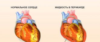
Pericardial puncture For diagnostic or therapeutic purposes, mainly for effusion pericarditis. De Larre technique. Using a thin trocar or thick needle on the left at the level of attachment of the VII rib to the sternum, a puncture of the pericardium is made at an angle of 45 to the surface of the body to a depth of 11.5 cm.
Then the needle is tilted downwards, positioned almost parallel to the sternum, and advanced into the anterior inferior part of the pericardial cavity. The sensation of pulsation indicates the proximity of the needle tip to the heart. Exudate is removed from the pericardial cavity using a syringe.
Opening the pericardium (pericardiotomy) Indication. Purulent pericarditis. Technique. The operation is performed from a lateral or anterolateral approach into the 4th intercostal space. The lung is retracted dorsally, and the pericardium is usually opened dorsal to and parallel to the phrenic nerve. The second incision is perpendicular to the first along the projection of the atrioventricular groove - provides drainage of the pericardial cavity in the postoperative period.
The pericardium is opened with longitudinal and perpendicular to the phrenic nerve incisions. An intraoperative examination of the heart is performed. With severe stenosis of the left atrioventricular valve, there is an enlarged left atrium with an enlarged appendage.
Ligation of the patent ductus arteriosus 2 types of interventions: 1) Crossing and suturing the duct 2) Inserting a plug blocking the duct into the duct The operation is indicated at the age of 3 to 15 years
Operations for coarctation of the aorta Indication: circulatory failure Technique. Lateral or posterolateral thoracotomy along the 4th intercostal space. The mediastinal pleura is incised, the aorta is mobilized above and below the coarctation and clamped with Craford clamps.
The most physiological method is resection of the narrowed area with the formation of an end-to-end anastomosis of the aorta.
When the aorta is narrowed over a large area and aneurysms are present, it is necessary to resort to aortic replacement with synthetic grafts.
Complications. The most severe complication during correction of aortic coarctation is bleeding. To prevent injuries and ruptures of blood vessels during aortic cross-clamping, controlled hypotension is used.
Src="https://present5.com/presentation/147079386_437011314/image-27.jpg" alt="Surgical treatment of coronary artery disease Atherosclerotic lesion of the coronary arteries -> coronary insufficiency "> Surgical treatment of coronary artery disease Atherosclerotic lesion of the coronary arteries -> coronary insufficiency
Implantation of the internal mammary artery into the myocardium (Weinberg operation) Creation of an anastomosis between the internal mammary artery and the anterior interventricular branch of the left coronary artery.
Src=»https://present5.com/presentation/147079386_437011314/image-29.jpg» alt=»Fieschi Operation Ligation of the internal thoracic arteries on both sides -> increased blood flow along»> Fieschi Operation Ligation of the internal thoracic arteries on both sides - > increased blood flow through the pericardial phrenic artery. The operation gives a satisfactory effect, but cannot stop the progressive course of coronary atherosclerosis. With multiple lesions of the coronary arteries, complete revascularization of the myocardium using the internal mammary artery is not always possible.
Coronary artery bypass grafting Technique. Longitudinal sternotomy (soft tissues are cut layer by layer along the midline along the entire sternum) with a sternotomy. The edges of the sternum are spread apart with a dilator.
In most cases, segments of the great saphenous vein of the thigh are used as a vascular graft.
After CABG using one's own vein within 3 to 5 years, thrombosis or occlusion of shunts is observed in 28-35% of cases, which leads to persistent relapse of angina.
Performing such an operation is difficult in patients with venous disease of the lower extremities (varicose veins, thrombophlebitis)
It is performed for multi-vessel lesions of the coronary arteries - 3 or more - proximal coronary arteries, and for trunk stenosis of the left artery. Up to 4 coronary arteries can be bypassed at the same time. The results of CABG are determined by: 1) the patency of the shunts, 2) the condition of the peripheral bed of the coronary artery in the recipient
The esophagus (oesophagus) is a hollow tubular organ. The length of an adult is 25 -27 cm. Pars cervicalis. 5 -7 cm. Pars thoracica. 16 -18 cm. Pars abdominalis. 1.5 -4 cm.
Skeletotopy of the esophagus. It begins at the level of the VI cervical vertebra and flows into the stomach at the level of the XII thoracic vertebra.
Surgical approaches to the esophagus Cervical access v Collar-shaped incision on the anterior surface of the neck v Oblique Razumovsky incision along the anterior edge of the left sternocleidomastoid muscle Indications. Removal of foreign bodies, pharyngoesophageal diverticula, paraesophageal abscesses and upper sternal mediastinitis.
Technique. The patient is placed on his back with a bolster placed under his shoulders, his head is thrown back and turned to the right. A skin incision is made along the anterior edge of the sternocleidomastoid muscle from the sternal notch to the level of the upper edge of the thyroid cartilage. The skin, subcutaneous tissue, superficial fascia of the neck, subcutaneous muscle of the neck are dissected in layers, the sheath of the sternocleidomastoid muscle is opened near the anterior edge, the muscle is pulled laterally with a Farabeuf hook. The internal plate of the sternocleidomastoid muscle and the scapuloclavicular fascia of the neck are dissected.
The left lobe of the thyroid gland, together with the trachea and muscles (sternohyoid, sternothyroid), is pulled back and pushed upward and to the right. The inferior thyroid artery is ligated, the scapular-hyoid muscle is pushed upward (if the neck is short, it is crossed). The leaf of the intracervical fascia is bluntly separated, exposing the tissue of the esophageal-tracheal groove, where the left recurrent laryngeal nerve passes. The wall of the esophagus is determined by its red color and longitudinal striations.
Transpleural access To access the thoracic region, a transpleural anterolateral right thoracotomy is used, since the arch and descending thoracic aorta are located on the left. Indications: bifurcation diverticula of the esophagus, mediastinitis.
Technique. The patient is placed on his left side with a small bolster with his right arm raised upward. An incision is made in the 5th or 6th intercostal space. The skin and subcutaneous tissue are dissected from the edge of the sternum to the scapular line. In women, an incision is made under the mammary gland along the lower fold.
The skin, superficial fascia, pectoralis major muscle, serratus anterior muscle, partly the latissimus dorsi muscle, intercostal muscles (along the upper edge of the underlying rib) are dissected, the pleura is opened, the lung is retracted anteriorly and medially, and the mediastinal pleura is dissected. The esophagus is isolated from the mediastinal tissue with a gauze strip that serves as a holder.
After the manipulations, rare interrupted catgut sutures are applied to the mediastinal pleura. A rubber drain is inserted into the 8th or 9th intercostal space along the posterior axillary line and the chest wall wound is sutured. To create a seal, catgut sutures are applied in layers to the muscles, fascia, subcutaneous fatty tissue and silk sutures to the skin.
Posterior extrapleural mediastinotomy according to Nasilov for access to the mid-thoracic esophagus. Technique. At the level of the abscess, a vertical incision 15-20 cm long is made along the outer edge of the long back muscles on the right. After this, the muscles are retracted to the spine, 2-3 ribs are resected, and the intercostal arteries are ligated. They simply peel off the pleura, approach the esophagus, and open the abscess. The abscess cavity is drained and a gastrostomy tube is applied.
Transabdominal access Indication. Perforation of the esophagus below the level of the bifurcation of the trachea, supradiaphragmatic, abdominal sections.
Technique. Position the patient on his back with a cushion placed at the level of the shoulder blades. An upper midline laparotomy is performed. After revision of the abdominal cavity, the hepatophrenic, esophageal-splenic, and gastroesophageal ligaments are dissected.
The esophagus is bypassed with the index finger and taken on a holder. A sagittal diaphragmotomy is performed, crossing the tendon part of the diaphragm anteriorly for 6 cm.
Before dissecting the diaphragm, it should be carefully separated from the pericardium to avoid damage to it. After this, the esophagus is stretched using a holder. A gastrostomy tube is inserted through a separate incision on the left.
Bougienage of the esophagus Cicatricial stenosis of the esophagus occurs after 1-2 months. If early bougienage was carried out, then the formation of stenosis occurs in only 4.2% of patients, without prophylactic bougienage - in almost 50%. Early bougienage begins 5-10 days (up to the 14th day) after the burn. The bougie inserted into the narrowing is left in the esophagus for 15-20 minutes, and if there is a tendency to narrowing - up to 1 hour. The next day, a bougie of the same diameter is introduced for a short time, followed by a bougie of the next number, leaving it in the esophagus for the required time . If a painful reaction occurs, signs of malaise, or an increase in body temperature, bougienage is postponed for several days. Previously, bougienage was carried out daily or every other day for a month, even in the absence of signs of narrowing of the esophagus, and then for 2 months 1-2 times a week
Early bougienage in children is aimed at preventing the development of narrowing of the lumen of the esophagus in the phase of reparative processes of scarring of its affected wall. According to the author, bougienage started in the first 3-8 days after a burn is not dangerous for the victim, since morphological changes during this period apply only to the mucous and submucosal layers, and therefore the danger of perforation is minimal. Indications for early bougienage are normal body temperature for 2-3 days and the disappearance of acute phenomena of general intoxication. Later than the 15th day from the moment of the burn, bougienage becomes dangerous for both the child and the adult, so the phase of scarring of the esophagus begins, it becomes rigid and less pliable, and the wall has not yet acquired sufficient strength.
Therapeutic bougienage should be used no earlier than 7 weeks after the burn, since when bougienage of the esophagus occurs within 2 to 6 weeks after the burn, the bougie destroys granulations and fresh connective tissue. During this period, perforation of the esophagus is most often noted.
During palpation of the bougie, the patient sits with his head slightly tilted forward. The doctor uses his left index finger to press the root of the tongue forward and down, and with his right hand, strictly adhering to the midline, inserts the bougie into the lower part of the pharynx, and then into the esophagus (Fig. 289). At the same time, at some point the doctor senses an obstacle corresponding to the upper limit of the narrowing. A mark is made on the bougie about the distance to the beginning of the narrowing. If the bougie does not fit into the stricture, smaller bougies are introduced successively until one of them passes into the narrowing. A - correct head position; B - incorrect head position.
Bougienage through the mouth (blind). With a slight narrowing of the esophagus. In case of formed cicatricial narrowings, a bougie of increasing size (up to 3840) should be injected daily or every 2 days. Then bougie 2 times a week and 1 time a month for a year.
There are two main surgical approaches to perform heart surgery: extra-pleural and transpleural.
EXTRA PLEURAL ACCESS
Extrapleural access penetrates into the mediastinum through the interpleural field (area interpleurica).
This access is used primarily for adhesive and effusion pericarditis, as well as for operations on a “dry” heart with opening of its cavity.
Technique.
Extrapleural access to the heart and large blood vessels is provided by a longitudinal dissection of the sternum along its entire length according to
Milton
and a T-shaped incision according to
Lefort
(Fig.
10-48).
Rice. 10-47. Lobectomy (removal of the upper lobe of the left lung),
a — the upper lobe vein, as well as part of the upper lobe arteries, are ligated and crossed, the place of division of the upper lobe bronchus is visible, b — the upper lobe is removed, the stump of the upper lobe bronchus is sutured.
(From: Kupriyanov P.A., Grigoriev M.S., Kolesov A.P.
Operations on the breast organs. - P., 1960.)
774 ♦ TOPOGRAPHIC ANATOMY AND OPERATIVE SURGERY ♦ Chapter 10
Rice. 10-48. Access to the heart
a -
Milton's access,
b - Lefort's access
.
(From:
Shabanov A.N., Kushkhabiev V.I., Veli-Zade B.K.
Operative surgery (atlas). - M., 1977.)
TRANSPLEURAL ACCESS
With a transpleural approach to the mediastinal organs, one or both pleural cavities are opened using an anterolateral incision along the second, third or fourth intercostal space on the left, with the intersection of one or two costal cartilages. Transpleural incisions create more extensive access to all parts of the heart and large vessels.
Technique. The incision is made from the sternum to the anterior axillary line. Sometimes transpleural access is used with transverse dissection of the sternum and opening of the right and left pleural sacs (for example, during operations for extensive adhesive pericarditis).
MINIMALLY INVASIVE ACCESS TO THE HEART
Attempts to reduce surgical trauma, alleviate patient suffering, and improve cosmetic results of surgery have led to the development of a trend toward minimally invasive surgery in cardiac surgery.
When considering various aspects of minimally invasive surgery of acquired heart defects, two main directions are distinguished:
Technique using video thoracoscopic
peak technology;
Open operations performed from minimal access.
Technique. Access to the heart is carried out by means of a median mini-sternotomy, when a skin incision 10 cm long is made, 2 cm from the jugular notch. The sternum is cut
along the midline from top to bottom using a power saw to the level of the third or fourth rib, then obliquely to the right to the level of the fourth intercostal space.
The mitral valve is approached through a longitudinal biatrial approach with dissection of the upper wall of the left atrium, and the aortic valve is approached through a transverse aortotomy. After spreading the sternum using a wound retractor, the thymus gland is resected and the pericardium is opened longitudinally. The wound exposes the base of the heart and the right atrium. At the end of the operation, the pericardial cavity and mediastinum are drained. The operation is completed by restoring the integrity of the sternum. Cosmetic stitches are applied to the skin.
Questions about surgical approaches are most controversial when studying the problems of minimally invasive surgery of acquired heart defects.
Doty method
D. Dots
in 1998, he proposed a technique that allows operations through a small incision to treat most acquired heart diseases.
Technique (Fig. 10-49, a). A vertical incision 10 cm long is made along the midline above the sternum, starting from the third intercostal space downwards. The sternum is dissected perpendicularly in the third intercostal space and then vertically along the midline from this point down through the xiphoid process. The upper half of the sternum remains intact. Using a special retractor, this part is raised by 2.5 cm, which significantly improves visibility. Cannulation and cross-clamping of the aorta can be performed either through the surgical field or through separate incisions, which significantly improves visualization. The incision can be easily converted into a complete sternotomy, while this is difficult with paramedian, transverse sternal and intercostal incisions.
Kasegawa method
Another option for accessing the mitral and aortic valves is the approach proposed by H. Kasegawa
and the “open door method” he called (Fig. 10-49, b).
A transverse sternotomy was made in the second intercostal space with an incision from the right border to
Rice. 10-49. Minimally invasive approaches to the heart.
A
- Dochi, b - Kasegawa.
pericardium at an angle of 45° to the surface of the body to a depth of 1-1.5 cm. Then the needle is tilted downwards, positioned almost parallel to the sternum, and advanced into the anterioinferior part of the pericardial cavity; the sensation of pulsation indicates the proximity of the needle tip to the heart. Exudate is removed from the pericardial cavity using a syringe.
1. Extrapleural access – is carried out by a longitudinal dissection of the sternum along its entire length and a T-shaped incision (for adhesive and effusion pericarditis, operations on a “dry” heart with opening of its cavity).
2. Transpleural access - carried out by an anterolateral incision along the second, third or fourth intercostal space on the left, with the intersection of one or two costal cartilages or a transverse dissection of the sternum with opening of the right and left pleural sacs (extensive access to all parts of the heart and large vessels).
Suturing a heart wound
1. operational access (usually along the wound channel);
2. longitudinal opening of the pericardium with a wide incision anterior to the phrenic nerve;
3. application of interrupted or U-shaped sutures to the wound;
4. freeing the pericardial cavity from blood clots;
5. suturing the pericardium with rare sutures.
Surgeries for heart defects
All operations used for damage to the valvular apparatus of the heart are divided into two groups: valve-sparing and valve-replacing.
The first group includes open commissurotomy and annuloplasty (restoration of the obturator function of the mitral valve using a rigid support ring). They are used for stenosis of the aortic and mitral valves.
The second group includes valve prosthetics (single-valve, multi-valve) - replacement of valves with mechanical or biological prostheses. It is used for insufficiency of the aortic and mitral valves.
Atrial septal defect
– suturing the defect in patients with pulmonary hypertension or plastic surgery of the septum with a patch from autopericardium (synthetic tissue) with a large diameter of the defect.
Ventricular septal defect:
1. radical surgery - closing the defect with separate sutures or a patch made of synthetic fabric or biological materials (for large defects);
2. palliative surgery - narrowing of the pulmonary artery with a cuff, as a result of which the discharge of blood through the defect is reduced and the volume of pulmonary blood flow and pressure in the pulmonary artery are reduced.
Patent ductus arteriosus
- X-ray endovascular occlusion (up to 80% of all operations); less often - thoracotomy and ligation of the duct with two ligatures.
Coarctation of the aorta
– resection of the aorta at the site of narrowing followed by prosthetics with synthetic grafts or creation of a bypass anastomosis of the subclavian artery with the aorta below the site of stenosis.
To touch, to touch, to feel, to feel, to feel, to take. See disturb, excite, excite, bully, touch, offend, embarrass, disturb, touch... don’t touch a finger... Dictionary of Russian synonyms and similar expressions. under... ... Dictionary of synonyms
reach the heart
- to take by the soul, to excite, to find a way to the heart, to grab the soul, to enter the soul, to find access to the heart, to touch, to enter the heart, to grab the heart, to excite, to take by the heart, to touch the living, to touch, to touch for the living,... ... Dictionary of synonyms
touch
- To pity, to move, to touch, to stir. Yes, as he says! It really takes away your soul. Turg. Whether a plowman sings a song in the distance, the long song touches the heart. Nekr. .. Wed ... Dictionary of synonyms
favor
- ▲ to attract favor to someone. dispose. cause favor. prepossessing (# appearance). predispose make a good impression. to be popular. | avant-garde. imposing. representative. find access... ... Ideographic Dictionary of the Russian Language
Pushkin, Alexander Sergeyevich
- - born on May 26, 1799 in Moscow, on Nemetskaya Street in Skvortsov’s house; died January 29, 1837 in St. Petersburg. On his father’s side, Pushkin belonged to an old noble family, descended, according to genealogies, from a person “from ... ...
Alexander II (part 2, XIII-XIX)
- XIII. Internal Affairs (1866-1871). On April 4, 1866, at four o'clock in the afternoon, Emperor Alexander, after a routine walk in the Summer Garden, was sitting in a carriage when an unknown person shot him with a pistol. At that moment, standing in... ... Big Biographical Encyclopedia
Alexander II (part 2, I-VII)
- PART TWO. Emperor Alexander II (1855-1881). I. War (1855). The highest manifesto announced to Russia the death of Emperor Nicholas and the accession of his successor. In this first act of his reign, the young Sovereign took before the face of... ... Great Biographical Encyclopedia
Battlefield 3
— Russian cover of the expanded edition of the game Developer ... Wikipedia
Myst III: Exile
— Myst 3: Exile Developer Presto Studios Publisher Ubisoft Localization ... Wikipedia
Batyushkov, Konstantin Nikolaevich
- genus. in Vologda May 18, 1787, d. there on July 7, 1855; came from an ancient noble family. His father, Nikolai Lvovich († 1817), even in his youth was involved in the investigation of the case of his uncle, Ilya Andreevich, who in 1770... ... Great Biographical Encyclopedia
Kochubey, Prince Viktor Pavlovich
- State Chancellor of Internal Affairs, b. November 11, 1768, d. in Moscow, on the night of June 2-3, 1834. He was the son of Pavel Vasilyevich Kochubey and the great-grandson of the famous Vasily Leontyevich Kochubey; his mother, Ulyana Andreevna, was his own... ... Large biographical encyclopedia
Surgical intervention is required in cases of diseases of the cardiovascular system quite often. This happens in cases where conservative treatment becomes ineffective and the disease progresses, or when the patient seeks help too late, when only cardiovascular surgery can help him. Diseases that often require cardiovascular surgery include coronary heart disease, myocardial infarction and atherosclerosis.
Types of heart surgery
Open heart surgery. Open heart surgery is any type of surgery in which the surgeon opens the breastbone. Although they say “open heart surgery,” most often the breastbone is opened, but not the heart. But depending on the type of operation, the surgeon may also open the heart. Coronary artery bypass surgery, heart valve replacement, and heart transplantation are all open-heart surgeries. Operations on the beating heart. Surgeons can perform coronary artery bypass surgery on a beating heart. This method is similar to open heart surgery in that the sternum is also opened to access the heart. However, the heart is not stopped and a heart-lung machine is not used. Minimally invasive cardiac surgery. During minimally invasive surgery, small incisions are made between the ribs. Minimally invasive surgeries are needed for some types of bypass and labyrinth-type surgeries.
Is it possible to do without surgery to remove a cardiac aneurysm?
There is no medical treatment for cardiac aneurysm. It is possible that this formation may be accidentally detected in patients without symptoms of cardiac pathology. This type of aneurysm without signs of disease and threat of rupture can be considered an indication for dynamic monitoring of the patient.
You need to understand that the presence of any type of protrusion of the heart wall poses a potential threat to life, so in most cases its removal is recommended.
Indications for mandatory surgery are:
- large saccular and mushroom-shaped formations, “aneurysm within an aneurysm”;
- threat of rupture for any type;
- the danger of thromboembolism due to a loose blood clot in the cavity;
- progressive increase in size;
- arrhythmia and/or heart failure that cannot be treated with medications;
- the presence of reduced contractile function of the left ventricle or immobile areas with an increase in its volume;
- false and congenital forms.
We recommend reading the article about aortic aneurysm. From it you will learn about the indications for surgery, types of surgical intervention, rehabilitation period, consequences and prognosis for patients. And here is more information about the aneurysm of the sinus of Valsalva.
Operative access to the heart
The main requirements when choosing an operative approach are anatomical accessibility and the technical ability to carry out all stages of the operation through the access. a) Anterolateral thoracotomy – an incision is made from the cartilage of the 3rd rib, slightly departing from the parasternal line, carried down to the lower edge of the 4th rib and, bordering the nipple, continued along the fourth intercostal space to the posterior axillary line (if necessary, expand access to the pleural cavity or mediastinum resort to intersection of the third or fourth costal cartilages). b, d) Transverse sternotomy - incision at the level of the fourth intercostal space. c) Longitudinal sternotomy - an incision along the sternum 2-3 cm above its manubrium and continuing 3-4 cm below the xiphoid process. Lateral thoracotomy - the chest cavity is opened along the V-VI ribs from the paravertebral to the midclavicular line. Posterolateral thoracotomy - an incision from the spinous processes of the III-IV thoracic vertebrae, going around the angle of the scapula from below, continues along the VI intercostal space to the anterior axillary line (to expand surgical access, resection of the neck of two adjacent ribs is used)
Heart removal
After completing the “Meeting with the Family” task, we will receive this task, or rather the opportunity to infiltrate the kett base. Having gone to the indicated coordinates, we will find ourselves in a cave leading to the enemy base; by hacking the shield code through the console, we get inside, continuing to move through the cave and destroying enemies. Having climbed to the surface, we climb up the path to the buildings of the kett base. Having entered the premises of the base, we bypass the security system by turning off three Kett generators. After which you will need to destroy the kett security terminal, thus opening the way to the base core in the central tower. As a result, having hacked the next panel, we jump into the opened hatch and clear the next room from the kett. Continuing to move down the corridor we will find ourselves in the hangar, where we will need to destroy three landing platforms. Once on one of the platforms, we find out that the controls are blocked and to bypass the blocking you need to find and activate the terminal. SAM will find several additional terminals in the hangar, our task is to find the one we need while fighting off the Kett attacks. After you destroy all the terminals, all that remains is to eliminate the arriving Prefect and the remaining kett defenders, as well as get to the top of the tower in the Zenith observation room. As a result, having found ourselves in the right place, using the console, we neutralize the kett base and then create an outpost on Voeld in the marked place. And to complete the task “Removing the Heart” we return to Baksil.
Attack on the Kett base
To complete this small task, we scan the security matrix for vulnerabilities. After which we arrange sabotage and gain access to a safe storage facility from which we take out everything that is not nailed down (do not forget to inspect both the upper and lower rooms).
In the dark
After completing the “Removing the Heart” task, the force field covering the excavation site will be turned off and we will be able to check the damaged signal. Having penetrated under the disconnected dome, we descend into the ice cave, where SAM will find a communication device nearby. Our task is to find a device that will be located on a corpse lying in a small dead end on the right side of the cave. After checking the data block, we return to senior intelligence officer Kaas.
Searching for the past
Having descended deeper into the ice cave, we destroy all the kett we meet along the way, after which we will need to free the captured hangars and talk with one of the liberated ones. Next, going to the partition, we scan it and then move away to a safe distance to blow up the wall. After the wall is destroyed, we go into the hall, where we will find a huge computer with ancient AI. After talking with the computer, you will need to decide to let this AI live or destroy it, thus saving one of the liberated hangars.
Missing scientists
We go to the specified coordinates and scan the found debris and bodies, you will also need to read the audio recording. Having finished at the crash site, we set off along the route planned by scientists and, having reached the indicated point, we will find the Architect of Relics. To defeat the Architect, wait until he falls to the ground, then, taking up defense in one of the buildings nearby, we shoot at the enemy’s legs and in his mouth (depending on what is indicated as the target). Also, in the battle with the Architect of Relics, you will periodically need to change your position, moving to other buildings and shooting small Relics. As a result, having damaged three limbs and shot the mouth of a giant robot (destroying the contours of the head and legs), we approach the defeated enemy and connect to him, after which we talk with Priya Blake.
Restoring the world
To complete this task, we will need to activate three monoliths located on this planet. Having driven to the indicated place, we scan three glyphs, after which we approach the relic console and activate the monolith by placing the missing glyphs, which should not be repeated in rows of columns and selected forms, as shown in the screenshots. Next, we repeat the procedure on two more monoliths, having previously scanned three glyphs on each. After successfully activating the three monoliths, we go to the entrance to the vaults, where we go down the gravity well. Having gone downstairs, we turn on the emergency generator, to do this we activate the console and then remove the lock from the storage, moving from one thermal generator to another so as not to freeze. Having killed all the relics along the way and eventually reached the desired place, we activate the next console, after which we leave the purification zone and exit the storage facility, thus improving the living conditions on another planet.
To be continued…
Minimally invasive approaches to the heart
Attempts to reduce surgical trauma, alleviate patient suffering, and improve cosmetic results of surgery have led to the development of a trend towards minimally invasiveness in cardiac surgery. When considering various aspects of minimally invasive surgery of acquired heart defects, two main directions are distinguished: a technique using video thoracoscopic technology; open operations performed from minimal access. Technique. Access to the heart is carried out by median ministernotomy, when a skin incision 10 cm long is made, 2 cm from the jugular notch. The sternum is dissected along the midline from top to bottom using a power saw to the level of the third or fourth rib, then obliquely to the right to the level of the fourth intercostal space. The mitral valve is approached through a longitudinal biatrial approach with dissection of the upper wall of the left atrium, and the aortic valve is approached through a transverse aortotomy. After spreading the sternum with a retractor, the thymus gland is resected and the pericardium is opened longitudinally. The wound exposes the base of the heart and the right atrium. At the end of the operation, the pericardial cavity and mediastinum are drained. The operation is completed by restoring the integrity of the sternum. Cosmetic stitches are applied to the skin.
How cardiac surgeons save our lives by transplanting hearts
I will tell you how a heart transplant is done in a hospital, away from the worries, adventures and trials that befall donor teams.
First of all, we cut the sternum with a circular saw. After this, the usual steps of almost all open heart surgery are performed. To connect the patient to the heart-lung machine, we insert a tube into the aorta, and two more into the large veins through which venous blood enters the right atrium.
During other operations, we insert just one tube directly into the right atrium. We then wrap the two tubes tightly with bandages to securely separate the veins.
The point of this modification is to ensure that when the right atrium is cut off, air does not enter the tubes and block blood flow through the lines of the artificial circulation machine. Once connected, we turn on the machine and clamp the aorta, isolating the heart from the circulatory system.
In this situation, naturally, there is no point in protecting the heart with a cold solution of potassium chloride, because the patient will no longer need it. We then dissect the organ through four chambers - two atria and two great vessels leaving the heart (aorta and pulmonary artery).
While we do this, the heart slowly dies from lack of oxygen.
The saddest thing about transplantation is watching how the doomed organ continues to stubbornly, gradually weakening, shrink while we cut it out. It beats for several minutes after it is removed from the chest and placed on a table with instruments. And then it still stops.
After extraction, a terrifying emptiness gapes in the chest between the lungs - in the very place where the heart was previously located. There are only three tubes left.
If everything goes according to plan, then immediately after removing the unnecessary organ, the donor organ is brought into the operating room in a large container with ice. There is a bag of cold saline solution there. The heart itself is in a separate bag, immersed in a solution.
All it takes is four rows of stitches to place the new heart where it needs to be. None of them require special delicacy: the left atrium of the donor heart is sutured to the stump of the left atrium of the old heart, the right atrium is sutured similarly, the pulmonary artery of the new heart is sutured to the stump of the recipient's pulmonary artery, and the same is done with the aorta.
All these sutures must be sealed, like other vascular anastomoses (connections), cardiac surgeons usually do a good job of this task. True, suturing takes a long time - and suturing the right atrium is especially labor-intensive. When all the sutures are ready, all the air is washed out of the new heart and the clamp is removed from the aorta.
Once the clamp is removed, blood flow begins in the coronary arteries, and two events occur.
The first seems like a miracle: after removing the donor from the chest and several hours of travel, the heart begins to contract in its new home. The second event cannot be called so joyful: the recipient’s blood cells instantly recognize the donor heart as foreign, and the immune system begins to methodically destroy this new heart.
To prevent this from happening, the patient is given immunosuppressants - they enable the donor heart and the recipient’s cells to “get to know” each other better. After this, the heart-lung machine is turned off, the chest is closed and stitches are placed on the wound. The operation is over.
For the next few days, the patient balances on a knife's edge - insufficient immune suppression can lead to a rejection reaction and failure of the new heart, and too much can make the patient a victim of infection.
Having many years of experience in transplantation, the professionals working in the hospital help the patient walk safely along the knife's edge. They adjust the dosage of immunosuppressants so that rejection does not occur and the patient does not die from a minor infection.
It is a tradition at Papworth that when a patient can walk around the hospital's duck pond on his own, he can be discharged.
Now let's look back to fully experience the magic of a heart transplant. Cardiac surgery was born in the 1950s, when the heart-lung machine was invented, and since then it has continuously advanced by leaps and bounds. It continues to develop today.
Over the past 50 years, the list of heart diseases that can be cured with a scalpel has expanded incredibly. Sometimes we heart surgeons even act as if there are no heart diseases that we cannot cure.
We place bypasses on blocked coronary arteries, can replace or reconstruct all four heart valves, and are ready to correct any abnormalities in the structure of the aorta. We close any holes in the heart that appear where they should not be, and create holes where they belong, but for some reason they are not there.
Our inventive surgical solutions can correct congenital heart defects, including those that are hard to imagine, such as a congenital absence of a ventricle and a lack of normal communication between arteries or veins.
Not long ago, a surgical method was developed to correct heart rhythm disorders such as atrial fibrillation, when the normal propagation of an electrical impulse is disrupted and the heart begins to beat chaotically. Contractions become less effective and the risk of stroke increases.
Now this problem can be easily solved surgically - that is, we are now not only plumbers, but also electricians.
Unfortunately, there remains one more problem that stubbornly defies the efforts of cardiac surgeons - heart failure. Of course, if it is caused by coronary heart disease or valve disease, then we can eliminate it, because the pump itself works normally.
But there is no such operation with which it would be possible to force a weakened heart to contract more strongly and better pump blood throughout the body. Failure of the heart's pumping function is very bad news for the patient: 30–40% of patients die within a year of diagnosis. The mortality rate is even higher than for cancer.
Why does heart failure develop? There are many reasons, and the first is coronary heart disease. When a coronary artery is completely blocked, a small portion of the heart muscle dies (this death is called a myocardial infarction).
Each time this happens, the heart loses more and more of its pumping ability, until it becomes so weak that it can no longer pump blood around the body properly. We can place a bypass on a blocked coronary artery to prevent this from happening, but a bypass will not bring the dead myocardium back to life.
Heart failure may have another cause: stenosis (narrowing) or insufficiency (incomplete closure of the valves). These abnormalities force the heart to work harder to maintain normal blood flow while counteracting the valve's narrowing or backflow of blood into it. The heart has to work in this mode constantly.
The load can be the same as when running a marathon or climbing mountains, although in fact the person is sleeping or sitting quietly in a chair at this time.
The heart can withstand such stress for a short time, such as during a marathon, but cannot do so for weeks, months and years. Sooner or later, if the valve defect is not corrected, the pump will fail, and heart failure will become irreversible.
Other causes are possible, although they are less common. These could be, for example, viral infections or autoimmune diseases, when a person’s immune system mistakenly attacks his own heart.
Whatever the reason, when the pump itself breaks down, cardiac surgeons are powerless, and the prognosis is the saddest. Currently, we do not have any means to help the damaged heart muscle recover.
Intensive research is currently underway regarding the possibility of using stem cells for this purpose, but so far they have been inconclusive and there are no clinically suitable methods for treating heart failure. The only way to cure heart failure is a heart transplant.
But no matter how brilliant and exciting this method may be, it cannot be considered an ideal method of treatment for three main reasons.
We have already discussed the first reason: the new heart is foreign to the recipient’s immune system, and it will never take root unless the patient receives medications that suppress the immune system. When the immune system is suppressed, the dangers of infectious diseases and some rare forms of malignant tumors, especially blood and bone marrow, come to the fore.
The result is that a failing heart becomes a heart with transplant disease. This is better than insufficiency, but it is still a problem that will only get worse over the years.
The second reason is that the number of donor hearts is limited. Thus, transplantation is the lot of the lucky few, that is, not too old and not suffering from numerous concomitant diseases.
This is not ageism or discrimination, but simply an attempt to get the most out of a limited resource. But even with this limitation, there are still not enough donor hearts. This means that many patients who would really benefit from a transplant may not wait their turn and die earlier.
Heart failure is widespread and affects many people. Unfortunately, it cannot be expected that transplantation will somehow reduce the severity of the problem, because it is available only to a small number of patients. There is an aphorism that can be formulated in different ways, and I will give only one of the possible options:
A transplant is as much a cure for heart failure as the lottery is a cure for poverty.
Finally, the unpleasant truth about heart transplantation (and indeed all other organs) is that this treatment requires the death of a young and healthy person so that the old and sick one can live. Behind every triumph of transplantation there is certainly a human tragedy hidden.
However, there is hope on the horizon for a solution to this problem. If the heart can be called just a pump that must pump five liters of blood per minute, then making a spare one is just a matter of human ingenuity.
Work on its creation has been going on for many years, and the design of the devices is being improved every year. They can already maintain a patient’s relatively safe existence for weeks while he waits in line for a transplant.
Such devices have already become quite widely used in clinical practice. We call them “bridges to transplant,” and they have allowed many patients to wait safely for a donor heart.
Pumps that can be used for a lifetime were also invented, and many people are already using them. These devices are imperfect, their use is accompanied by damage to blood cells and require recharging all the time, but the most important thing is that they actually work. Improvement in design and performance is only a matter of time.
There will come a time when these pumps will be better than transplanted live hearts, and then they will definitely take pride of place in every cardiac surgery operating room.
When this happens, transplantation can be forgotten, because living hearts will be replaced by mechanical ones.
Instruments for surgery on the heart and great vessels:
18 — scarifier; 19 — flexible turnstile; 20 — forceps for grasping the ear of the heart; 21 — two-bar dilator 1 and 2 - wound dilators for the chest cavity (1 — large, 2 — medium); 3 - sternotome; 4 — blunt-ended curved scissors; 5-7 - valvulotomes (5 - with diamond-shaped knives, 6 - with petal knives, 7 - with two parallel knives); 8 and 9 — punchers (8 — semi-circular, 9 — circular); 10-clamp for the ear of the heart; 11 - needle clamp for wounds and cardiac aneurysms; 12 — clamps for partial lateral pressure of vessels (right and left); 13 — frame clamp for aortic and cardiac aneurysms; 14 and 15 — vascular dissector clamps (14 curved, 15 — with a straight handle); 16 — needle for cardiac puncture; 17 - vascular scissors (from left to right - curved, curved along the plane, curved along the edge, straight);.