The following stages of thrombus formation are distinguished:
Platelet agglutination. Adhesion of platelets to the damaged area of the vessel intima occurs due to platelet fibronectin and collagen types III and IV, which are part of the exposed basement membrane. This causes the binding of von Willebrand factor produced by endothelial cells, which promotes platelet aggregation and factor V. Destroyed platelets release thromboxane A2, which has a vasoconstrictor effect and helps slow blood flow and increase the aggregation of blood platelets, the release of serotonin, histamine and platelet-derived growth factor. Hageman factor (XII) and tissue activator (factor III, thromboplastin) are activated, triggering the coagulation cascade. Damaged endothelium activates proconvertin (factor VII). Prothrombin (factor II) is converted into thrombin (factor IIa), which causes the development of the next stage.
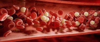
Fibrinogen coagulation. Further platelet degranulation and release of adenosine diphosphate and thromboxane A2 are noted. Fibrinogen is transformed into fibrin and an insoluble fibrin bundle is formed, capturing the formed elements and components of the blood plasma with the development of subsequent stages.
Agglutination of red blood cells.
Precipitation of plasma proteins.
Morphology of the thrombus.
White, red, mixed and hyaline thrombi are distinguished.
A white thrombus, consisting of platelets, fibrin and leukocytes, forms slowly, with rapid blood flow, usually in the arteries, between the trabeculae of the endocardium, on the leaflets of the heart valves in endocarditis. A red thrombus, which consists of platelets, fibrin and red blood cells, occurs quickly in vessels with slow blood flow, and therefore is usually found in the veins.
A mixed thrombus includes platelets, fibrin, erythrocytes, leukocytes and is found in any part of the bloodstream, including the cavities of the heart and aneurysms.
Hyaline thrombi are usually multiple and form only in the vessels of the microcirculatory bed during shock, burn disease, severe trauma, disseminated intravascular coagulation syndrome, and severe intoxication. They contain precipitated plasma proteins and agglutinated blood elements.
In relation to the lumen of the vessel, thrombi are divided into parietal and occlusive (usually red). In the first case, the tail of the thrombus grows against the blood flow, while in the second it can spread in any direction.
Depending on the characteristics of their occurrence, arrowroot blood clots are also isolated, usually mixed in composition, occurring during exhaustion, dehydration of the body, in the superficial veins of the lower extremities, sinuses of the dura mater; tumor thrombi formed when a malignant neoplasm grows into the lumen of a vein and grows there along the bloodstream or when a conglomerate of tumor cells clogs the lumen of microvessels; septic thrombi - infected mixed thrombi in the veins, developing with purulent vasculitis, sepsis.
A special type of thrombus is a spherical one, which is formed when the left atrium of a patient with mitral stenosis is torn off from the endocardium.
Other types
In addition to the main classifications of blood clots, there are several separate types that are specific to certain groups of patients. These varieties include the following types:
- Mirantic. It occurs in weakened elderly people with long-term dehydration. The thrombus is localized mainly in the superficial veins.
- Tumorous. Formed as a result of metastasis, that is, the formation of secondary foci of a malignant tumor. Often such a clot gradually grows towards the right lobes of the heart.
- Septic. Occurs as a result of a local inflammatory process caused by infection. Localized in the veins and on the valve flaps of the heart.
Thus, there are several types of blood clots, each of which will require a specific approach during treatment. The type of blood clot is determined by the doctor during diagnosis.
Thrombus in human vessels
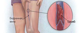
Thrombi are blood clots that appear intravitally as a result of blood thickening and platelet aggregation (sticking together). The reasons are activation of the blood coagulation system. The latter helps maintain homeostasis (constancy of the internal environment). Blood clots most often form in the veins, since blood flows slower there.
These formations may be a consequence of the body's defensive reaction. This occurs with injuries accompanied by bleeding. In this situation, clots prevent blood loss and infection. When the bleeding stops, the clot dissolves. Thrombosis is a pathological condition, as blood flow is disrupted.
The following types of blood clots are distinguished:
White ones are formed mainly in the arteries. They develop very slowly and often cause atherosclerosis. The composition of such blood clots includes leukocytes, fibrin and the blood cells themselves (platelets). Red clots are distinguished by the fact that they arise mainly in the veins.
They differ in the content of red blood cells. In humans, blood clots also form in the smallest vessels (capillaries). Blood clots are parietal and occlusive. In the latter case, blockage of blood vessels occurs.
Causes of blood clots
Among the causes of thrombosis are:
- Damage to the vascular wall;
- Changes in the functioning of the coagulation and anticoagulation systems;
- Changes in the nature and speed of blood flow.
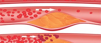
atherosclerosis is a process that promotes thrombus formation in the arteries
A healthy, smooth vascular wall is the key to good blood flow, however, with various damage to it, the coagulation system is activated and thrombosis occurs. On the one hand, this is a protective mechanism in case of injury, on the other hand, it is a pathological condition with various kinds of changes in the internal lining of the vessel. Thus, inflammatory processes (vasculitis) and the very common atherosclerosis are often the causes of pathological thrombus formation outside of traumatic injuries. Surgical interventions, infectious diseases, and malignant neoplasms are also accompanied by thrombosis.
A change in the coordinated functioning of the coagulation and anticoagulation systems provokes the activation of a number of enzymes and protein fractions, causes aggregation of formed elements, and the consequence is thrombosis in a variety of tissues and organs. Such conditions are often accompanied by autoimmune disorders, severe infections, tumors of the hematopoietic system, shock and even genetic defects.
A change in the nature of blood flow through a vessel affects the state of the inner lining (endothelium), which can be damaged, causing thrombosis. These phenomena can be observed especially clearly in the areas of branching of large vessels, where laminar blood flow is replaced by turbulent one, and blood under high pressure and at high speed seems to hit the vascular wall at the mouths of other vessels, damaging the endothelium (inner layer). If there are any changes in such areas (atherosclerosis, for example), then thrombus formation will occur more intensely.
To a large extent, the appearance of blood clots in the vessels is facilitated by a slowdown in blood flow and its stagnation, which can be observed with varicose veins of the legs (venous insufficiency), chronic heart failure, prolonged immobilization of the patient after surgical operations, and in bedridden patients.
Heart rhythm disturbances (atrial fibrillation, various types of blockades, etc.) lead to thrombosis not only of peripheral vessels, but also of the chambers of the heart. In addition, intracardiac thrombi often accompany valve lesions in rheumatic or atherosclerotic diseases; they form after implantation of artificial valves or other cardiac interventions. Often in such cases, a blood clot breaks away from the site of its formation and circulates with the blood, reaching other organs and causing dangerous consequences in them.
An increased tendency to thrombosis is found in pregnant women, as well as when taking contraceptives. This possibility must be taken into account, and a coagulogram will help to identify a blood clotting disorder in time.
Smoking, a sedentary lifestyle, the presence of cardiovascular pathology, autoimmune diseases, as well as hereditary predisposition are risk factors for possible thrombosis.
Venous thromboses occupy a special place during air travel, and according to some data, at least one passenger on each flight develops them during the journey. This is facilitated by pressure fluctuations, stagnation of blood in the vessels of the legs, and prolonged exposure to an uncomfortable position, so if you have varicose veins or heart problems, you need to be extremely careful when planning long trips by plane.
How blood clots form
The formation of a blood clot is a complex process. Physiological clots form as a result of damage to the vessel. At the same time, substances (thrombin and thromboplastin) are released into the bloodstream, which activate coagulation processes. This occurs as a result of the breakdown of platelets. The following phases of clot formation are distinguished:
Upon activation, the formation of prothrombinase occurs. With its help, the protein thrombin appears. Next comes the coagulation phase. Under the influence of the protein thrombin, fibrinogen is converted into fibrin. The latter is the basis of the resulting blood clot. A mesh is formed in the damaged area, which receives red blood cells, white blood cells and platelets.
This is how a dense fibrin clot is formed. This is the retraction phase. When hemodynamic parameters stabilize, the thrombus resolves. This is a normal blood clotting process in any person. The formation of blood clots, which cause blockage of blood vessels, is no different. Subsequently, such blood clots do not resolve on their own and cause thrombosis.
In the first days, the clots are still poorly fixed. They may break off and enter the main arteries or veins with the development of thromboembolism. This is an even more dangerous condition. The following factors play an important role in the development of thrombosis:
- decrease in circulating blood volume;
- increased blood viscosity;
- valve dysfunction;
- slowing blood flow;
- platelet tendency to aggregation;
- mechanical damage to the walls of blood vessels.
There are people whose blood clotting process is impaired as a result of a lack of special factors. They practically do not form blood clots, which is fraught with large blood loss at the slightest damage. An example would be hemophilia.
Parietal thrombi
They are located in the lumens of large veins and arteries, for example, in the chamber or on the valves of the heart, and are formed during inflammatory processes (thrombophlebitis, thromboendocarditis).
Wall thrombi are dangerous because over time they layer on top of each other, resulting in complete blockage of the vessel, which can lead to organ failure or even death.
However, the very location of the blood clot along the vessel wall does not block the blood flow, so it can be considered less dangerous. A type of wall thrombus can be considered an extended thrombus, which is attached to the wall along its entire length. It also interferes with blood flow, but does not fill the entire lumen of the vessel.
Clinical manifestations and causes of vascular blockage
You need to know not only the mechanism of blood clot formation, but also the reasons for blockage of blood vessels by blood clots. The veins are most often affected. Most often, this pathology is detected in adults who lead an unhealthy lifestyle and suffer from varicose veins. The following reasons for the development of venous thrombosis are distinguished:
- congenital anomalies;
- varicose veins;
- surgical interventions;
- severe dehydration of the body;
- hormonal disbalance;
- mechanical injuries (bruises, fractures);
- long-term compartment syndrome;
- DIC syndrome;
- septic conditions;
- paralysis;
- physical inactivity;
- bed rest.
Less common is arterial thrombosis. It develops during a stroke, against the background of atherosclerosis, atrial fibrillation, and during organ transplantation. Experienced doctors know not only what a blood clot is, but also the symptoms of vascular blockage. Most often, the process involves the superficial and deep veins of the lower extremities. With thrombosis of the leg veins, the following symptoms are observed:
- swelling;
- heaviness in the legs;
- convulsions;
- bursting pain;
- numbness;
- tingling;
- pale skin;
- fever (when combined with phlebitis).
Sometimes the place where blood clots form is in the veins of the upper extremities and eyes. In the latter case, vision loss may occur. The consequences of the formation of blood clots can be pulmonary embolism, stroke, acute cardiac ischemia, gangrene of the limb, atherosclerosis, and impaired organ function (kidneys, liver, lungs). The most dangerous is blockage of the deep veins.
Indications
Prevention of the development of pulmonary embolism is mainly carried out with anticoagulant drugs. Conservative therapy is used in patients with recurrent thrombosis. Although the use of a vena cava filter for thrombosis is a simple, minimally invasive surgical procedure, drug therapy is preferred. A vena cava filter for deep vein thrombosis is prescribed if conservative treatment is ineffective, strictly according to indications. It can also be prescribed if there are other factors in which the use of a filter for thrombosis is the most favorable solution to the problem.
The following indications for installing a vena cava filter are distinguished:
- deep vena cava thrombosis;
- extended floating thrombi inside the deep veins;
- thrombosis in heart failure;
- thrombosis in cancer;
- a history of pulmonary embolism;
- ineffectiveness of anticoagulant agents for thrombosis;
- contraindications to the use of anticoagulant agents;
- recurrence of thrombosis after conservative therapy;
- complications that arose during conservative therapy;
- surgical operations in patients prone to thromboembolism (in this case, a vena cava filter is installed temporarily).
A vena cava filter can be installed for life or temporarily. With temporary implantation, the device is removed on average six months after eliminating the pathology that can cause pulmonary embolism. The appointment of a temporary or lifelong vena cava filter is carried out by the attending physician, based on the individual characteristics of the patient. Cardiologists at the Yusupov Hospital perform all the necessary examinations to obtain the most accurate data on the patient’s condition, which is necessary for selecting adequate therapy. The patient is provided with the most effective treatment that will show maximum results for this pathology.
The vena cava filter also has a number of contraindications. It cannot be installed in patients with an excessively narrowed lumen of the vessel into which the device is planned to be implanted. Implantation of a vena cava filter is not performed in children with septic conditions when thrombosis has spread above the adrenal glands.
How to get rid of a blood clot
Experienced doctors know not only the stages of blood clot formation, but also methods of treating patients. Before this, an examination is required (Dopplerography, ultrasound, general tests, coagulogram, angiography, functional tests). To treat patients, drugs are used that thin the blood, dissolve formed blood clots and eliminate swelling and pain.
Direct and indirect anticoagulants will help against thrombosis.
These include Warfarin, Fragmin, Clexane, Heparin. Direct anticoagulants are administered intravenously or subcutaneously. The dose is determined by the attending physician. Thrombolytics (Streptokinase) are effective against blood clots. Drugs that increase circulating blood volume (Reopoliglucin) are often prescribed.
If pain appears at the stage of thrombosis, then anti-inflammatory drugs from the NSAID group (Ketoprofen, Diclofenac) are used. For superficial thrombosis, the treatment regimen includes various local agents (ointments, gels, solutions). Troxevasin gel has proven itself well. If thrombophlebitis develops, antipyretic drugs may be needed.
If there are symptoms of not only thrombosis, but also varicose veins, medications are prescribed that improve the condition of the vein wall. With the development of severe thrombophlebitis, the progression of a blood clot and the risk of its rupture, radical treatment (surgery) is required.
Diagnosis of pathology
To detect thrombosis of the lower extremities, the following diagnostic methods are used:
- Ultrasound combined with Doppler method. The procedure is also called duplex scanning.
- Scanning using a special component - fibrogen.
- Use of radionuclide venography. This method is prescribed by doctors if the patient is intolerant to various substances used in certain studies.
The doctor will usually perform a visual inspection of the affected area. As a result, the patient is diagnosed with a disease of the corresponding classification (left or right type). Based on this, the course of necessary treatment is selected.
Prevention methods
It is important to know not only the mechanisms of thrombus formation, but also preventive measures. To prevent the formation of blood clots, you must adhere to the following recommendations:
- rationally organize your daily routine;
- move more;
- spend less time in a static position (sitting, lying or standing);
- drink more fluids and wear compression stockings after surgery;
- monitor body weight;
- consult a doctor promptly and treat varicose veins, as well as hemorrhoids;
- stick to a diet;
- eliminate hard work;
- exercise;
- treat somatic diseases;
- take vitamins and minerals.
Hormonal disorders are a risk factor for the development of thrombosis, so you need to avoid hormonal medications. People who stand or sit in one place for a long time while working need to do a warm-up. If possible, then you just need to walk around. When working on a computer at home, you need to take breaks. During them, it is recommended to give your legs a horizontal or elevated position.
Females should avoid wearing high-heeled shoes. This is bad for the veins. Thrombosis is prevented by maintaining a healthy lifestyle. Smoking and alcoholism are risk factors for the development of this pathology. Vascular surgeons know how to prevent the formation of blood clots.
Phlebotonics (Venarus, Detralex, Phlebodia-600) are used for preventive purposes. They need to be taken for several months. The doctor should tell the patient how to avoid thrombosis. It is necessary to exclude all possible risk factors. Be sure to follow a drinking regime (at least 1.5-2 liters of water per day).
It is necessary to exclude possible injuries (fractures) and observe safety precautions. A risk factor for developing blood clots is obesity. In this regard, prevention includes weight normalization. Some foods are also useful (mussels, shrimp, sea fish, nuts, herbs, vegetables, cranberries, grains, green tea, blueberries, onions, garlic, ginger). They strengthen blood vessels and reduce blood viscosity. Thus, a blood clot poses a danger to any person.
Prevention measures
The following categories of patients are at risk:
- Having excess body weight.
- Those who have reached old age.
- Women during pregnancy and the postpartum period.
- All those who are forced to spend a long time either in a standing or sitting position.
Thrombotic changes require the use of preventive measures to prevent them. First of all, these include work to eliminate stagnant phenomena. In this case, they recommend:
- Use of elastic bandages and compression stockings.
- Engage in moderate physical activity. This will help normalize microcirculation in the vessels.
- Quitting bad habits (smoking, drinking alcohol).
- Normalization of the daily diet. It is necessary to include fresh herbs, vegetables, fruits, and grains.
- Sometimes blood thinners based on heparin or warfarin are prescribed for prevention purposes. They come in the form of tablets for oral use, as well as gel or ointment for external use.
If timely medical care is not provided, the pathology can develop into serious complications that can be life-threatening. To receive qualified advice, you should contact a phlebologist immediately at the first symptoms.
It is recommended to avoid self-medication , and as therapy progresses, strictly follow all instructions prescribed by the doctor.
Thrombus in human vessels
Thrombi are blood clots that appear intravitally as a result of blood thickening and platelet aggregation (sticking together). The reasons are activation of the blood coagulation system. The latter helps maintain homeostasis (constancy of the internal environment). Blood clots most often form in the veins, since blood flows slower there.
These formations may be a consequence of the body's defensive reaction. This occurs with injuries accompanied by bleeding. In this situation, clots prevent blood loss and infection. When the bleeding stops, the clot dissolves. Thrombosis is a pathological condition, as blood flow is disrupted.
The following types of blood clots are distinguished:
White ones are formed mainly in the arteries. They develop very slowly and often cause atherosclerosis. The composition of such blood clots includes leukocytes, fibrin and the blood cells themselves (platelets). Red clots are distinguished by the fact that they arise mainly in the veins.
They differ in the content of red blood cells. In humans, blood clots also form in the smallest vessels (capillaries). Blood clots are parietal and occlusive. In the latter case, blockage of blood vessels occurs.
Morphology and types of blood clots
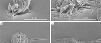
A thrombus is a blood clot attached to the wall of a blood vessel at the site of damage, usually of dense consistency, dry, easily crumbles, layered, with a corrugated or rough surface. It must be differentiated at autopsy from a post-mortem blood clot, which often follows the shape of the vessel, is not connected to its wall, is moist, elastic, homogeneous, with a smooth surface.
Depending on the structure and appearance, they are distinguished:
-white thrombus;
-red thrombus;
- mixed thrombus;
- hyaline thrombus.
A white thrombus consists of platelets, fibrin and leukocytes with a small amount of red blood cells; it forms slowly, often in the arterial bed, where there is a high blood flow rate.
The red thrombus is composed of platelets, fibrin and a large number of red blood cells, which fall into the fibrin network as if trapped. Red blood clots usually form in the venous system, where slow blood flow helps trap red blood cells.
A mixed thrombus is the most common, has a layered structure, and contains blood elements that are characteristic of both white and red thrombus. Layered thrombi form more often in the veins, in the cavity of the aortic aneurysm and heart. In a mixed thrombus there are:
- the head (has the structure of a white blood clot) - this is its widest part,
-body (actually mixed thrombus),
—tail (has the structure of a red blood clot).
The head is attached to an area of destroyed endothelium, which distinguishes a thrombus from a post-mortem blood clot.
Hyaline thrombus is a special type of blood clot. It consists of hemolyzed erythrocytes, platelets and precipitating plasma proteins and contains virtually no fibrin; the resulting masses resemble hyaline. These thrombi occur in the microvasculature. Sometimes blood clots are found that are composed almost entirely of platelets. They usually form in patients who are treated with heparin (its anticoagulant effect prevents fibrin formation).
In relation to the lumen of the vessel there are:
— parietal thrombus (most of the lumen is free);
- obstructing or clogging thrombus (the lumen of the vessel is almost completely closed).
Localization of blood clots
Arterial thrombosis:
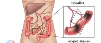
Thrombi in arteries are much less common than in veins, and usually form after damage to the endothelium and a local change in blood flow (turbulent blood flow), for example, with atherosclerosis. Among the arteries of large and medium caliber, the most commonly affected are the aorta, carotid arteries, arteries of the circle of Willis, coronary arteries of the heart, arteries of the intestines and extremities.
Less commonly, arterial thrombosis is a complication of arteritis, for example, with periarteritis nodosa, giant cell arteritis, thromboangitis obliterans and Henoch-Schönlein purpura and other rheumatic diseases. In hypertension, arteries of medium and small caliber are most often affected.
Cardiac thrombosis:
Blood clots form within the chambers of the heart under the following circumstances:
1. Inflammation of the heart valves leads to endothelial damage, local turbulent blood flow and deposition of platelets and fibrin on the valves. Small blood clots are called warts (rheumatism), large ones are called vegetations. Vegetations can be very large and loose, crumbling (for example, with infective endocarditis). Fragments of a blood clot often break off and are carried through the bloodstream in the form of emboli.
2. Damage to the parietal endocardium. Endocardial damage can occur during myocardial infarction and the formation of ventricular aneurysms. Blood clots that form on the chamber walls are often large and may also crumble to form emboli.
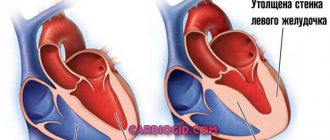
3. Turbulent blood flow and stasis in the atria. Thrombi often form in the atrial cavity when turbulent blood flow or blood stasis occurs, for example, with mitral stenosis and atrial fibrillation. The blood clots can be so large (ball-shaped) that they obstruct blood flow through the atrioventricular opening.
Venous thrombosis:
1. Thrombophlebitis.
With thrombophlebitis, venous thrombosis occurs secondary, as a result of acute inflammation of the veins. Thrombophlebitis is a common occurrence with infected wounds or ulcers; The superficial veins of the extremities are more often affected. The damaged vein has all the signs of acute inflammation (pain, redness, feeling of warmth, swelling). This type of blood clot tends to become firmly attached to the vessel wall. Emboli rarely form from it.
Sometimes thrombophlebitis develops in numerous superficial veins of the legs (migratory thrombophlebitis) in patients with malignant neoplasms, most often with cancer of the stomach and pancreas (Trousseau's sign), since mucins and other substances formed by tumor cells have thromboplastin-like activity.
2. Phlebothrombosis
is a vein thrombosis that occurs in the absence of obvious signs of inflammation. Phlebothrombosis is observed mainly in the deep veins of the legs (deep vein thrombosis). Less commonly, the veins of the pelvic venous plexus are affected. Deep vein thrombosis is quite common and is of medical importance because the large blood clots that form in these veins are quite loosely attached to the vessel wall and often break off easily. They migrate with the bloodstream to the heart and lungs and close the lumen of the pulmonary arteries (thromboembolism of the pulmonary trunk and its branches).
Causes of phlebothrombosis:
the factors causing deep vein phlebothrombosis are typical of thrombosis in general, but endothelial damage is usually subtle and difficult to detect. The most important causative factor in the occurrence of phlebothrombosis is decreased blood flow. In the venous plexus of the leg, blood flow is normally maintained by contraction of the leg muscles (muscle pump). The development of blood stasis and the development of thrombosis is facilitated by prolonged immobilization in bed and heart failure. The second factor - an increase in the adhesive and aggregation ability of platelets, as well as acceleration of blood clotting due to an increase in the level of some coagulation factors (fibrinogen, factors VII and VIII) - occurs in the postoperative and postpartum period, when using oral contraceptives, especially with high doses of estrogens , in cancer patients. Sometimes several factors can act together.
Clinical manifestations:
deep vein thrombosis of the legs can be mild or asymptomatic. When examining the patient, moderate swelling of the ankles and pain in the calf muscles when plantar flexing the foot are detected (Homan's symptom). In most patients, pulmonary embolism is the first clinical manifestation of phlebothrombosis. Deep vein thrombosis can be detected by phlebography, ultrasound, radiological methods, and comparative measurement of the legs with a centimeter tape.
Outcome of thrombosis
The formation of blood clots triggers a response from the body that aims to remove the clot and restore blood flow to the damaged blood vessel. There are several mechanisms for this:
Clot lysis (fibrinolysis),
leading to complete destruction of the blood clot is an ideal favorable outcome, but is very rare. The fibrin that makes up the clot is broken down by plasmin, which is activated by Hageman factor (factor XII) when the intrinsic coagulation cascade is activated (that is, the fibrinolytic system is activated simultaneously with the coagulation system; this mechanism prevents excessive thrombosis). Fibrinolysis prevents the formation of excess fibrin and the breakdown of small blood clots. Fibrinolysis is less effective at breaking up large blood clots found in arteries, veins, or the heart. Some substances, such as streptokinase and tissue plasminogen activator, which activate the fibrinolytic system, are effective inhibitors of thrombus formation when used immediately after thrombosis and cause thrombus lysis and restoration of blood flow. They are used with success in the treatment of acute myocardial infarction, deep vein thrombosis and acute peripheral arterial thrombosis.
Organization and recanalization
usually occur in large blood clots. Slow lysis and phagocytosis of the thrombus is accompanied by the proliferation of connective tissue and collagenization (organization). Cracks can form in the thrombus - vascular channels that are lined with endothelium (recanalization), due to which blood flow can be restored to some extent. Recanalization occurs slowly over several weeks and, although it does not prevent acute manifestations of thrombosis, it may slightly improve tissue perfusion in the long term.
Petrification of thrombus—
This is a relatively favorable outcome, which is characterized by the deposition of calcium salts in the thrombus. In the veins, this process is sometimes pronounced and leads to the formation of vein stones (phleboliths).
Septic thrombus breakdown
- an unfavorable outcome that occurs when a blood clot or vessel wall becomes infected.
Meaning
thrombosis is determined by the speed of development, localization, prevalence and its outcome.
In some cases, we can talk about the positive significance of thrombosis, for example, with an aortic aneurysm, when the organization of a blood clot leads to strengthening of the thinned vessel wall.
In most cases, thrombosis is a dangerous phenomenon. In arteries, occluding blood clots can cause heart attacks or gangrene. Mural thrombi in arteries are less dangerous, especially if they form slowly, since during this time collaterals can develop that will provide the necessary blood supply.
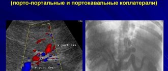
Obstructing blood clots in the veins cause local venous congestion and in the clinic give different manifestations depending on the location. For example, thrombosis of the dural sinuses leads to fatal cerebrovascular accident, portal vein thrombosis leads to portal hypertension, and splenic vein thrombosis leads to splenomegaly. With thrombosis of the renal veins, in some cases, either nephrotic syndrome or venous renal infarction develops, with thrombophlebitis of the hepatic veins - Chiari disease. The clinical significance of venous thrombi in the systemic circulation also lies in the fact that they serve as a source of pulmonary embolism and are thus fatal complications of many diseases.
Embolism
Embolism is the transfer of foreign particles by the bloodstream and their blockage of the lumen of the vessel. The particles themselves are called emboli. Most often, emboli are individual fragments of blood clots that are carried through the bloodstream (thromboembolism). Less commonly, embolic material is other substances (Table 7.2).
Depending on the direction of movement of the embolus, there are:
· ordinary (orthograde) embolism (
movement of the embolus through the bloodstream);
· retrograde embolism
(movement of the embolus against the blood flow under the influence of gravity);
· paradoxical embolism
(if there are defects in the interatrial or interventricular septum, the embolus from the veins of the systemic circle, bypassing the lungs, enters the arteries).
Pathogenesis of embolism.
It cannot be reduced only to mechanical closure of the lumen of the vessel. In the development of embolism, reflex spasm of both the main vascular line and its collaterals is of great importance, which causes severe discirculatory disorders. Arterial spasm can spread to the vessels of a paired or some other organ (for example, the reno-renal reflex during vascular embolism of one of the kidneys, the pulmonary coronary reflex during pulmonary embolism).
Localization of embolism
depends on the location and size of the embolus.
Formation of an embolus in the veins of the systemic circulation. Emboli that form in the veins of the systemic circulation (as a result of venous thrombosis) or in the right side of the heart (for example, in infective endocarditis of the tricuspid valve) clog the arteries of the pulmonary circulation, unless they are so small (for example, fat droplets, cells tumors) that can pass through the pulmonary capillary. The location of the blockage in the pulmonary vessels depends on the size of the embolus. Very rarely, an embolus that occurs in the veins of the systemic circulation can pass through a defect in the interatrial or interventricular septum (thus bypassing the pulmonary circle) and cause embolism in the arteries of the systemic circulation (paradoxical embolism).
Emboli that occur in the branches of the portal vein cause circulatory disorders in the liver.
Formation of an embolus in the heart and arteries of the systemic circulation: emboli occurring in the left half of the heart and arteries of the systemic circulation (as a result of thrombosis of the heart or arteries) cause embolism in the distal parts of the systemic circle, i.e. in the brain, heart, kidneys, limbs, intestines, etc.
Types and location of embolism (Table 7.2)
Thromboembolism:
separation of a fragment of a thrombus and its transfer by the bloodstream is the most common cause of embolism.
1. Pulmonary embolism (PE)
Causes and prevalence:
The most serious complication of thromboembolism is pulmonary embolism, which can cause sudden death. Approximately 600,000 patients per year experience pulmonary embolism in the United States; approximately 100,000 of them die. In more than 90% of cases, emboli occur in the deep veins of the legs (phlebothrombosis). More rarely, the source of blood clots is the pelvic venous plexus. Pulmonary embolism is most often observed in the following conditions that predispose to the occurrence of phlebothrombosis: 1) approximately 30-50% of patients after surgical interventions develop deep vein thrombosis in the early postoperative period. However, signs of pulmonary embolism occur in only a small proportion of these patients; 2) early postpartum period; 3) long-term immobilization in bed; 4) heart failure; 5) use of oral contraceptives.
Clinical manifestations and significance of pulmonary embolism:
The size of the embolus is the most significant factor determining the degree of clinical manifestations of pulmonary embolism and its significance.
1) massive emboli:
large emboli (several centimeters long and with a diameter similar to the femoral vein) can stop at the exit from the right ventricle or in the trunk of the pulmonary artery, where they create an obstruction of blood circulation and sudden death as a result of the pulmonary-coronary reflex. Obstruction by an embolus of large branches of the pulmonary artery can also cause sudden death as a result of severe vasoconstriction of all vessels of the pulmonary circulation, which occurs reflexively in response to the appearance of a thromboembolus in the vessel, or spasm of all bronchi.
2) medium-sized emboli:
in healthy people, the bronchial artery supplies blood to the lung parenchyma, and the function of the pulmonary artery is mainly gas exchange (not local tissue oxygenation). Therefore, a moderate-sized pulmonary embolus will result in an area of the lung that is ventilated but does not participate in gas exchange. This causes impaired gas exchange and hypoxemia, but pulmonary infarction does not always develop. More often, a heart attack occurs in patients with chronic left ventricular heart failure (against the background of chronic venous congestion) or with pulmonary vascular diseases, in whom the blood supply through the bronchial arteries is also impaired, as a result of which the lung receives oxygen and nutrients, mainly from the pulmonary vessels. In these patients, disruption of blood flow in the pulmonary artery leads to pulmonary infarction.
3) small emboli:
obstruct small branches of the pulmonary artery and can occur without clinical symptoms - this depends on the extent of the embolism. In most cases, emboli disintegrate under the influence of fibrinolysis. If numerous small emboli enter the pulmonary circulation for a long time, there is a risk of developing pulmonary hypertension.
2. Thromboembolism of vessels in the systemic circulation
Causes:
thromboembolism in the vessels of the systemic circulation occurs when an embolus forms in the left half of the heart or a large-caliber artery. Thromboembolism of vessels in the systemic circulation usually occurs:
– in patients suffering from infective endocarditis with thrombotic lesions on the mitral and aortic valves;
– in patients who have suffered a left ventricular myocardial infarction with parietal thrombosis;
– in patients with rheumatism and coronary artery disease with severe cardiac arrhythmias (atrial fibrillation, atrial fibrillation), which leads to the formation of a blood clot in the heart cavity, most often in the left atrium;
– in patients with aortic and left ventricular aneurysms, in which parietal thrombi often form. Thromboemboli from any of these locations are transferred to the arteries of various organs. Due to the anatomy of the aorta, cardiac emboli tend to penetrate more often into the lower extremities or into the bed of the right internal carotid artery than into other arteries of the systemic circle.
Clinical manifestations and significance of thromboembolism of the systemic circulation
determined by the size of the affected vessel, the development of collateral circulation and the sensitivity of the tissue to ischemia. Infarctions of the brain, heart, kidneys and spleen may occur. Infarction in the intestines and lower extremities develops only when large arteries are occluded or when the collateral circulation is damaged.
3. Air embolism:
An air embolism occurs when enough air (approximately 150 ml) enters the bloodstream.
Causes:
Surgery or injury to the internal jugular vein.
When the internal jugular vein is damaged, negative pressure in the chest leads to air being sucked into it. This does not happen with injuries to other veins because they are separated by valves from the negative pressure in the chest cavity.
Childbirth and abortion.
Very rarely, air embolism can occur during childbirth or abortion, when air can be forced into ruptured placental venous sinuses during uterine contractions.
Embolism during blood transfusion, intravenous infusions (droppers), X-ray contrast angiographic studies.
Air embolism occurs only when the manipulation technique is violated.
In case of inadequate mechanical ventilation under conditions of hyperbaric oxygenation.
Clinical manifestations.
When air enters the bloodstream, it passes through the right ventricle, where a foamy mixture arises, which greatly impedes the blood flow; closing 2/3 of the capillaries of the lungs with air causes death.
4. Nitrogen gas embolism (decompression syndrome)
Causes.
Decompression syndrome is observed in divers during rapid ascent from great depths, in pilots and astronauts when the cabin is depressurized. By inhaling air at high underwater pressure, an increased volume of air, mainly oxygen and nitrogen, dissolves in the blood and accordingly penetrates the tissues.
With rapid decompression, the gases that are in the tissues pass from a dissolved state to a gaseous state. Oxygen is quickly absorbed by the blood, but nitrogen cannot be absorbed quickly and forms bubbles in the tissues and blood that act as emboli.
Clinical manifestations and significance.
Platelets adhere to nitrogen bubbles in the bloodstream and activate the blood clotting mechanism. The resulting disseminated intravascular thrombosis worsens the ischemic condition of tissues caused by blockage of capillaries by gas bubbles. In severe cases, necrosis of brain tissue occurs as nitrogen dissolves in lipid-rich tissues, leading to death. In less severe cases, the muscles and nerves that innervate them are primarily affected; this causes severe muscle spasms with intense pain. Nitrogen gas embolism in the lungs causes respiratory failure and is accompanied by alveolar edema and hemorrhage.
5. Fat embolism
Causes.
A fat embolism occurs when fat droplets enter the bloodstream. When large bones (such as the femur) are broken, particles of yellow bone marrow enter the bloodstream. Rarely, extensive damage to subcutaneous fat results in fat embolism. Although fat droplets are detected in the bloodstream in 90% of patients with severe fractures, clinical signs of fat embolism are much less common.
Although the mechanism of fat droplets entering the bloodstream when fat cells rupture seems simple, there are several other mechanisms that influence the clinical manifestations of fat embolism. It turned out that fat droplets in the bloodstream can increase in size. This explains the fact that small particles of fat, passing freely through the pulmonary capillaries, can then cause embolism in the capillaries of the systemic circulation. It is assumed that the release of catecholamines as a result of injury leads to the mobilization of free fatty acids, due to which a progressive increase in fat droplets occurs. Adhesion of platelets to fatty particles leads to their further increase in size, which also leads to thrombosis. When this process occurs in a generalized manner, it is equivalent to disseminated intravascular coagulation syndrome.
Clinical manifestations and significance.
Circulating fat droplets initially enter the capillary network of the lungs. Large fatty particles (> 20 µm) remain in the lungs and cause respiratory failure (dyspnea and impaired gas exchange). Smaller fat globules pass through the capillaries of the lungs and enter the systemic circulation. Typical clinical manifestations of fat embolism: the appearance of a hemorrhagic rash on the skin and the occurrence of acute disseminated neurological disorders.
The possibility of developing fat embolism should be taken into account when respiratory disorders, brain disorders and hemorrhagic rash appear 1-3 days after injury. The diagnosis can be confirmed by detecting fatty droplets in the urine and sputum. Approximately 10% of patients with clinical signs of fat embolism die. During autopsy, fat drops can be found in many organs, which requires special staining of preparations for fat.
6. Bone marrow embolism:
Bone marrow fragments containing fats and hematopoietic cells can enter the bloodstream after traumatic bone marrow injury and can be found in the pulmonary arteries of patients who experience rib fractures during resuscitation. Bone marrow embolism has no clinical significance.
7. Atheromatous embolism
(cholesterol embolism): When large atheromatous plaques ulcerate, very often cholesterol and other atheromatous substances can enter the bloodstream. Embolism is observed in small arteries of the systemic circulation, most often in the brain, which leads to transient ischemic attacks, with the transient development of neurological symptoms corresponding to acute cerebrovascular accidents.
8. Amniotic fluid embolism:
the contents of the amniotic sac can rarely (1:80,000 births) penetrate through uterine ruptures into its venous sinuses during myometrial contraction during childbirth. Although rare, amniotic fluid embolism is associated with a high mortality rate and is the leading cause of maternal mortality in the United States (approximately 80%).
Amniotic fluid contains a large amount of thromboplastic substances, which lead to the development of DIC syndrome. Amniotic fluid also contains fetal keratinizing epithelium (sloughed from the skin), fetal hair, fetal fat, mucus and meconium. All of these substances can cause pulmonary embolism, and their detection at autopsy confirms the diagnosis of amniotic fluid embolism. Women in labor usually die from bleeding caused by fibrinolysis due to “consumptive coagulopathy” in DIC.
9. Tumor embolism:
Cancer cells, destroying blood vessels, often penetrate into the bloodstream.
This process underlies metastasis (from the Greek metastasis
- movement) of malignant tumors. Typically, these individual cells or small groups of cells are too small to interfere with blood flow to the organs. However, sometimes large tumor fragments can form large (several centimeters) emboli (tissue embolism), for example, in kidney cancer, the inferior vena cava can be affected, and in liver cancer, the hepatic veins.
10. Microbial embolism
occurs when microbes circulating in the blood obstruct the lumen of the capillaries. Sometimes these can be lumps of stuck together fungi, animal parasites, protozoa (parasitic embolism). Most often, bacterial emboli are formed during the septic disintegration of a blood clot. At the site of blockage of the vessel, metastatic ulcers form: with embolism of the vessels of the pulmonary circulation - in the lungs, with embolism of the vessels of the systemic circulation - in the kidneys, spleen, heart and other organs.
11. Embolism by foreign bodies
occurs when bullets, shell fragments and other bodies enter the lumen of large vessels. The mass of such bodies is high, so they pass through small sections of the bloodstream, for example, from the superior vena cava to the right heart. More often, such bodies descend in the vessels against the blood flow (retrograde embolism).
Meaning.
The meaning of embolism is ambiguous and is determined by the type of embolus, the prevalence of emboli and their location. Thromboembolic complications and especially pulmonary embolism, leading to sudden death, are of great clinical importance. Thromboembolism of the arteries of the systemic circulation is a common cause of infarction of the brain, kidneys, spleen, gangrene of the intestines, and limbs. No less important for the clinic is bacterial embolism as a mechanism for the spread of purulent infection and one of the most striking manifestations of sepsis.
Local anemia, or ischemia
Anemia or ischemia (from the Greek ischo
- obstruct, delay) - a decrease or cessation of arterial blood flow to an organ, tissue or part of the body.
Depending on the causes and conditions of occurrence, the following types of anemia are distinguished:
- angiospastic;
— obstructive;
- compression;
- ischemia as a result of blood redistribution.
Angiospastic ischemia occurs due to spasm of the arteries due to the action of various irritants. Angiospasms are observed with any injury (domestic, surgical, gunshot), especially if it is accompanied by a feeling of pain and fear. Anemic conditions can occur in remote areas of an organ or organs and tissues, for example, post-traumatic cortical necrosis of the kidneys with limited damage, anuric conditions during operations on the bladder, the formation of acute ulcers of the stomach and duodenum during injuries of the central nervous system, burns. Angiospasms may occur with the administration of drugs (for example, adrenaline). Angiospastic processes may have an allergic basis. Jenson and Smith (1956) caused intestinal infarctions by repeated injections of horse serum into dogs. Infarctions did not develop with preliminary extirpation of the ganglia of the autonomic nervous system or with intravenous administration of cortisone. In humans, infarctions can also be associated with angiospastic anemia, and not due to mechanical closure of blood vessels, for example, non-occlusive intestinal ischemia during a hypertensive crisis. Angiospastic ischemia also appears with negative emotional affects (“angiospasm of unreacted emotions”).
Obstructive ischemia occurs as a result of blockage of the lumen of the arteries and is most often associated with either thrombosis or embolism of the arteries, as well as with the proliferation of connective tissue in the lumen of the artery during inflammation of its wall (obliterating endarteritis) or narrowing of the lumen of the artery by an atherosclerotic plaque. Often obstructive ischemia is combined with angiospastic one.
Compressive ischemia is observed as a result of compression of the artery when applying a tourniquet, when ligating the arteries during operations with a ligature, as well as when they are compressed by inflammatory effusion (exudate), tumor, scar or enlarged organ.
Ischemia as a result of blood redistribution. For example, cerebral ischemia after rapid removal of ascitic fluid from the abdominal cavity, where a large mass of blood rushes.
Morphological changes in organs and tissues in all types of ischemia are, one way or another, associated with hypoxia or anoxia, that is, with oxygen starvation. Depending on the cause of anemia, the suddenness of its occurrence, the duration and degree of reduction in arterial blood flow, acute and chronic ischemia are distinguished.
Acute ischemia is a complete, sudden cessation of arterial blood flow to an organ or tissue. Microscopically, glycogen disappears in tissues, the activity of redox enzymes decreases, and mitochondria are destroyed. Macroscopically, such an area or an entire organ is pale and dull. When treated with tetrazolium salts, which allow histochemical determination of the degree of dehydrogenase activity, ischemic areas remain unstained (enzyme activity is either reduced or absent), while adjacent areas of tissue are stained gray or black (dehydrogenase activity level is high). Acute ischemia should be considered as a pre-necrotic (pre-infarction) condition.
Chronic ischemia - a long-term, gradual decrease in arterial blood flow leads to the development of atrophy of parenchyma cells and stromal sclerosis as a result of increased collagen-synthesizing activity of fibroblasts. For example, the development of cardiosclerosis in chronic ischemic heart disease.
How blood clots form
The formation of a blood clot is a complex process. Physiological clots form as a result of damage to the vessel. At the same time, substances (thrombin and thromboplastin) are released into the bloodstream, which activate coagulation processes. This occurs as a result of the breakdown of platelets. The following phases of clot formation are distinguished:
Upon activation, the formation of prothrombinase occurs. With its help, the protein thrombin appears. Next comes the coagulation phase. Under the influence of the protein thrombin, fibrinogen is converted into fibrin. The latter is the basis of the resulting blood clot. A mesh is formed in the damaged area, which receives red blood cells, white blood cells and platelets.
This is how a dense fibrin clot is formed. This is the retraction phase. When hemodynamic parameters stabilize, the thrombus resolves. This is a normal blood clotting process in any person. The formation of blood clots, which cause blockage of blood vessels, is no different. Subsequently, such blood clots do not resolve on their own and cause thrombosis.
Treatment methods
Treatment of thrombosis of the lower extremities of the ileofemoral type is carried out in a hospital setting. Conservative therapy is most often used, and surgery is used in rare cases.
Once such a diagnosis is made, therapy may include:
- Use of medications.
- Thrombolysis procedure. The essence of the method is to restore blood supply in a blood vessel by dissolving the blood clot.
- If the doctor suspects a high risk of blood clot detachment (which can lead to life-threatening blockage of the vessel), then special filters are used. Their purpose is to “catch” a detached blood clot. But such therapy is not completely safe and its appropriateness should be determined by the attending physician after a visual examination of the patient.
When making a diagnosis confirming the disease, the following groups of medications can be used:
- Anticoagulants. This group is designed to inhibit the blood coagulation system. They are prescribed for both indoor and outdoor use.
- Pharmacological group of blood thinning medications.
- Anti-inflammatory. The process of inflammation is often accompanied by severe pain.
- If the disease has recently arisen, then methods can be used to dissolve thrombotic formations.
Prescription of radical therapy
In some cases, radical therapy is carried out. It involves the removal of thrombotic fractions. Such operations are prescribed in cases where:
- There is a high probability of venous gangrene.
- The lesion began to spread to the inferior vena cava region.
If necessary, the surgeon recommends retrograde elimination of thrombosis.
In this case, a hole is made in the thigh. Often such treatment is impossible due to the enormous pressure in the right iliac vein. Another contraindication to this method is the presence of adhesions in the lumen of the vein. Patients, as a rule, are not recommended to delay therapy due to the high risk of complications with the pulmonary arteries.
Preventive measures designed to strengthen the walls of blood vessels also play an important role. The use of funds aimed at the general improvement of the body also helps to stop the development of a dangerous process.
Clinical manifestations and causes of vascular blockage
You need to know not only the mechanism of blood clot formation, but also the reasons for blockage of blood vessels by blood clots. The veins are most often affected. Most often, this pathology is detected in adults who lead an unhealthy lifestyle and suffer from varicose veins. The following reasons for the development of venous thrombosis are distinguished:
- congenital anomalies;
- varicose veins;
- surgical interventions;
- severe dehydration of the body;
- hormonal disbalance;
- mechanical injuries (bruises, fractures);
- long-term compartment syndrome;
- DIC syndrome;
- septic conditions;
- paralysis;
- physical inactivity;
- bed rest.
Less common is arterial thrombosis. It develops during a stroke, against the background of atherosclerosis, atrial fibrillation, and during organ transplantation. Experienced doctors know not only what a blood clot is, but also the symptoms of vascular blockage. Most often, the process involves the superficial and deep veins of the lower extremities. With thrombosis of the leg veins, the following symptoms are observed:
- swelling;
- heaviness in the legs;
- convulsions;
- bursting pain;
- numbness;
- tingling;
- pale skin;
- fever (when combined with phlebitis).
Sometimes the place where blood clots form is in the veins of the upper extremities and eyes. In the latter case, vision loss may occur. The consequences of the formation of blood clots can be pulmonary embolism, stroke, acute cardiac ischemia, gangrene of the limb, atherosclerosis, and impaired organ function (kidneys, liver, lungs). The most dangerous is blockage of the deep veins.
If a thrombus breaks off and enters the lumen of the pulmonary artery, thromboembolism develops. Its symptoms include pain, decrease or disappearance of the pulse, loss of sensitivity, pallor of the skin in the affected area, cyanosis, and tissue swelling. Against this background, the function of the limb is impaired. In the case of thromboembolism in the mesentery, symptoms of an “acute abdomen” appear.
?Death from a blood clot: symptoms, causes of rupture and treatment features
Knowing the first signs of a blood clot in the body can prevent a potentially fatal situation.
A blood clot usually forms in the veins of the legs and leads to deep vein thrombosis. The danger of a blood clot is that it often goes unnoticed
, but can suddenly come off and cause death.
A thrombus is a blood clot that has changed from a liquid state to a thick or semi-solid state.
In general, you need to understand that blood clotting is a necessary process that prevents large amounts of blood loss in certain situations, such as when you receive an injury or cut.
When a blood clot forms in one of the veins, it does not always dissolve, which can later lead to a dangerous and even fatal situation.
A stationary blood clot is usually harmless, but if it breaks off and travels through the veins to important organs such as the heart or lungs, it can cause death.
Here are some signs that a dangerous blood clot has formed in your body.
How to prevent death from a blood clot?
Thrombosis is accompanied by the formation of a blood clot in the vessels, blocking the outflow of blood. The pathological process develops as a result of a blood clotting disorder.
The danger of the disease lies in the high risk of a blood clot breaking off and moving through the circulatory system. Death from a blood clot occurs due to blockage of the arteries of vital organs - the brain, intestines, heart, lungs, etc.
A few minutes are allocated to save a person. It is not easy to take action in this short period. In this article we will tell you:
Preventive actions
It is impossible to eliminate genetically determined thrombogenesis or atherogenesis, therefore the following recommendations are the basis for prevention:
- active life position (you need to go in for sports and physical education);
- mandatory and unconditional cessation of smoking;
- control of blood sugar with selection of diet and treatment at the first signs of endocrine pathology;
- adherence to the principles of rational and balanced nutrition;
- fight against excess body weight;
- control of blood pressure with timely initiation of continuous therapy for hypertension;
- taking any medications only as prescribed by a doctor;
- complete therapy of infectious diseases;
- following the doctor's advice in preparation for any surgical intervention;
- wearing compression hosiery for varicose veins of the legs;
- regular preventive visits to the doctor.
The presence of congenital changes in the blood coagulation system against the background of existing risk factors will be the main cause of blockage of arteries or veins anywhere in the body. Knowing what thrombosis is and how the disease manifests itself, you can do everything possible to prevent deadly situations associated with occlusion of the coronary and main arteries.
A thrombus is a blood clot that forms in the lumen of a vessel when the blood coagulation system is disrupted in the direction of its thickening.
Cholesterol plaque, the result of changes in fat metabolism, the harmful product of which is cholesterol, which is deposited under the inner lining of the vessel, forming a streamlined, roll-like formation of an oblong shape. It is quite dense to the touch, yellowish in color.
The inner wall of the vessel above the plaque may rupture. Atheromatous plaque masses then enter the bloodstream and serve as a source of blood clot formation. Blood clots in the veins of varicose veins are not as dangerous as blood clots formed from atherosclerotic plaques.
Namely, these blood clots can be carried anywhere in the body.
A thrombus is a blood clot that forms in a vessel. As a rule, it has a leg with which it is attached to the wall of the vessel. Thinning medications, prescribed in time when visiting a medical facility, allow it to gradually dissolve. Then the blood flow is restored to the organ where the vessel went.
By the way, when a blood clot clogs a vessel, severe pain begins. For example, when a vessel in a limb is damaged, the limb “swells” very quickly and very severe pain begins. Under no circumstances should the patient move or strain, only rest in bed.
Otherwise, under stress, the blood clot may break off and travel further through the circulatory system, and this can lead to death.
Cholesterol plaque is deposited on blood vessels gradually as a result of improper metabolism and poor nutrition. Smoking also makes it worse. The process is long. The disease is called atherosclerosis.
But sometimes these plaques also begin to move through the vessel. As soon as the size of the plaque is smaller than the vessel, the vessel becomes blocked. And the result will be the same.
Damage to the blood vessels of the brain leads to an ischemic stroke, and damage to other organs leads to the consequences of what functions this organ performed. Moreover, all this is not always diagnosed in time.
Sometimes it is confused with pneumonia (if a lung vessel is blocked) and so on.
So a blood clot and a cholesterol plaque are different concepts, but the outcome may be the same. Three years ago, a friend of mine had an ischemic stroke, and the doctors said that there might be a blood clot, or maybe a plaque.
Vascular atherosclerosis has become a real “pandemic” problem in countries with low quality of medical care and disdainful attitude of the population towards compliance with the rules of a healthy lifestyle.
With this chronic disease, which can affect the vessels of the entire body (brain, lower extremities, heart and other organs), there is a deposition of cholesterol and other fats on the inner vascular wall.
They form a kind of plaque that interferes with normal blood flow.
Blood clot formation
A blood clot forms as a result of increased blood clotting. In medicine, this pathology is called hypercoagulation. The reasons for its development include alcohol or smoking abuse, long-term use of certain medications, hereditary diseases of the vascular system, etc.
After the age of 40, the risk of developing the disease increases. This is due to the natural slowdown of metabolic processes in the body, which affects the condition of blood vessels. These processes can be slowed down at a younger age.
Source: https://pb17.ru/veny/otlichiya-tromba-ot-posmertnogo-sgustka.html
How to get rid of a blood clot
Experienced doctors know not only the stages of blood clot formation, but also methods of treating patients. Before this, an examination is required (Dopplerography, ultrasound, general tests, coagulogram, angiography, functional tests). To treat patients, drugs are used that thin the blood, dissolve formed blood clots and eliminate swelling and pain.
Direct and indirect anticoagulants will help against thrombosis.
These include Warfarin, Fragmin, Clexane, Heparin. Direct anticoagulants are administered intravenously or subcutaneously. The dose is determined by the attending physician. Thrombolytics (Streptokinase) are effective against blood clots. Drugs that increase circulating blood volume (Reopoliglucin) are often prescribed.
If pain appears at the stage of thrombosis, then anti-inflammatory drugs from the NSAID group (Ketoprofen, Diclofenac) are used. For superficial thrombosis, the treatment regimen includes various local agents (ointments, gels, solutions). Troxevasin gel has proven itself well. If thrombophlebitis develops, antipyretic drugs may be needed.
If there are symptoms of not only thrombosis, but also varicose veins, medications are prescribed that improve the condition of the vein wall. With the development of severe thrombophlebitis, the progression of a blood clot and the risk of its rupture, radical treatment (surgery) is required.
Prevention methods
It is important to know not only the mechanisms of thrombus formation, but also preventive measures. To prevent the formation of blood clots, you must adhere to the following recommendations:
- rationally organize your daily routine;
- move more;
- spend less time in a static position (sitting, lying or standing);
- drink more fluids and wear compression stockings after surgery;
- monitor body weight;
- consult a doctor promptly and treat varicose veins, as well as hemorrhoids;
- stick to a diet;
- eliminate hard work;
- exercise;
- treat somatic diseases;
- take vitamins and minerals.
Hormonal disorders are a risk factor for the development of thrombosis, so you need to avoid hormonal medications. People who stand or sit in one place for a long time while working need to do a warm-up. If possible, then you just need to walk around. When working on a computer at home, you need to take breaks. During them, it is recommended to give your legs a horizontal or elevated position.
Females should avoid wearing high-heeled shoes. This is bad for the veins. Thrombosis is prevented by maintaining a healthy lifestyle. Smoking and alcoholism are risk factors for the development of this pathology. Vascular surgeons know how to prevent the formation of blood clots.
Phlebotonics (Venarus, Detralex, Phlebodia-600) are used for preventive purposes. They need to be taken for several months. The doctor should tell the patient how to avoid thrombosis. It is necessary to exclude all possible risk factors. Be sure to follow a drinking regime (at least 1.5-2 liters of water per day).
It is necessary to exclude possible injuries (fractures) and observe safety precautions. A risk factor for developing blood clots is obesity. In this regard, prevention includes weight normalization. Some foods are also useful (mussels, shrimp, sea fish, nuts, herbs, vegetables, cranberries, grains, green tea, blueberries, onions, garlic, ginger). They strengthen blood vessels and reduce blood viscosity. Thus, a blood clot poses a danger to any person.