Special attention should be paid to combined defects. In this case, not only one, but several valves are affected. In medical practice, pathologies occur when two defects are observed in one heart valve. Thus, symptoms will manifest depending on the prevalence of the defect.
A commentary from a cardiologist about heart disease can be found here:
Reference! CHD is divided into two atypical types - blue and white. In the blue form, venous and arterial blood is mixed, and in the second form, there is no mixing of blood.
Consequences of vices
As the disease progresses, it threatens with irreversible morphological changes in the heart.
Complications of the defect:
- Heart failure develops over time
- conduction disturbance,
- The consequence is pulmonary edema.
One of the early signs of complications of the defect is shortness of breath during exercise. At night, patients experience suffocation due to increased blood flow to the heart from the lower extremities. Before an attack, bronchospasm, cough and hemoptysis sometimes appear.
A harbinger of pulmonary edema is swelling of the neck veins, swelling of the face and enlargement of the liver. Overload of the right ventricle causes fluid retention. If there is an excess amount, it also collects in the pleural and abdominal cavities (ascites).
In newborns and infants, heart failure occurs in the form of shortness of breath. At the same time, the respiratory and heart rate increases at rest. Sucking is difficult, the wings of the nose swell. Swelling appears on the face and ankles.
Drug therapy is used for complications and heart failure. In critical cases, empty heart syndrome may develop when blood pressure drops sharply. When treating shock, doctors know that to prevent it, they need to immediately inject a solution of glucose or saline in a stream.
Consequences of acquired heart defects
The consequences of acquired heart defects are very life-threatening and can lead to disability or premature death:
- Heart failure is a pathological condition in which the heart cannot supply blood to organs and tissues in the required volume.
- Arrhythmias are any abnormal heart rhythms that differ from the normal rhythm of your heart.
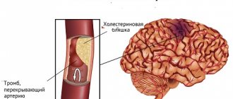
- Thrombosis - can lead to sudden death
If you have any of the symptoms we've outlined above, don't try to cure yourself or live with the expectation that "it will go away." It’s better to make an appointment with our cardiologist, who will thoroughly understand your situation, determine exactly whether you have a heart threshold, and tell you in detail what to do next.
What is the reason?
Congenital defects of the heart and large vessels form during the first 12 weeks of pregnancy, when all organs and systems of the unborn baby are actively developing. This occurs under the influence of the following factors:
- chromosomal mutations, which arise as a result of a structural or quantitative change in DNA segments, for example, due to the duplication or deletion of chromosomes;
- heredity (presence of heart defects in one of the parents or close relatives);
- unreasonable use of medications (antibiotics, non-steroidal anti-inflammatory drugs, etc.);
- bad habits of parents (smoking, taking alcohol or drugs);
- infectious or viral diseases in the mother during pregnancy (influenza, ARVI, measles, rubella, herpes, chickenpox, etc.);
- adverse effects of chemicals (paints, varnishes, nitrates, gasoline, acetone, etc.);
- age of women over 35 years old;
- exposure to radiation.
Classification
Conventionally, all congenital heart defects are divided into two types: the so-called “blue” and “white”.
The first are characterized by the discharge of blood from right to left with mixing of venous and arterial blood. They include 2 groups:
- defects with depletion of the pulmonary circulation (tetralogy of Fallot, combining four anomalies);
- with enrichment of the pulmonary circulation (for example, complete transposition of large vessels, when the aorta and pulmonary artery change places).
The second (pale) are distinguished by the discharge of blood from left to right without mixing venous and arterial blood. They include 4 groups:
- congenital defects with impoverishment of the systemic circulation (segmental narrowing of the aorta);
- with depletion of the pulmonary circulation (isolated narrowing of the pulmonary artery);
- with enrichment of the pulmonary circulation (patent ductus arteriosus, ventricular or atrial septal defects, etc.);
- without significant disruption of systemic circulation (change in the position of the heart, for example, dextroposition - when it is located on the right).
Dr. Komarovsky advises: “Always watch your child. A pediatrician may not always notice changes in health status. The main criteria for a child’s health are: how he eats, how he moves, how he sleeps.”
Clinical manifestations of congenital heart defects
The symptoms of this anomaly directly depend on the type and severity of the defect. They often appear in the very first stages of a newborn baby's life, but some of them may remain undetected throughout life. Some children, for example, may experience fainting, cyanosis, shortness of breath, poor appetite, short stature, underdevelopment of muscles and limbs, frequent acute respiratory viral infections, and hear heart murmurs, while others may not experience any symptoms.
Cyanosis is a bluish discoloration of the skin and mucous membranes, which is caused by a decrease in hemoglobin content in the blood (general or local). It is often a sign of diseases of the cardiovascular system. Acrocyanosis is also isolated, which is expressed in areas of the body most distant from the heart (fingers and toes, nasolabial triangle, ears).
All symptoms of congenital heart disease can be combined into four syndromes
- Cardiac (pain in the chest region, rapid heartbeat, shortness of breath, interruptions in heart function, etc. are noted; during the examination, cyanosis or pallor, swelling and pulsation of the vessels of the neck are observed, the chest may be deformed like a heart hump, changes in blood pressure and characteristics of the pulse in the peripheral arteries; during auscultation, that is, listening with a phonendoscope, changes in the strength, rhythm, timbre of heart sounds, and the appearance of noise characteristic of any defect are heard).
- Heart failure syndrome with characteristic manifestations (can be either acute or chronic, right or left ventricular, manifested by attacks of shortness of breath and cyanosis).
- Chronic systemic oxygen starvation syndrome (the child has a delay in growth and development, as well as the so-called drumstick symptom, which is manifested by a flask-shaped thickening of the terminal phalanges of the fingers in combination with deformation of the nail plates in the form of watch glasses).
- Syndrome of respiratory disorders (often with defects with changes in the pulmonary circulation).
There are three phases of symptom manifestation
The first phase is characterized by adaptation, that is, the body’s adaptation to disturbances in the functioning of the heart and the entire circulatory system. Symptoms appear extremely rarely or in a mild form. However, this is observed only before the onset of hemodynamic disturbances, resulting in a more frequent development of cardiac decompensation (depletion of compensatory forces). This moment is the most dangerous because a person can die during this period. If this phase passes, then after a couple of years health is restored.
The second phase is characterized by an improvement in the patient’s condition, which is caused by the development of compensation of the cardiovascular system and stabilization of heart function. But this period lasts relatively short, and soon after it symptoms characteristic of the next one appear.
The third (terminal) phase begins, during which compensatory forces are depleted and the body begins to experience severe stress. As a result, dystrophic and degenerative disorders of the heart muscle and other organs develop. As a result, clinical manifestations worsen and become more frequent, which leads to death.
Mothers of newborn children need to constantly and carefully monitor their baby, how he behaves, how he sucks the breast and how active he is. If the baby’s nasolabial triangle turns blue during feeding or if the baby’s arms and legs become blue when crying a lot, you should immediately consult a doctor and have an examination.
Symptoms
The first symptoms of a hole in the heart such as ASD appear in adulthood, despite the fact that this disease is congenital. In children, the pathology is almost impossible to diagnose.
A cardiologist diagnoses the presence of a hole in the heart based on the following signs:
- Pathological murmurs in the left and right atrium;
- Complaints of weakness, fatigue that does not go away even after a long rest;
- Dyspnea;
- Increased heart rate;
- Blue skin tone;
- Pathological swelling of the abdomen and lower extremities;
- History of stroke.
Symptoms of VSD pathology make themselves felt in the first year of a child’s life. An experienced pediatrician or cardiologist may already suspect a VSD in the first few days/weeks after the birth of a baby by a blue tint of the skin, cyanosis of the fingers and toes, loss of appetite, swelling of the lower torso and limbs, as well as pronounced heart murmurs.
How do heart defects work in the body?
Two mechanisms are leading.
For example, with defects such as valve insufficiency, narrowing of the openings and large vessels of the heart, its parts are overloaded with blood volume or resistance, which leads to depletion of the compensatory mechanisms involved, resulting in thickening of the muscle wall and stretching of the heart parts. As a result, heart failure occurs.
Disruption of systemic blood flow due to defects that are accompanied by plethora or anemia of the pulmonary and systemic circulation leads to general oxygen starvation of the body.
Clinical picture in children
A compensated form of congenital heart disease is identified when there are no symptoms, since the body is still coping with the pathology itself and the newborn is developing normally. Outwardly, a child’s heart defect does not manifest itself in any way. A sign of a heart defect can be determined later, when by the age of three it is clear that his physical development lags behind the norm, he is not active, and has breathing problems. The birth of such a baby is always a test for parents.
If the type is not compensated, then a significant symptom that people pay attention to is cyanosis of the lips, palms, nose, and in case of a severe defect, cyanosis may be observed throughout the body. The reason for this condition lies in the lack of blood circulation, as a result of which the tissues do not receive a sufficient amount of oxygen (hypoxia). In addition, it is noted:
- shortness of breath (rest or stress);
- cardiac cough;
- change in heart rate.
The baby sleeps poorly, shudders in his sleep, is agitated, and a severe form of the defects leads to fainting and suffocation.
Patent ductus arteriosus
Patent ductus arteriosus is a congenital anomaly of the heart in which the ductus arteriosus, which functioned in the prenatal period, does not close after birth. During normal fetal development, it connects the aorta to the pulmonary artery, protecting the right ventricle from overload by discharging excess blood, which in turn, due to the high resistance in the compressed lungs, cannot pass through their arteries, bypassing the pulmonary circulation into the aorta.
When a newborn baby takes his first breath, at this moment his lungs expand and the resistance of the pulmonary vessels decreases. After birth, the smooth muscles in the walls of the ductus arteriosus contract and blood flow through it decreases. The main stimulus for its closure is an increase in the amount of oxygen in the child’s blood. The ductus botallus, as a rule, narrows and completely closes in the first day of life; in rare cases, this can last up to three months. Eventually a scar is formed, called the ligamentum arteriosus, which remains in the normal healthy adult heart for life.
Symptoms of patent ductus arteriosus:
- rapid heartbeat (so-called tachycardia);
- shortness of breath (rapid shallow breathing);
- fast fatiguability;
- high pulse pressure;
- slow growth and development;
- an increase in the size of the heart, which can be detected by percussion (tapping fingers on the surface of the chest) or on an x-ray;
- long-lasting so-called “machine” noise, which is heard during auscultation in the second or third intercostal spaces.
With this anomaly, expectant management is acceptable if there are no symptoms of life-threatening conditions. We wait until the child is six months old for the ductus arteriosus to close on its own. However, if the infection does not occur for up to a year or the clinical picture is very pronounced, then this pathology requires removal through surgery.
Acquired heart disease in an adult
Also, the attention of cardiology to this problem is not only due to the need to eliminate the causes, but also due to the excessive disability of patients who suffer from heart disease. Therefore, acquired heart disease in adults is an urgent problem that requires an understanding of the mechanisms of formation and treatment of disorders.
Content
The human heart is a collection of muscle tissues. which ensures the smooth functioning of the entire body.
In one day it distills seven liters of blood. This is a huge job for such a small person.
an important “motor” weighing 300 grams. This organ has four chambers.
consisting of two atria and two ventricles. which are separated by a thin partition.
It is this that prevents mixing of arterial blood with venous blood.
In recent years, the incidence of heart disease has increased significantly. One common one is heart disease.
In the modern world, developed medical technologies detect this disease in the womb. This allows you to find out about the disease in time and prevent its development.
Every person should know. what is heart defect.
signs of heart disease. methods and means of preventing this disease.
Modern medicine can detect congenital heart defects in the womb. at 1 - 2 months of fetal development. Many parents try to cure this disease at an early age of the child through surgery. But there are also those. who is accompanied by this disease throughout their life.
Acquired heart disease can present with a variety of symptoms in adults. This is a consequence of complicated viral diseases. autoimmune reactions. heart injuries. The following are signs of heart disease:
- lethargy
- fatigue
- bad dream
- sore breasts
- swelling of the arms and legs
- ischemic heart disease
- unsteadiness of gait, dizziness
Acquired heart disease in an adult can manifest itself as:
- Mitral stenosis - characterized by strong or weak shortness of breath. regular cough. ascites. enlarged veins
- Mitral regurgitation - weakness, poor performance, shortness of breath
- aortic stenosis - dizziness. headache. fainting. nocturnal shortness of breath.
- aortic insufficiency - often characterized by asymptomatic progression. manifested by pulsation of blood vessels in the neck and head. tachycardia
- tricuspid insufficiency - shortness of breath, tachycardia, bluish-purple necks, chest pain. asthma, swelling of the airways, arrhythmia, pain/heaviness in the right hypochondrium, swelling. yellowish skin. dysfunction of the kidneys, liver and stomach
- combined defects are a pathological phenomenon characterized by the manifestation of several diseases simultaneously.
There is no single method of treating both congenital and acquired defects in adults. But this disease is worth fighting. There are ways and recommendations. which are worth sticking to.
Coarctation of the aorta
Coarctation of the aorta is a congenital heart defect that manifests itself as segmental (partial) narrowing of the aortic lumen and requires mandatory surgical treatment.
Clinical manifestations:
- an increase in blood pressure in the upper half of the body and a decrease in the arteries of the lower extremities;
- headache, much less often nausea and vomiting, as well as visual impairment;
- numbness, heaviness in the legs, weakness when walking as a result of insufficient blood supply in the lower half of the body;
- signs of enlargement and dilation of the left ventricle;
- systolic murmur upon auscultation in the second to fourth intercostal spaces, on the left at the edge of the sternum and between the shoulder blades at the back.
The first sign of coarctation of the aorta in children may be a weakening or complete absence of pulsation in the inguinal fossa (on the femoral artery).
The presence of this anomaly can be detected by electrocardiography (ECG). However, the most informative method confirming this disease is cardiac ultrasound (EchoCG). Magnetic resonance imaging (MRI) and angiography can also be used for diagnosis. When measuring blood pressure in patients at the hip, it will be significantly lower than at the shoulder.
Modern treatment of the disease is exclusively surgical, since conservative (drug) therapy only alleviates the course of the disease, but does not eliminate the causes and its further development.
What is a heart defect?
Heart disease is a group of diseases in which there is a change in the structure and disruption of the heart valve.
Heart failure includes combined (affecting several valves) and combined (on one valve) defects. Pathologies of this type lead to changes in the circulatory system inside the heart. Heart disease ¾ is an anomaly in the structure of any structure of the heart, leading to heart failure and, as a consequence, to insufficient blood supply in general.
Dextrocardia
This developmental anomaly is manifested in the fact that most of the heart is located in the chest to the right relative to the midline of the human body, symmetrically to its normal position in it. This condition is also accompanied by a “mirror” arrangement of all large blood vessels entering and leaving the heart.
Dextrocardia should be distinguished from dextroposition of the heart, in which it is mechanically displaced to the right half of the chest due to compression by any extracardiac factors. This defect is often combined with a mirror arrangement of all unpaired internal organs (the stomach and spleen are on the right side of the abdominal cavity, and the liver and gall bladder are on the left) - organ transposition. In the case of such a pathology, if there are no other concomitant heart defects, as a rule, deviations in the state of human health are not observed.
Main reasons and types
Rheumatism is the main cause of acquired heart defects. With its long course and frequent attacks, stenosis of the mitral and aortic orifices, as well as damage to the tricuspid valve, most often occurs. Often the role of etiological factors is played by the following pathologies:
- endocarditis caused by infection;
- some types of degenerative processes and connective tissue dystrophy (Marfan and Barlow syndromes);
- heart attack affecting areas of the heart with a valve;
- atherosclerosis and calcification;
- syphilis;
- chest injury.
Vice happens:
- By origin: rheumatic, syphilitic, atherosclerotic, etc.
- By the number of affected areas: single (local), combined (stenosis and insufficiency) and combined (the disorder affects several structures simultaneously).
- According to the degree of hemodynamic disturbance: no change, transient with increased load and decompensated.
Ventricular septal defect
VSD is a congenital heart defect in which there is a defect between the right and left ventricles of the heart.
The interventricular septum has three sections: the upper part (membranous), the middle (muscular) and the lower (trabecular). Accordingly, they are also called anomalies. By size they are divided into large, medium and small. To correctly assess the value, their size is compared with the diameter of the aorta. Small defects (1-2 mm in size) located in the muscular part of the septum are called Tolochinov-Roget disease. According to the pronounced auscultatory picture and the absence of hemodynamic disturbances, the expression is used to characterize them: “Much ado about nothing.”
Intracardiac hemodynamic disorders develop in a child some time after birth, usually on the third to fifth day. During the early neonatal period, that is, in the first days of a child’s life, there may be no heart murmur. The gradual decrease in pressure in the right ventricle and in the pulmonary artery system creates a difference between the pressures in the ventricles, resulting in a left-to-right shunt. The additional volume of blood that enters the pulmonary artery and the right ventricle of the heart leads to overfilling of the vessels of the pulmonary circulation, resulting in an increase in pressure in the vessels of the lungs (so-called pulmonary hypertension).
Clinically, VSD is manifested by heart failure syndrome, which, depending on the size of the defect, usually develops in the first to third months of life. As a result of blood stagnation in the pulmonary circulation, early and severe pneumonia can develop. During the examination of the child, it is possible to detect rapid heartbeat and shortness of breath, expansion of the borders of the heart, as well as movement of the apical impulse (it manifests itself as pulsation when palpated in the area of the apex of the heart) to the left and down. In some cases, the so-called symptom of “cat purring” is determined (vibration (or trembling) of the chest wall, which resembles the purring of a cat when stroking it). As a rule, a systolic murmur is detected; it is quite intense and is heard over the entire region of the heart. When palpating the abdomen, an increase in the size of the liver and spleen is determined.
There are three stages in the development of pulmonary hypertension:
- hypervolemic (stagnation of blood, which leads to pulmonary edema and frequent infection, resulting in the development of pneumonia, which manifests itself in the early stages of life, has a severe course and is difficult to treat);
- transitional stage, when the child stops getting sick, begins to gain weight and becomes more active; a stable condition in this phase is the best period for surgical treatment;
- so-called high pulmonary hypertension occurs when surgical correction has not been carried out; this process has no reverse development, and it mainly leads to a strong increase in pressure in the pulmonary artery. The most characteristic sign of this stage is the gradual increase in cyanosis (first peripheral, and then general); this occurs due to the shunting of blood through a defect in the septum from right to left. The presence of stage three in patients often becomes the main reason why cardiac surgeons refuse surgical intervention.
The most informative methods for identifying this pathology are electrocardiography and echocardiography (ultrasound of the heart, echocardiography).
Treatment of ventricular septal defect collectively includes conservative (drug) therapy for heart failure and surgical correction of the defect. Surgical interventions are divided into palliative (alleviating the course of the disease - surgery to narrow the pulmonary artery) and radical (plasty of the defect with a patch under conditions of artificial circulation and low temperature).
Mitral valve: anatomy and functions
The valve separates the left atrium and the left ventricle. It consists of:
- Atrioventricular ring. In its center there is a hole with a diameter of about seven centimeters.
- Valve leaf. They are necessary to close the hole in the ring.
- Tendon chords. They are attached over the entire surface of the valves, and the other end is connected to the papillary muscles. These tendons allow the valve to hold in place during contractions and prevent the valves from opening and allowing blood into the atrium.
- Papillary and papillary muscles. They are a continuation of the muscle tissue of the heart. During contraction, they provide tension on the tendon strands, which prevent the valve flaps from opening.
Thanks to the joint efforts of all the component parts, the valve does not open into the atrium. If the structure of one section of the valve is disrupted, this is accompanied by a malfunction of the entire apparatus.
Tetralogy of Fallot
Tetralogy of Fallot is a combined congenital cardiac anomaly that includes four defects:
- ventricular septal defect;
- right ventricular outflow stenosis;
- dextroposition of the aorta (when it partially departs from the right ventricle or the blood flow in it is mainly maintained due to the work of the left ventricle);
- hypertrophy of the right ventricular myocardium (the growth of its muscle component develops with age).
Depending on the time of appearance of cyanosis, five clinical forms are distinguished and, accordingly, the same number of periods of clinical manifestation of the defect:
- early cyanotic form (the appearance of a bluish color from the first months or first year of life);
- classical (in the second or third year of life);
- severe (occurs with attacks of shortness of breath and cyanosis);
- late cyanotic (the appearance of cyanosis by 6–10 years);
- acyanotic (pale) form.
A bluish discoloration of the lips and skin in severe forms of tetralogy of Fallot appears from three to four months of a child’s life and becomes stably expressed by about a year. It may worsen with crying, feeding, straining, or emotional stress. Any physical activity (outdoor play, walking, running) is accompanied by the rapid onset of weakness, shortness of breath, rapid heartbeat, and dizziness. After exercise, patients with this pathology assume a characteristic squatting position.
An extremely severe manifestation of this pathology are seizures, which usually appear between the ages of 2 and 5 years. They develop suddenly, accompanied by the child’s anxiety, rapid heartbeat, severe weakness, increased shortness of breath and cyanosis, and even loss of consciousness. Respiratory arrests and convulsions may develop.
Children with tetralogy of Fallot are retarded in physical development (they have II-III degree malnutrition), they often develop acute respiratory diseases, chronic tonsillitis and sinusitis, and recurring pneumonia. In adult patients with such a heart defect, pulmonary tuberculosis may be associated.
Surgical treatment includes two stages
First stage. Up to three years, operations are carried out that significantly facilitate the life of patients with this defect. The purpose of their implementation is to increase blood flow to the pulmonary circulation.
Second phase. It is carried out a couple of months after the first one. The operation is radical and is carried out under conditions of artificial circulation using cardiac arrest and cooling. A general decrease in temperature to 21.6 degrees. After clamping the aorta, the cavity of the right ventricle is opened and, under visual control, surgical techniques are performed to eliminate the narrowing of its outlet section. The ventricular septal defect is repaired by suturing the patch with U-shaped sutures. Then a patch is sewn into the anterior surface of the right ventricle to expand the outflow tract.
The course of the defect depends significantly on the degree of pulmonary narrowing. Approximately 25% of children with severe tetralogy of Fallot die in the first year of life, half of them in the newborn period. The average life expectancy without surgery is approximately twelve years; less than five percent of patients live to be forty years old. The cause of death in patients with this developmental anomaly is most often ischemic stroke due to cerebral vascular thrombosis.
The long-term results of surgical correction of the defect are quite favorable: patients tolerate physical activity well, are socially active and able to work. But the later in life the operation is performed, the worse the long-term results. Absolutely all patients with this defect require dynamic monitoring by a cardiologist and cardiac surgeon, and prophylactic antibiotics before undergoing any surgical or dental procedures that have a potential risk of developing a bacterial infection.
Drugs for the treatment of heart disease in older people
The modern range of pharmacies is quite diverse. It is represented by various drugs both for the prevention of cardiovascular diseases and for their treatment - both current and intensive. The medications are suitable for both older people and those who encountered the problem at a younger age.
Do not forget that the heart is a very important organ, and you should make a decision on a course of any drug only after consulting a doctor.
Medicines to prevent heart disease and strengthen blood vessels:
- "Venoton". Contains vitamin C, garlic juice and buckwheat extract, lactose.
- "Ascorutin". Increases the strength of blood vessels, which is very important for the heart of an elderly person.
- "Piracetam." Improves blood circulation in the central nervous system. If the first two drugs are “vitamins”, then this one can be used only after consultation with a specialist.
- "Riboxin" Increases oxygen delivery to almost all organs. Prescribed for coronary heart disease and liver problems. Stimulates the generation of protein cells in muscles, including the heart of an elderly person.
- "Cavinton". Strengthens blood vessels, including those of the brain and heart, eliminates hypoxia. Prescribed to reduce the risk of myocardial infarction and stroke.
- "Asparkam." Contains magnesium and potassium. Normalizes the supply of electronic heart impulses, improves the rhythm and quality of contractions of the heart muscle of an elderly person.
- "Hawthorn forte." Non-alcoholic preparation of hawthorn flowers. Tones blood vessels, normalizes heart rhythm.
- "Rhodiola rosea." Also a herbal remedy. Slows down myocardial wear. An elderly person feels an improvement in health almost at the very beginning of the course of the drug. The frequency of administration, dosage and dilution ratio are prescribed by the doctor. Self-medication with this medicine (as with all others) is highly discouraged.
Read material on the topic: High blood pressure in older people - causes, symptoms and treatment methods
Lifestyle with congenital heart disease
It is extremely important for patients to adhere to the following recommendations regarding their lifestyle:
- compulsory stay in the fresh air;
- proper and balanced nutrition;
- diet (limiting salt and drinking fluids in order to reduce volume overload of the heart and blood vessels);
- complete and long sleep;
- Strong physical activity should be limited and sports should be completely eliminated;
- regular observation by doctors - a cardiologist and a cardiac surgeon, as well as carrying out the necessary set of diagnostic and therapeutic measures;
- for women with “blue” type defects, pregnancy is strictly contraindicated, however, if surgical correction has been performed, the possibility of maintaining the pregnancy will be determined individually in each case, along with the management of the pregnant woman by an obstetrician-gynecologist together with a cardiac surgeon and cardiologist in a specialized hospital; in this case, delivery is usually performed by caesarean section.
Diagnostic methods
This diagnosis can be established by the pulse; the rhythm on the left and right hands may differ. Diagnosis in patients with suspected heart disease begins with checking their well-being at rest and their tolerance to physical activity.
The reasons are determined using the medical history and patient complaints. The method of palpation and examination is used to identify cyanosis, pulsation of peripheral veins, shortness of breath, and edema.
The presence of cardiac hypertrophy is determined, cardiac murmurs and tones are heard.
They check the functioning of the lungs and determine the size of the liver. Using an ECG, the heart rhythm, type of arrhythmia, blockade and signs of ischemia are established.
Phonocardiography is used to record heart murmurs and sounds and determine heart valve defects. Accuracy of diagnosis is also achieved using cardiac X-ray, echocardiography, MSCT or cardiac MRI.
Laboratory tests - rheumatoid tests, determination of sugar levels and the presence of cholesterol. Clinical blood and urine tests are required.
Heart disease is a very serious disease, and it can only be cured by conducting a special diagnosis and eliminating all associated complications.
A cardiologist treats such patients. He carefully talks with the patient, studying his complaints, examines him visually, listens (auscultation) and taps (percussion) the heart.
If a displacement of the borders of the heart is detected, as well as characteristic noises identified during the initial examination, the doctor can diagnose a heart defect, and to confirm this, prescribe an additional examination.
Hardware research methods include:
- Electrocardiography. It can detect cardiac arrhythmias and myocardial ischemia.
- Echocardiography. Able to visualize and evaluate various lesions of the heart valve apparatus.
- X-ray of the chest organs. It can reveal signs of stagnation of venous blood in the lungs, clarify the presence/absence of effusion in the pleural cavity.
- Computed or magnetic resonance imaging. Helps to study cardiac disorders in detail and determine the tactics of therapeutic management of the patient.
- Laboratory research. They give a complete picture of the blood composition and help prescribe appropriate treatment.
Only a comprehensive examination will help organize the correct treatment of a patient with heart disease.
The diagnosis of heart disease can be assumed during a clinical examination of the patient with mandatory auscultation of the chest organs, when listening to which pathological tones and noises caused by improper functioning of the heart valves are revealed; The doctor may also hear wheezing in the lungs due to stagnation of blood in the vessels of the lungs.
Attention is drawn to pallor of the skin, the presence of edema, determined by palpation (by palpating the abdomen) and enlarged liver.
This is, for example, what a heart looks like with hypertrophy of the atria and ventricles due to heart defects on an x-ray.
Of the listed research methods, echocardiography helps to reliably confirm or refute the diagnosis, as it allows you to visualize the heart and its internal structures.
In case of mitral stenosis, ultrasound of the heart determines the severity of stenosis over the area of the atrioventricular opening, compaction of the valve leaflets, hypertrophy (increase in mass) of the left atrium, turbulent (not unidirectional) blood flow through the atrioventricular opening, increased pressure in the left atrium.
Mitral valve insufficiency according to ultrasound is characterized by a break in the echo signal from the leaflets at the moment of valve closure; the severity of regurgitation (backflow of blood into the left atrium) and the degree of hypertrophy of the left atrium are also determined.
With stenosis of the aortic mouth, ultrasound determines the severity of the stenosis, hypertrophy of the left ventricular myocardium, a decrease in the ejection fraction and stroke volume of blood (indicators characterizing the flow of blood into the aorta per one heartbeat).
Aortic insufficiency is manifested by deformation of the aortic valve leaflets, their incomplete closure, regurgitation of blood into the cavity of the left ventricle, and left ventricular hypertrophy.
For defects of the tricuspid valve and pulmonary valve, similar indicators are identified and assessed, only for the right parts of the heart.