- Procedure for providing medical care
ACVA (acute cerebrovascular accident) is a concept that combines a transient ischemic attack and a pre-stroke condition. ACVA is characterized by sudden development and is very dangerous for human health and life, therefore, when its first signs appear, urgent medical attention is necessary. Timely adequate treatment can reduce the severity of the consequences of an attack. To receive qualified assistance for acute stroke, you can contact the Yusupov Hospital, which operates around the clock and provides the necessary assistance in this situation.
What is ONMK?
The diagnosis of stroke (and the resulting stroke) is established in the event of disturbances in the functioning of the cerebral vessels. When blood circulation is disrupted in a certain area of the brain, part of the nervous tissue dies. This can lead to serious disability or death. A stroke is not yet a stroke, but a condition that can lead to it. The development of stroke indicates that a person urgently needs the help of a qualified neurologist, since a full-fledged stroke or cerebral infarction may soon occur, when the consequences will be much more severe. Decoding the diagnosis of stroke will depend on the type of disorder in the vessels: hemorrhage, blockage or narrowing of the vessel, etc. The name of the disease of acute stroke is deciphered by the attending physician based on symptoms and examination data.
It is important to know about the diagnosis of stroke, that this is the most dangerous condition. According to WHO, approximately 12 million people worldwide die from strokes every year. The disease affects both poor and rich, men and women. People with obesity, diabetes mellitus, alcohol abusers, and smokers are most susceptible to this condition. In women, the risk of stroke increases after menopause. Recently, cases of stroke and subsequent strokes have begun to occur in young people (25-40 years old), which is associated with an unhealthy lifestyle and constant stress.
Classification and code according to ICD 10
ACVA code according to ICD 10 is included in the class of cerebrovascular diseases (I60-I69). The consequences of acute stroke according to ICD 10 codes refer to various hemorrhages, heart attacks, strokes, blockages and stenosis of arteries, as well as other lesions of cerebral vessels. The consequences of acute stroke in ICD 10 can be classified as follows:
- subarachnoid hemorrhage;
- intracerebral hemorrhage;
- non-traumatic hemorrhages;
- cerebral infarction;
- unspecified stroke;
- blockage and stenosis of the precerebral and cerebral arteries.
Also, stroke code according to ICD 10 in adults is divided according to the nature of vascular damage:
- ischemic type;
- hemorrhagic type.
ACVA of ischemic type
Acute ischemic cerebrovascular accident is a brain lesion resulting from the formation of an obstruction in a vessel. Most often, this obstacle is a blood clot or cholesterol plaque. An obstruction interferes with the flow of blood to any part of the brain, resulting in oxygen starvation. Nerve tissue needs a constant, continuous supply of nutrients, since the metabolism in nerve cells is very intense. When the access to oxygen and nutrients transported by the blood is stopped, the functioning of the nerve cells is disrupted, and after a short period of time they begin to die. In the case of an ischemic type of circulatory disorder, a certain obstacle interferes with the normal flow of blood, provoking a cerebral infarction. This type of violation is quite common and accounts for up to 80% of cases. ICD 10 codes for stroke of ischemic type include:
- I63 cerebral infarction;
- I65 obstruction and stenosis of the precerebral arteries;
- I66 blockage and stenosis of the cerebral arteries.
stroke of hemorrhagic type
Stroke of the hemorrhagic type is classified as a pathological condition caused by a violation of the integrity of the vessel, resulting in hemorrhage. Depending on the location of the disorder and its extent, the result of hemorrhage is a hematoma in the brain tissue or penetration of blood into the space surrounding the brain. ACVA of the hemorrhagic type in ICD 10 includes:
- I60 subarachnoid hemorrhage;
- I61 intracerebral hemorrhage;
- I62 other non-traumatic hemorrhage;
The condition after an acute stroke, related to any code according to ICD 10, is severe and requires urgent intervention from a specialist. The consequence of stroke is the death of nerve cells, which occurs very quickly. The consequences of acute cerebrovascular accident can be stopped if the person receives help within 4-5 hours after the attack.
Stroke in the vertebrobasilar region
The incidence of cerebral infarctions in the vertebrobasilar system, compared with the carotid system, is 5 times less. A stroke in the VBB, like any other stroke, can be of two types: ischemic and hemorrhagic. The subtype of ischemic stroke in the vertebrobasilar region is determined depending on the patient’s risk factors:
- Atherothrombotic (40−45%) - blockage of blood vessels due to atherosclerosis, thrombosis or arterio-arterial thromboembolism. Closure of the lumen can be caused by a plaque gradually increasing in size, on which thrombotic masses are layered, or by an embolus that has broken off from atherothrombotic formations.
Cardioembolic (25−30%) - the source of emboli can be pathological formations on the heart valves, intracardiac thrombi. This is facilitated by the presence of artificial valves, atrial fibrillation, and intracardiac shunts.- Lacunar (5−10%) - formation of a lesion up to 1.5 cm against the background of changes in small vessels. The most likely cause of such changes is arterial hypertension.
- Hemodynamic (up to 5%) develops against the background of a sudden drop in blood pressure, for example, during myocardial infarction.
- Unspecified - in this case, the cause has not been found, there is the presence of 2 or more risk factors, for example, atherosclerosis and atrial fibrillation.
In case of hemorrhagic stroke, the localization of the lesion is indicated, as determined by computed tomography.
Symptoms of a stroke in the VBS include:
- Dizziness is a feeling of rotation of a person or the surrounding space. It occurs abruptly, suddenly, most often in the morning, and is combined with gait disturbances. The cause is damage to the stem structures of the vestibular analyzer or the connections between the analyzer and the cerebellum.
- Visual impairment. It occurs when there is damage to the occipital cortex, where the cortical part of the visual analyzer is located. Violations manifest themselves in the form of loss of visual fields. The image of the right or left parts disappears in both eyes.
- Strabismus appears due to damage to the structures of the oculomotor system. The muscles that rotate the eye in all directions are innervated by three pairs of cranial nerves: the third is the oculomotor nerve, the fourth is the trochlear nerve, and the sixth is the abducens. If one of the nerves is damaged, the innervation of a certain muscle disappears, and the eye stops moving in the direction of the lesion.
- Paresis and paralysis are caused by damage to the cortico-muscular pathway, which is responsible for the innervation of the muscles of the limbs and torso. The degree of damage is directly proportional to the severity of motor disorders, therefore in some cases there is a decrease in muscle strength and range of active movements, that is, paresis, and in others - complete immobility, or plegia. These paths on the right and left intersect, therefore, if the focus appears on the right, then paresis or paralysis will appear on the left, and vice versa.
- Sensitivity disorders - the development mechanism is similar to movement disorders. There is a crossover of beams.
- Speech and swallowing disorders are combined under the term “bulbar palsy.” Occurs when cranial nerves such as the glossopharyngeal (9th pair), vagus (10th pair), and hypoglossal (12th pair) are damaged. They innervate the soft palate, tongue, and pharyngeal muscles, so if damaged, difficulty swallowing occurs, the timbre of speech changes, and the voice becomes nasal.
- Gait disturbances are associated with infarction in the cerebellum, the main organ responsible for the sense of balance and equilibrium, as well as its connections with the vestibular system. A person cannot move independently; he always needs additional support.
If such symptoms suddenly develop, emergency medical care is required.
Causes and symptoms
To assess the degree of brain damage, the Rankin scale is often used for stroke and subsequent stroke. Cerebrovascular diseases (CVD) and stroke can significantly reduce a person’s performance and lead to disability. Therefore, conditions such as acute coronary syndrome (ACS) and stroke, associated with disruption of blood vessels in vital organs (heart and brain), require urgent treatment to the hospital.
The Rankin scale presents six degrees of disability after stroke and stroke:
0. There are no clinical symptoms; 1. The vital systems are not significantly impaired, there are minor symptoms, but the person can perform all daily activities; 2. Mild disturbances in vital activity systems: the performance of some actions is limited or inaccessible, a person can take care of himself without outside help; 3. Moderate disability: some assistance is required, the person can walk independently; 4. Severe disabilities: a person is unable to walk independently, requires care and assistance in everyday life; 5. Severe disabilities: complete immobility, urinary and fecal incontinence, the person requires constant assistance from specialized medical personnel.
Each grade of the Rankine scale has its own symptoms, which make it possible to determine clinically how affected the brain is. With minor lesions of the 1st degree, a person has no signs of disability, he is able to care for himself and perform daily work. However, slight muscle weakness, speech disorders, and loss of sensitivity may occur. These disorders are mild and do not lead to restrictions on daily life.
In grade 2, mild signs of activity impairment are observed: the person is unable to perform previous work involving complex manipulations or fine motor skills. However, he can take care of himself independently, without the help of others.
At grade 3, moderate signs of brain dysfunction are observed:
- the person needs some outside help in performing hygiene procedures;
- he cannot prepare food or dress himself;
- speech impairments are pronounced (difficulties arise in communication, expressing one’s thoughts);
- It is possible to use a cane or other walking aids.
Symptoms of acute cerebrovascular accident of the 4th degree are clearly expressed, there are clear signs of disability. A person cannot walk independently, take care of himself, he needs round-the-clock assistance.
With the 5th degree of disability, a person is bedridden, he cannot speak, cannot eat independently, and does not control bowel movements. A person needs constant help and supervision.
One of the most clinically striking and health-threatening strokes is damage to the VBB (vertebrobasilar region). In this case, the pathological process affects parts of the brainstem, thalamus, cerebellum and occipital lobes of the brain. ACVA in the vertebrobasilar region manifests itself as follows:
- partial facial paralysis;
- impaired motor activity of the hands;
- difficulty moving the leg and arm on one side of the body;
- impaired coordination of movements;
- the appearance of muscle weakness in the lower extremities;
- mild arm paresis;
- swallowing disorder;
- nausea, vomiting;
- hearing and speech impairment;
- headache and dizziness.
When developing stroke, it is important to consult a doctor as soon as possible. To do this, you need to pay attention to the first symptoms of pathology:
- severe acute sudden headache;
- sudden loss of consciousness;
- sudden muscle weakness;
- sudden disturbance of speech and its understanding;
- sudden visual impairment;
- sudden numbness of the limbs or areas of the face;
- impaired coordination of movements;
- nausea, vomiting.
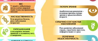
The severity of symptoms will depend on how severely the brain is damaged. ACVA occurs spontaneously and cannot be predicted. But you can try to exclude factors that increase the risk of developing strokes and strokes:
- smoking;
- alcohol abuse;
- unhealthy diet;
- lack of physical activity;
- chronic fatigue and stress.
People with diabetes, arrhythmia, and overweight need to be especially responsible about their health. These conditions quite often become the causes of the development of circulatory disorders in the brain.
Characteristics of VBB
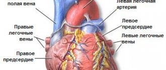
The cerebral circulatory system , which provides a continuous supply of oxygen and nutrients to nerve cells, consists of 2 pools.
2/3 of the total blood volume enters the carotid system, internal carotid arteries and its branches. They form the Circle of Willis. This system nourishes the anterior and middle parts of the brain - the frontal, parietal, temporal lobes, thalamus, hypothalamus, structures of the limbic system. 1/3 of the volume enters through the vertebrobasilar system. It is formed from 2 vertebral arteries, the unpaired basilar artery and their branches.
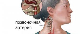
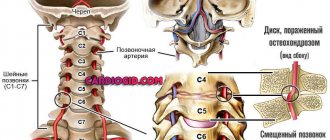
The vertebral arteries arise from the right and left subclavian arteries. They go in the bone canal formed by the transverse processes of the cervical vertebrae from the sixth to the second. Then they leave the canal, enter the cranial cavity and merge into the main artery.
The main, or basilar, artery lies on the anterior surface of the brain stem (pons). At the border of the pons and midbrain, it divides into 2 posterior cerebral arteries. The main branches of the vertebrobasilar system are the superior, inferior posterior, and inferior anterior cerebellar arteries. The VBB vessels supply blood to the brain stem (medulla oblongata, pons, midbrain), cerebellum (vermis and hemispheres), as well as the occipital lobe cortex.
Diagnosis of the disease
When the first signs of cerebrovascular accident appear, you must call an ambulance or go to the hospital yourself (if your condition allows). The doctor will perform an examination and collect anamnesis (description of the patient’s condition and related data). The doctor must be provided with the following information:
- main complaints (headache, disturbance in the functioning of the sensory organs, nausea, etc.);
- when the condition worsened;
- under what conditions;
- the presence of risk factors for stroke (smoking, alcoholism, chronic diseases, taking medications).
A simple test can detect the development of stroke or stroke (provided that the patient is conscious):
- It is necessary to ask the patient to smile (with stroke, the smile will be skewed);
- It is necessary to ask the patient to stretch his arms forward and then raise them up (with a stroke he will not be able to do this or will only raise one arm);
- Ask the patient to repeat any simple sentence (with stroke, this will cause difficulties);
- Ask the patient to stick out his tongue (with stroke, the tongue will be clearly displaced from the center).
The doctor assesses the general and local status of stroke. General status represents the general condition of the patient, clinical manifestations of cerebrovascular accident. Local status is described in the presence of head trauma. The collected data gives the doctor an idea of the patient’s condition, on the basis of which he prescribes examinations to obtain a complete picture of what is happening.
Diagnosis of stroke is carried out using visualization of nerve tissue using CT and MRI. These are the most informative diagnostic methods that allow you to identify the lesion. To provide timely assistance in case of acute stroke, the examination is carried out urgently. In some cases, the patient will need emergency surgery.
At the Yusupov Hospital you can undergo examinations of any complexity for acute stroke and stroke. The hospital is equipped with the latest technology, which allows for a quick and high-quality examination of the patient. High-precision technology will help establish an accurate diagnosis and the extent of brain damage.
Diagnostics
Brain stem structures are best visualized using MRI. Magnetic resonance imaging allows you to see the smallest foci of ischemic lesions. Computed tomography images clearly show the nature of the stroke. Other instrumental methods:
- X-ray contrast and magnetic resonance angiography (study of elements of the circulatory system). Shows areas of arterial damage as a result of thrombosis, atherosclerosis, and mechanical stress.
- Triplex scanning. Ultrasound examination in combination with Dopplerography. The extracranial (external) arteries are clearly visible, while the intracranial (internal, located in the cranial cavity) elements of the circulatory system are worse.
- Audiometry. Study of hearing acuity and auditory sensitivity.
A blood test shows changes characteristic of diabetes mellitus, atherosclerosis, and inflammatory processes. When making a diagnosis, the medical opinions of highly specialized doctors - neurologist, otoneurologist, ophthalmologist - are taken into account.
Treatment
Treatment of stroke will include first emergency care and subsequent therapy. Further therapy consists of a number of measures to normalize and support brain function. The doctor informs the patient about how to take nootropics for stroke and other medications, nutritional habits of patients with stroke, clinical recommendations for stroke. Treatment of acute cerebrovascular accidents includes basic and specific therapy. Basic therapy contains the following measures:
- Restoring and maintaining respiratory function.
- Maintaining optimal blood pressure and cardiovascular activity. The patient is given intravenous medications (labetalol, nicardipine, sodium nitroprusside) according to indications; anaprilin, enaprilin, captopril, esmolol are used to correct blood pressure. Antihypertensive therapy depends on the type of stroke - hemorrhagic or ischemic.
- Treatment of cerebral edema.
- Fighting seizures, intracranial hypertension, various neurological complications.
Specific therapy includes:
- Carrying out intravenous or intra-arterial thrombolysis depending on the time of onset of the first symptoms of the disease. Aspirin is prescribed, and anticoagulants are prescribed according to indications.
- Maintaining optimal blood pressure.
- In certain cases, surgery is performed to remove the brain hematoma, and the hemicraniectomy method is used to decompress the brain.
Treatment of stroke is carried out in a hospital setting. The faster the patient receives medical assistance, the higher the chance of recovery.
Procedure for providing medical care
The scope of medical care for stroke or stroke will depend on the severity of the patient’s condition. It is important to get to the hospital as soon as possible. If the cause of the stroke is a blood clot, then it is necessary to take an antithrombotic drug within 3 hours after the onset of cerebrovascular accident to reduce the consequences.
Treatment of stroke occurs in a hospital, its duration is from two weeks (for mild damage). The patient is prescribed infusion therapy, medications to stabilize blood pressure, and medications to normalize the functioning of nerve cells. In the future, the patient will need a rehabilitation course to restore lost skills or adapt to new living conditions. Rehabilitation is a very important part of treatment. It is rehabilitation measures, when performed regularly, that contribute to the restoration of working capacity.
At the Yusupov Hospital you can undergo a full course of treatment for acute stroke and stroke, including emergency care and rehabilitation. The hospital employs the best neurologists, cardiologists, surgeons in Moscow, doctors of science, and doctors of the highest category who have extensive experience in successfully treating these conditions. The hospital is equipped with everything necessary for a speedy and high-quality recovery of patients.
Artificial ventilation for acute stroke
When a patient is admitted with stroke or stroke, the doctor assesses the adequacy of spontaneous breathing and the level of oxygen in the blood. If the patient has a low level of consciousness, there is a risk of aspiration, high rates of intracranial hypertension, he requires artificial ventilation (ALV).
Ventilation is also performed for:
- Violation of central regulation of breathing;
- Obstruction of the tracheobronchial tree;
- Pulmonary embolism.
Treatment with a dropper (infusion therapy)
Infusion therapy begins from the moment a patient is admitted with stroke or stroke. A 0.9% sodium chloride solution is prescribed. With stroke, hypovolemia (decreased blood volume) quite often occurs, which can be eliminated by infusion therapy. Infusion is also necessary to control the water balance in the body. Infusion therapy is canceled gradually, after confirmation of normalization of the level of electrolytes and other elements in a blood test.
Normalization of blood pressure
The first three days are critical after stroke. During this period, repeated violations or the development of a major stroke are possible. Now it is necessary to stabilize the patient's condition and respond to any changes. Some important indicators are intracranial pressure and blood pressure. Pressure level indicators should not exceed the permissible norm or be below the norm. Therefore, pressure monitoring is carried out constantly. To normalize the indicators, special drugs are first administered intravenously, and then switched to tablet form of drugs.
Elimination of convulsive syndrome
With stroke, there is a high risk of seizures. However, there is no prevention for this condition. Anticonvulsants are prescribed immediately when a seizure occurs. The drugs are administered orally or intravenously.
Use of neuroprotectors and nootropics
An important direction in the treatment of stroke and stroke is the restoration of damaged nerve tissue and the protection of healthy tissue from the spread of “vascular catastrophe”. Treatment is performed with the help of neuroreparants and neuroprotectors.
Nutritional Features
If swallowing is impaired, the patient is prescribed feeding through a tube. At the beginning of treatment, food contains the necessary elements to maintain the functioning of the body, combined with infusion therapy. The calorie content of food increases gradually. In the future, the method of eating will depend on the severity of the brain damage. The course of rehabilitation of patients after acute stroke and strokes includes the restoration of self-care skills, so with the proper effort and capabilities of the patient, he can feed himself again. Food should be varied, contain all the necessary microelements and vitamins, that is, comply with the principles of rational nutrition.
Treatment of vertebrobasilar insufficiency
A neurologist will tell you how to treat the disease after receiving the results of a diagnostic examination. Therapy is carried out taking into account the prevailing symptoms, nature and causes of brain tissue damage. If VBS develops due to blockage of the vascular lumen by a thrombus, drugs are prescribed to prevent the formation of edema. Mainly osmotic diuretics (Glycerol, Mannitol). When VBI provoked a TIA or ischemic stroke, long-term use of antiplatelet agents (Clopidogrel, Dipyridamole, Ticlopidine), which prevent platelet aggregation, is indicated.
Drug therapy
Thrombolytics are rarely used in the treatment of vertebrobasilar insufficiency due to the high risk of hemorrhage and other complications. If the patient has pronounced atherosclerotic changes in the vascular wall, indirect-acting anticoagulants (prevent the formation of blood clots) (Dicoumarin, Warfarin) are indicated. During treatment, the prothrombin index is monitored. Other groups of drugs:
- Low molecular weight dextrans (Reomacrodex, Reopoliglucin). Reduce viscosity and improve the rheological properties of blood.
- Correctors for impaired blood supply to the brain (Vinpocetine). Regulate microcirculation, normalize venous outflow.
- Sedatives (Betagistin, Bellataminal). Reduce neurological symptoms, eliminate dizziness.
- Nootropic drugs (Piracetam). Normalize metabolic processes in brain structures at the cellular level.
- Calcium antagonists (Cinnarizine, Verapamil). They reduce the excitability of the vestibular apparatus, have a vasodilating effect, and help improve cerebral blood flow.
If signs of oxygen starvation of tissues are detected, antihypoxants (Actovegin, Cytoflavin) are prescribed in parallel. The drugs and regimen are selected individually, taking into account the factors and diseases that provoked the pathological condition.
Surgery
In emergency cases, when disruption of blood flow threatens the patient’s life, surgical decompression of brain structures or elements of the circulatory system is performed. When drug therapy does not produce results, surgical intervention is indicated:
- Microdiscectomy. Minimally invasive surgery to remove a herniated intervertebral disc.
- Endarterectomy. An operation to restore normal blood flow by removing a section of the vascular wall with atherosclerotic damage.
- Reconstruction of intervertebral discs.
- Angioplasty of the artery. Installation of a stent inside a damaged vessel to artificially expand the lumen.
Mechanical thrombus extraction involves the removal of blood clots that impede the movement of blood in the vessels. The type of operation is chosen by a neurosurgeon based on the results of the examination, taking into account the causes of the pathology.
Physiotherapy
The comprehensive treatment program includes therapeutic exercises, massage and physiotherapy:
- photochromotherapy (for visual impairment);
- electrophoresis;
- magnetic therapy;
- laser therapy;
- transcranial micropolarization.
Traditional medicine recipes
Treatment with folk remedies is effective in the early stages. Traditional healers recommend tinctures and decoctions prepared from medicinal plants with vasodilating, anti-inflammatory, immunomodulatory, and antihypertensive properties. Homemade preparations based on the fruits of hawthorn, rose hips, lemon balm leaves, speedwell, motherwort, valerian, periwinkle, and mint expand the vascular lumen, tone and strengthen the walls of blood vessels.
Providing emergency assistance
Acute cerebrovascular accident requires emergency care, since it will not be possible to normalize the patient’s condition on his own. The standard for emergency medical care for acute stroke and stroke states that the patient should be taken to the hospital within 3-5 hours after the onset of the attack. In this case, it is possible to stop the spread of the pathological condition and minimize the severity of the consequences. Help for a person with a stroke can only be provided in a hospital. At home you can do the following:
- Call an ambulance;
- Lay the person on a flat surface (floor, bed), placing a pillow, blanket or folded sweater under the head;
- Turn the person on their side if they feel sick;
- Open the windows to let in fresh air;
- Unfasten clothing that impedes blood flow and air flow (belt, collar, scarf, tight buttons);
- While waiting for doctors, collect documents and personal belongings.
In case of an emergency, it is necessary to provide assistance to the patient before the medical team arrives. If you lose consciousness, you should check your breathing and pulse, and place the person in a position that will not interfere with breathing. If there is no breathing or pulse, it is necessary to begin mouth-to-mouth artificial respiration and chest compressions. If convulsions occur, the patient should be protected from injury: remove nearby sharp and blunt hard objects. You should not try to restrain the patient or unclench his teeth. It is better to wait until the attack ends and check the airway.
If stroke develops, you can go to the Yusupov Hospital, whose emergency department is open 24 hours a day, seven days a week. The hospital has an ambulance, so the patient will receive all necessary medical care in a timely manner. In the intensive care unit of the Yusupov Hospital, the patient will be able to provide the required assistance to stabilize his condition.
The procedure for providing medical care to patients with stroke after admission to the hospital emergency department is as follows:
- Medical examination, ECG, blood tests;
- Examination by specialized specialists: neurologist, cardiologist, neurosurgeon, resuscitator;
- Performing a computed tomography scan of the brain;
- Evaluation of survey results;
- Start of therapy.
After the patient is admitted to the hospital and before the start of therapy, no more than an hour should pass. If necessary, the patient is sent to the intensive care unit immediately, after which the necessary examinations are performed.
Instrumental diagnostic methods
Recently, the information capabilities of instrumental diagnostic methods in clinical practice have increased. Ultrasound methods for studying the vascular system of the brain have become the most accessible and safe. Doppler ultrasound allows you to obtain data on the patency of the vertebral arteries, linear speed and direction of blood flow in them. Compression-functional tests make it possible to assess the condition and resources of collateral circulation, blood flow in the carotid, temporal, supratrochlear and other arteries. Duplex scanning demonstrates the condition of the arterial wall, the nature and structure of stenotic formations. Transcranial Doppler ultrasound with pharmacological tests is important for determining cerebral hemodynamic reserve. Doppler ultrasound (USDG) - detection of signals in the arteries gives an idea of the intensity of microembolic flow in them, cardiogenic or vascular embologenic potential. Data on the condition of the main arteries of the head obtained by MRI angiography are extremely valuable. When deciding on thrombolytic therapy or surgical intervention on the vertebral arteries, contrast X-ray panangography becomes of decisive importance. Indirect data on the vertebrogenic effect on the vertebral arteries can also be obtained from conventional radiography performed with functional tests.
Otoneurological research occupies a special place, especially if it is supported by computer electronystagmographic and electrophysiological data on auditory evoked potentials characterizing the state of brain stem structures, as well as their MRI.
The sequence of application of the listed instrumental research methods is determined by the peculiarities of determining the clinical diagnosis.
Treatment of atherosclerosis is carried out according to generally accepted regimens with an emphasis on lifestyle and diet and with the use of statins. The selection of antihypertensive drugs and the procedure for their administration are determined based on the results of 24-hour Holter ECG and blood pressure monitoring.
Therapy in the acute period is carried out in two main directions: maintaining systemic hemodynamics, normalizing blood light levels and hemoangiocorrection. A positive effect of antioxidants and certain neurotrophic drugs was noted.
Drug treatment
Drugs that optimize cerebral circulation are used:
- vinpocetine under ECG monitoring of heart rhythm;
- nicergoline;
- aminophylline;
- instenon';
- cinnarizine;
- ginkgo biloba;
- fezam;
- encephabol;
- collected the vase;
- pyrroxane;
- Betahistine;
- Emoksipina;
- Mildronate;
- Mexidol;
- acetylsalicylic acid;
- pentoxifylline;
- low molecular weight heparins;
- non-steroidal anti-inflammatory drugs (NSAIDs);
Surgical methods of treatment
Surgical treatment methods have proven to be very effective. These include endarterectomy, reconstructive operations for replantation of vertebral arteries, arteriolysis and their redressing during adhesions in surrounding tissues. Surgeries are also performed to eliminate arterial obstructions (stabilization of excessively mobile segments of the cervical vertebrae, removal of uncovertebral osteophytes, etc.).
In cases where, for some reason, operations are impossible, therapeutic drug blockades and orthopedic treatment methods are used, the Shants collar, special sets of physical exercises are used, but without sharp jerking turns of the head, flexion and extension of the neck, and long-term fixation in these positions.
Consequences
The consequences of stroke and stroke can be very severe, even death. Residual effects of stroke may be present throughout life, even after the end of the main therapy. Therefore, it is very important to undergo a rehabilitation course and, if necessary, repeat it over time. A person after a stroke needs willpower, as well as the support of loved ones to restore lost functions. Regular implementation of rehabilitation measures allows you to achieve good results in eliminating the consequences of stroke. Professionals at the Yusupov Hospital, using specialized techniques, will help you get the best effect in this difficult work.
The consequences of stroke will depend on the area of brain damage and the extent of the disorders. The degree of their severity can vary greatly: from imperceptible changes in behavior to complete paralysis. The consequences of stroke and stroke include:
- Complete or partial paralysis;
- Speech impairment;
- Impaired coordination of movements;
- Visual and hearing impairment;
- Impaired perception of space and time.
It is difficult for a person to move around, do the same job, or take care of himself. In severe cases, a person remains bedridden after a stroke. After a moderate stroke, the patient's speech is impaired; he cannot speak clearly or control the timbre and volume of his voice. Communication usually occurs through gestures and facial expressions. Memory impairment and the development of dementia are often noted. Another serious consequence of a stroke is depression. This condition should be taken seriously as a positive mental attitude is important for a person's continued recovery.
After a stroke, it is very important to undergo rehabilitation. With its help, you can recover after a stroke, albeit not completely, but significantly. The brain also needs training, like the rest of our body. A damaged brain requires special training under the supervision of professionals. The sooner rehabilitation measures are started, the greater the chances of maximum recovery after a stroke.
Rehabilitation after stroke
In rehabilitation after stroke, the Yusupov Hospital uses an integrated approach to ensure the best recovery for patients. Physiotherapists, speech therapists, massage therapists, exercise therapy instructors, and occupational therapists work with the patient. Physiotherapy and exercise therapy can help restore motor functions. The massage therapist eliminates muscle spasms and normalizes their tone. The speech therapist's task is to restore speech and swallowing. An ergotherapist helps to adapt to new living conditions and teaches everyday skills.
The human brain has a unique property - neuroplasticity - the ability to regenerate. New connections between neurons are formed in the brain, due to which lost functions are restored. Neuroplasticity can be stimulated, which is what happens during the rehabilitation process. Regular exercises, which are selected individually depending on what function is to be restored, must be performed constantly, every day until the desired effect is obtained. Regularity is a key factor in achieving your goal; without it, it is impossible to achieve any results.
During the rehabilitation process, various elements of breathing exercises and intellectual exercises are used. All this helps the brain work better and better. Also in rehabilitation, various simulators can be used to help learn how to walk again or perform any action (for example, alternately bending and straightening fingers), provoking its implementation.
An important part of rehabilitation is moral and psychological support. The development of post-stroke depression significantly worsens the patient's condition. This condition can be caused by social isolation, lack of desired results in treatment, and certain medications.
Causes of cerebral infarction in the VVB
Cerebral infarction in the VVB develops against the background of insufficient blood circulation to tissues through the vertebral or basilar arteries.
Factors contributing to the development of pathology are conventionally divided into 2 groups:
- atherosclerotic lesions;
- stenosis and occlusion of the subclavian arteries;
- abnormalities of these arteries (this may be pathological tortuosity or the presence of multiple hypoplasias).
- embolisms of various nature;
- extravasal compression of the subclavian artery.
Manifestations of pathology may vary depending on where the lesion is located, its extent, general indicators of the body, including hemodynamics, blood pressure, and the degree of development of collateral circulation.
The disease can manifest itself as general cerebral symptoms and neurological disorders.
Among them it should be noted:
- Dizziness accompanied by disturbances in the perception of reality.
- Loss of stability during movement (can also be observed in a static state - the patient cannot keep the body in an upright position).
- Intense pain localized in the occipital region (radiating to the neck, parietal and temporal regions, eyes).
- Visual impairment.
- Drop attacks are sudden falls caused by weakness in the legs. In this case, there are no abnormal phenomena in health or behavior preceding the fall. Patients' legs simply give way.
- Deterioration or loss of memory.
Prevention of stroke
Prevention of pre-stroke and stroke conditions are measures to improve overall health and reduce the negative impact on the circulatory system. First of all, you need to quit smoking. Statistics for smokers are not favorable, and smoking negatively affects not only blood vessels, but also the condition of the lungs, heart muscle, liver, and skin.
You need to reconsider your diet. Eat more fruits and vegetables, foods with fiber (oatmeal, bran, beans, lentils). Reduce the amount of salt and salty foods you consume (salted fish, pickles, ready-made frozen meals, instant foods). Limit consumption of fatty foods (fatty meats, poultry skin, rendered pork and lamb fat, heavy cream and butter).
An effective way to prevent cerebrovascular accidents is moderate physical activity. Physical education should be done for at least 30 minutes three times a week. The intensity of exercise should correspond to the level of physical fitness and increase gradually, without overdoing it.
At the clinic you can get advice on individual methods of preventing strokes and strokes. Here they not only perform treatment, but also talk about measures to prevent pathology. You can make an appointment with a neurologist, cardiologist, or rehabilitation specialist by calling the Yusupov Hospital.
Author
Konstantin Yurievich Kazantsev
Neurologist, leading specialist of the neurology department