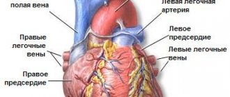
Venous blood - what is it?
Venous – blood that returns to the heart and lungs from organs and tissues. It circulates through the pulmonary circulation. The veins through which it flows lie close to the surface of the skin, so the venous pattern is clearly visible.
This is partly due to a number of factors:
- It is thicker, rich in platelets, and if damaged, venous bleeding is easier to stop.
- The pressure in the veins is lower, so if a vessel is damaged, the amount of blood loss is lower.
- Its temperature is higher, so it additionally prevents rapid heat loss through the skin.
The same blood flows in both arteries and veins. But its composition is changing. From the heart it enters the lungs, where it is enriched with oxygen, which it transfers to the internal organs, providing them with nutrition. The veins that carry arterial blood are called arteries. They are more elastic, blood moves through them in spurts.
Arterial and venous blood do not mix in the heart. The first passes along the left side of the heart, the second - along the right. They are mixed only in case of serious heart pathologies, which entails a significant deterioration in well-being.
Why are veins blue and not red?
Veins carry burgundy blood. They appear blue due to many factors. Primarily due to the color perception of the human eye.
Color is the wavelength of light emitted by an object or reflected by an object from another light source. Red light has the longest wavelength (700 nm). This means that it passes through objects and is not reflected from them. It also passes through the skin and, reaching the veins, is absorbed by hemoglobin. If you shine a red light on your hand, it will reflect everywhere except where the veins are. There it will turn black as it is absorbed. With this trick, doctors can find hard-to-reach veins.
Violet light is the shortest wavelength (400 nm). Blue has approximately the same indicators - 475 nm. It disperses easily and does not penetrate deep into the skin, but is reflected from it. If you look at your hand under a blue light, you won't be able to find any veins.
Attention! This trick is often used against those who like to go to clubs or other places. Violet or blue light in the toilet leaves no chance of revealing your veins.
Place your hand under regular white light. There are other colors in it, not just white. The veins will be blue, as it will be reflected from it, and the red will go deep into the skin and be absorbed there.
Why don't we see other vessels through which blood flows?
A person does not see the vessels because they are too deep under the skin. The light doesn't reach there. If the blood vessel is closer than 0.5 mm, it already absorbs all the blue light. Red is partially reflected, so people see this part of the skin as ruddy and pinkish. Veins that are clearly visible are located at a distance of no more than 0.5 mm from the surface of the skin.
Why can't we see arteries under the skin?
Most of the blood is in the veins, so they are much larger than arteries and vessels. Because blood puts more pressure on the arteries, they have thicker walls. Because of this, they are not as transparent and cannot be seen through the skin. If they were visible, arteries would likely look exactly like veins. Although the liquid in them is bright red.
What is the systemic and pulmonary circulation?
From the left ventricle, the contents are pushed out and enter the pulmonary artery, where they are saturated with oxygen. It is then distributed throughout the body through arteries and capillaries, carrying oxygen and nutrients.
The aorta is the largest artery, which is then divided into superior and inferior. Each of them supplies blood to the upper and lower parts of the body, respectively. Since the arterial system “flows around” absolutely all organs and is supplied to them with the help of a branched system of capillaries, this circle of blood circulation is called large. But the arterial volume is about 1/3 of the total.
Blood flows through the pulmonary circulation, which has given up all the oxygen and “taken away” metabolic products from the organs . It flows through the veins. The pressure in them is lower, the blood flows evenly. It returns through the veins to the heart, from where it is then pumped to the lungs.
Signs of bleeding
First aid for bleeding is stopping or reducing blood loss before the ambulance arrives. It is necessary to distinguish between types of bleeding and correctly use the necessary means to stop them. It is important to have dressings in your home and car first aid kits.
The most dangerous types of bleeding are arterial and venous. The main thing here is to act quickly, but do no harm.
- During arterial bleeding, blood flows in bright scarlet intermittent fountains at high speed in time with the heartbeat.
- With venous, a continuous or weakly pulsating dark cherry stream of blood flows from the injured vessel. If the pressure is low, a blood clot forms in the wound and blocks the blood flow.
- With capillary, bright blood slowly spreads over the entire wound or flows in a thin stream.
What is venous blood rich in?
When the blood reaches the organs, it gives them oxygen, in return it is saturated with metabolic products and carbon dioxide, and acquires a dark red hue.
A large amount of carbon dioxide is the answer to the question why venous blood is darker than arterial blood and why veins are blue. It also contains nutrients that are absorbed in the digestive tract, hormones and other substances synthesized by the body.
Its saturation and density depend on which vessels the venous blood flows through. The closer to the heart, the thicker it is.
What does color depend on?
First of all, the color of blood depends on red blood cells. They produce hemoglobin. It contains iron cells. Thanks to them, she becomes scarlet.
It is responsible for the supply of nutrients, oxygen, water, glucose to the organs, monitors the body temperature and, if necessary, can cool it down a little. To do this, the liquid takes excess heat from, for example, the liver and transfers it to the skin, which quickly cools.
Some marine inhabitants do not have hemoglobin, but there is another substance that performs exactly the same transport function - chlorocruorin. Because of this, a green liquid flows through their vessels.
Attention! The shade depends on what metal is present in the protein.
Crabs, spider crayfish and octopuses have blue-green blood. Hemocyanin flows in it. Some types of fish contain transparent biomaterial that looks like water. It consists of hemovanadium and vanadium ions.
Why are tests taken from a vein?
This is due to the type of blood in the veins - saturated with metabolic products and vital functions of organs. If a person is sick, it contains certain groups of substances, remains of bacteria and other pathogenic cells .
In a healthy person, these impurities are not detected. By the nature of the impurities, as well as by the level of concentration of carbon dioxide and other gases, the nature of the pathogenic process can be determined.
The second reason is that venous bleeding when a vessel is punctured is much easier to stop. But there are times when bleeding from a vein does not stop for a long time. This is a sign of hemophilia, a low platelet count. In this case, even a minor injury can be very dangerous for a person.
How to distinguish venous bleeding from arterial bleeding:
- Assess the volume and nature of leaking blood. The venous flows out in a uniform stream, the arterial flows out in portions and even in “fountains”.
- Determine what color the blood is. Bright scarlet indicates arterial bleeding, dark burgundy indicates venous bleeding.
- Arterial is more liquid, venous is thicker.
How to determine the type of bleeding
Visually, this is quite easy to do: the blood from the vein is dark, thicker and flows out in a stream, while the arterial blood is more liquid, has a bright scarlet hue and flows out like a fountain.
External differences between venous and arterial bleeding
Venous bleeding is easier to stop; in some cases, if a blood clot forms, it may stop on its own. A pressure bandage placed below the wound is usually required. If a vein in the arm is damaged, it may be enough to raise the arm up.
As for arterial bleeding, it is very dangerous because it will not stop on its own, the blood loss is significant, and death can occur within an hour.
How to stop venous bleeding?
With minor damage to the veins of the extremities, it is often enough to create an artificial outflow of blood by raising an arm or leg above the level of the heart.
A tight bandage should be applied to the wound itself to minimize blood loss. If the injury is deep, a tourniquet should be placed above the damaged vein to limit the amount of blood flowing to the injury site.
In summer you can keep it for about 2 hours, in winter - for an hour, maximum one and a half. During this time, you need to have time to deliver the victim to the hospital. If you hold the tourniquet longer than the specified time, tissue nutrition will be disrupted, which threatens necrosis.
It is advisable to apply ice to the area around the wound. This will help slow down your blood circulation.
Blood gas composition
Just understanding what color venous blood is is not enough to understand its differences from arterial blood. To do this, you should study biochemical indicators, especially considering how many misconceptions regarding their differences are described in materials on the Internet. In venous blood, the partial pressure of oxygen is almost 40 mmHg, which is more than two times lower than in arterial blood (96 mmHg). For carbon dioxide in connection with hemoglobin, the difference is approximately 14%: in the venous 46 mmHg, and in the arterial - 39 mmHg.
This means that in the veins the hemoglobin is saturated with oxygen by 50%, and the proportion of carbon dioxide is not 100%. This also means that carbon dioxide is also present in arterial blood. Its scarlet color is provided by the reflection spectrum of oxyhemoglobin, which is 2 times more than in veins and 3 times more than carboxyhemoglobin. In venous blood, the proportion of carbon dioxide is only 12% greater than oxygen, although even this difference ensures its cherry color with a dark blue tint.
The main differences between venous blood and arterial blood - Many good and useful tips
Arterial blood flows in capillaries. These are the smallest points on the human body. Each capillary carries a certain amount of liquid. The entire human body is divided into veins and capillaries. There is a certain type of blood flowing there.
Capillary blood gives a person life and ensures the flow of oxygen throughout the body and most importantly to the heart.
Arterial blood is red and flows throughout the body. The heart pumps it to all remote corners of the body, so that it circulates everywhere.
Its mission is to saturate the entire body with vitamins. This process keeps us alive.
Venous blood is blue-red in color, contains metabolic products, and flows through veins with very thin walls.
It can withstand high pressure, because when the heart contracts, changes can form that the vessels must withstand. Veins are located above the arteries.
They are easy to see on the body and easier to damage. But venous blood is thicker than arterial blood and flows out more slowly.
Systemic and pulmonary circulation
The most severe wounds for humans are the heart and groin. These places must always be protected. All the blood in a person flows through them, so with the slightest damage a person can lose all the blood.
There is a large and small circle of blood circulation. In the small circle, the liquid is saturated with carbon dioxide and flows to the lungs from the heart. It leaves the lungs, saturated with oxygen, and enters a large circle. Blood, based on carbon dioxide, runs from the lungs to the heart; through the capillaries, the lungs carry blood based on vitamins and oxygen.
The role and functions of venous blood
Venous blood is often used for human research. It is believed that it speaks better about human diseases, because it is a consequence of the work of the body as a whole.
In addition, it is not difficult to take blood from a vein, because it flows worse than a capillary, so a person will not lose much blood during the operation.
The largest human arteries should not be damaged at all, and if it is necessary to do a study of arterial blood, it is taken from a finger in order to minimize the negative consequences for the body.
Venous blood is used by doctors to prevent diabetes. It is necessary that the level of sugar in the veins does not exceed 6.1. Arterial blood is a pure liquid that flows throughout the body, nourishing all organs. Venous absorbs the waste products of the body, cleansing it. Therefore, it is by this type of blood that human diseases can be determined.
Bleeding can be external and internal. Internal is more dangerous for the body and occurs when human tissue is damaged from the inside. Most often, this occurs after a very deep external wound or a malfunction in the body leading to tissue rupture from the inside.
Blood begins to flow into the crack, and the body feels oxygen starvation. The person begins to turn pale and loses consciousness. This occurs because too little oxygen reaches the brain.
Venous blood can be lost due to internal bleeding and this will be harmless to humans, but arterial blood is not. Internal bleeding quickly blocks brain function due to lack of oxygen.
This will not happen with external bleeding, because the connection between human organs is not disrupted. Although, the loss of a large amount of blood is always fraught with loss of consciousness and death.
Summary
So, the main difference between venous blood and arterial blood is this color. Venous is blue, and arterial is red. Venous is rich in carbon dioxide, and arterial is rich in oxygen.
Venous flows from the heart to the lungs, where it turns into arterial, saturated with oxygen. The arterial flows through the aorta from the heart throughout the body.
Venous blood contains metabolic products and glucose, arterial blood is saltier.
Both types of blood are very important for humans. One feeds it, and the other collects harmful substances. In the process of blood circulation, blood passes one into another, which ensures the functioning of the body and the structure of the body that is optimal for life.
The heart pumps blood at high speed and does not stop working, even during sleep. This is very difficult for him. The division of blood into two types, each of which performs its own functions, allows a person to develop and improve.
This structure of the circulatory system helps us remain the most intelligent among all creatures born on Earth.
The main differences between venous blood and arterial blood - all the differences and differences on BigSovets.ru
Share the link and your friends will come to you for advice! Thank youツ
Source: https://bigsovets.ru/osnovnye-otlichiia-venoznoi-krovi-ot-arterialnoi/
By color
Both biological fluids are involved in all vital processes and ensure the normal functioning of the body.
How does venous blood differ from arterial blood? The first type of blood flow solves two main problems - reservoir and transport, while the second provides only the delivery function.
Other differences include the principle of movement, chemical composition and shades of blood.
By color
The venous fluid is deep red, almost cherry in color. This tone is given to it by decomposition products and carbon dioxide, which the substance is enriched with as a result of tissue metabolism.
The fluid in the arteries is rich in hemoglobin and oxygen, which is why it takes on a scarlet hue.
By composition
The venous substance, in addition to carbon dioxide and waste products of the body, contains useful substances that are broken down in the gastrointestinal tract. The blood substance also includes reduced hemoglobin, colloidal components and hormones synthesized by endocrine systems.
Arterial blood is cleared of metabolic products and is rich in compounds important for the body, obtained in the gastrointestinal tract: oxyhemoglobin, methemoglobin, salts and proteins.
By movement
Arterial blood moves from the heart to the cells under high pressure. Ejecting from the left cardiac ventricle into the aorta, which breaks up into vessels and arterioles, the liquid substance penetrates the capillaries, where oxygen and beneficial compounds are released into the cells. From there, the blood receives metabolic products and carbon dioxide.
Venous fluid flows in the opposite direction - towards the heart. Its pressure is significantly less than arterial pressure, since the flow has to overcome gravity and flow through the valves. Balance with bright red blood in the heart and vascular system is achieved due to the greater width and number of veins and the presence of a portal trunk in the liver.
Thanks to the branched system, the venous substance enters the heart through 3 large vessels and several small ones, and flows out through the pulmonary artery.
By function
The blood in the veins performs a cleaning function, as it collects and removes decay products and other toxic substances from the body. At the same time, it serves as a kind of depot of nutritional compounds and enzymes.
Arterial blood plays a transport role. It passes through all the cells of the body, saturating them with oxygen, stimulating metabolism and regulating some functions: respiratory, nutritional, homeostatic, protective.
By bleeding
It is not difficult to determine the type of external leakage from the vascular system. With venous blood loss, the substance comes out in a thick, slow stream. It is dark, almost black, and stops on its own after a while.
During arterial bleeding, the fluid gushes out like a fountain or splashes out in powerful bursts, obeying the contractions of the heart. Coping with such an outflow is difficult, and sometimes impossible, without the help of doctors.
The patient's condition deteriorates sharply, the skin turns pale and becomes covered with sweat, and loss of consciousness is possible.
Other differences
Another difference is that to determine the disease and make a diagnosis, blood is often taken from a vein. She is the one who can tell you about all the problems in the body.
The transformation of one substance into another occurs in the lungs. At the moment of receiving oxygen and releasing carbon dioxide, the blood fluid becomes arterial and continues its path through the body.
Isolation of flows is achieved by a perfect system of valves operating in one direction, so the liquids never mix anywhere.
The division of blood into arterial and venous is carried out according to 2 characteristics - the mechanism of its movement and the physical properties of the substance itself. However, these two indicators contradict each other - arterial fluid moves through the veins of the pulmonary circle, and venous fluid moves through the arteries. Therefore, the properties and composition of blood should be considered the determining factor.
A.K. has a bright red or scarlet tint. This color is given to it by hemoglobin, which has added O2 and become oxyhemoglobin. V. contains CO2, so its color is dark red, with a bluish tint.
Cardiac cycle
The right half of the heart and the left work synchronously. For convenience of presentation, the work of the left half of the heart will be considered here. The cardiac cycle includes general diastole (relaxation), atrial systole (contraction), and ventricular systole. During general diastole, the pressure in the cavities of the heart is close to zero, in the aorta it slowly decreases from systolic to diastolic, normally in humans they are 120 and 80 mm Hg, respectively. Art. Because the pressure in the aorta is higher than in the ventricle, the aortic valve is closed. The pressure in the large veins (central venous pressure, CVP) is 2-3 mm Hg, that is, slightly higher than in the cavities of the heart, so that blood enters the atria and, in transit, the ventricles. The atrioventricular valves are open at this time. During atrial systole, the circular muscles of the atria compress the entrance from the veins to the atria, which prevents the reverse flow of blood, the pressure in the atria rises to 8-10 mm Hg, and the blood moves into the ventricles. At the next ventricular systole, the pressure in them becomes higher than the pressure in the atria (which begin to relax), which leads to the closure of the atrioventricular valves. The external manifestation of this event is the first heart sound. Then the pressure in the ventricle exceeds the aortic pressure, as a result of which the aortic valve opens and blood begins to be forced out of the ventricle into the arterial system. The relaxed atrium fills with blood at this time. The physiological significance of the atria lies mainly in the role of an intermediate reservoir for blood coming from the venous system during ventricular systole. At the beginning of general diastole, the pressure in the ventricle drops below the aortic (closing of the aortic valve, II tone), then below the pressure in the atria and veins (opening of the atrioventricular valves), the ventricles begin to fill with blood again. The volume of blood ejected by the ventricle of the heart for each systole is 60-80 ml. This quantity is called stroke volume. The duration of the cardiac cycle is 0.8-1 s, giving a heart rate (HR) of 60-70 per minute. Hence, the minute volume of blood flow, as it is easy to calculate, is 3-4 liters per minute (minute volume of the heart, MVR).
What to do in case of arterial bleeding?
Bleeding from an artery is much more dangerous than venous bleeding. Blood quickly leaves the bloodstream, so immediate qualified assistance is required. Otherwise, hypovolemic shock develops, which leads to multiple organ failure - the gradual failure of all internal organs. To avoid this, it is important to clearly know how to act and what to do in case of arterial bleeding.
How to determine arterial bleeding?
First you need to make sure that the arterial vessel is directly affected. This is easy to do if you know what arterial bleeding looks like and the signs of this disorder. The main ones include:
- bright, scarlet color of blood flowing from the vessel;
- the blood flows in a stream, pulsates in time with the heartbeat;
- Before our eyes, the patient becomes pale, lethargic, and loses consciousness.
How to stop arterial bleeding?
If arterial bleeding is diagnosed, first aid should be provided immediately. Depending on the location of the injury, different tactics are used:
- if the limbs are affected, a tourniquet is applied;
- if the wound is on the torso or neck, apply a splint.
The tactics for stopping bleeding, taking into account the damaged artery, look like this:
- if the carotid artery is damaged, the vessel is pressed against the spinous processes of the neck;
- the jaw artery is damaged - to the jaw muscle;
- temporal artery - anterior to the edge of the auricle;
- subclavian - press with a fist to the outer edge of the clavicle from the back side;
- shoulder - presses against the inside of the muscle;
- femur - to the pubic bone;
- popliteal - to the kneecap.
Structure of the circulatory system
The four-chamber structure of the heart helps to separate arterial and venous fluid. Thus, they do not mix, which is necessary for the adequate functioning of the body.
There are 2 circles of blood circulation: small and large. Thanks to the first, blood passes through the capillaries of the lungs, is enriched with oxygen in the alveoli, becoming arterial. Then it goes to the heart, which, with the help of the powerful walls of the left ventricle, pushes it into a large circle through the aorta.
After the body tissues have taken all the nutrients from the capillaries, the blood becomes venous and returns through the systemic vessels of the same name to the heart, which sends it through the pulmonary arteries to the small one to again saturate it with oxygen.
So how does arterial blood differ from venous blood? What are their features?
Difference between venous and arterial blood
The blood that constantly circulates in the body is not the same everywhere. In some parts of the vascular system it is venous, in others it is arterial. What is this substance in each case, and how does venous blood differ from arterial blood? This is discussed below.
The content of the article
Among the functions of blood, the most important is supplying nutrition and oxygen to the tissues, as well as freeing the body from metabolic products.
All this movement of vital fluid occurs along a closed trajectory. In this case, there is a division of the system into two sectors, called circulatory circles.
What is the difference between arterial bleeding and venous bleeding?
How is arterial bleeding different from venous bleeding? The answer to this question can be obtained by studying medical literature and textbooks on anatomy. The human body is fraught with many interesting and unusual things.
A person needs a system that supplies organs with blood; without it, life would be impossible. Therefore, the whole body is permeated with various vessels, which have their own purpose.
Capillaries, veins, and arteries must always be active to ensure blood flow and organs and tissues to receive the required amount of oxygen.
All vessels have their own functions. Arteries and arterioles from the human heart carry blood, delivering it to each given area, providing the necessary nutrition on time.
Veins and venules carry blood back to the heart. The walls of the arteries are thicker, but very elastic, and their diameter is quite impressive.
This is necessary so that you can withstand the pressure under which the blood rushes through the arteries.
Arterial blood is distinguished by high oxygen saturation; thanks to this component, the blood becomes bright scarlet.
All these facts, when there is significant damage to the arteries, cause a phenomenon such as blood ejecting from the wound in sharp jolts, reminiscent of fountain jets, and the color of the blood is red, very bright.
The pulsating bleeding at these moments fully corresponds to the work of the heart, which contracts just as rhythmically.
Such bleeding is extremely life-threatening. If blood loss exceeds the acceptable level, the following may occur:
- blackout, clouding of consciousness;
- cardiopalmus;
- weak pulse beat;
- pale skin that becomes too cold;
- severe shortness of breath;
- death.
The ability of arterial blood to flow like a fountain causes rapid blood loss, and if large vessels are damaged, and these include the carotid artery, brachial artery, femoral artery, then help is needed without delay, otherwise death occurs very quickly.
The walls of the veins are much thinner, but also elastic, so that it is more convenient for the blood to function, and the venous circulation has slightly different functions.
Through the veins, which are much more numerous than arteries, blood rushes to the heart, but some components are missing in its composition. There is no longer oxygen in it, since after tissue gas exchange the blood is filled with carbon dioxide.
Because of this, it differs from the arterial one, moreover, it already has almost no nutrients, including glucose, but it contains the end products of metabolism.
The blood in the veins is much warmer, and its pH level is different from that that flows in the arteries: it is lower. By the way, for laboratory tests and diagnosis, doctors use the patient’s venous blood, since it can tell a lot about a person’s state of health.
In the veins, the blood changes its color slightly. Now it is dark red, almost cherry, but with a bluish or purple tint.
By the color of the blood you can distinguish whether it comes from a vein or an artery.
Venous bleeding has a significant difference from that which occurs with arterial injury. The blood flows calmly and can only pulsate very weakly in time with the heart. It happens that when a blood clot forms, the bleeding stops, which cannot happen when an arterial injury occurs.
Damage to the veins is not so dangerous, but still, if the femoral vein, subclavian or jugular vein is damaged, the blood flows out too quickly, and air embolism can occur from venous bleeding, and if the patient is not helped, death can occur.
source
Venous and arterial blood
To properly help a person with bleeding, you need to know exactly how. For example, arterial and venous bleeding requires a special approach. Arterial and venous blood are different from each other.
How is arterial bleeding different from venous bleeding?
Bleeding is the release of blood from the bloodstream. It can be primary, when it occurs immediately after damage to the blood vessels, and secondary, if it appears after some time.
Source: https://sfmggu.ru/chem-otlichayutsya-arterialnoe-krovotechenie-ot-venoznogo/
Venous and arterial blood: features, description and differences
Blood performs an important function in the body - it supplies all organs and tissues with oxygen and various useful substances. It takes carbon dioxide and decay products from the cells. There are several types of blood: venous, capillary and arterial blood. Each type has its own function.
For some reason, almost all people are sure that arterial blood is the type that flows in arterial vessels. In fact, this opinion is wrong. Arterial blood is enriched with oxygen, which is why it is also called oxygenated.
It moves from the left ventricle to the aorta, then goes through the arteries of the systemic circulation. After the cells are saturated with oxygen, the blood turns into venous and enters the veins of the BC. In a small circle, arterial blood moves through the veins.
Different types of arteries are located in different places: some are deep in the body, while others allow you to feel the pulsation.
Venous blood moves through the veins into the CD and through the arteries into the MC. There is no oxygen in it. This liquid contains a large amount of carbon dioxide, decay products.
Differences
Venous and arterial blood are different. They differ not only in function, but also in color, composition and other indicators. These two types of blood have differences in bleeding. First aid is provided in different ways.
Blood has specific and general functions. The latter include:
- nutrient transfer;
- transport of hormones;
- thermoregulation.
Venous blood contains a lot of carbon dioxide and little oxygen. This difference is due to the fact that oxygen enters only the arterial blood, while carbon dioxide passes through all vessels and is contained in all types of blood, but in different quantities.
Venous and arterial blood have different colors. In the arteries it is very bright, scarlet, light. The blood in the veins is dark, cherry-colored, almost black. This is related to the amount of hemoglobin.
When oxygen enters the blood, it enters into an unstable combination with the iron contained in red blood cells. Once oxidized, iron colors the blood bright red. Venous blood contains many free iron ions, which is why it becomes dark in color.
In this part of the circulatory system, blood circulation is slow as the heart pushes fluid away from itself. Valves located in the vessels also affect the reduction in movement speed. This type of blood movement occurs in the systemic circulation.
In the pulmonary circle, arterial blood moves through the veins. Venous - through the arteries.
In textbooks, on schematic representations of blood circulation, arterial blood is always colored red, and venous blood is always colored blue. Moreover, if you look at the diagrams, the number of arterial vessels corresponds to the number of venous ones. This image is approximate, but it fully reflects the essence of the vascular system.
The difference between arterial blood and venous blood also lies in the speed of movement. The arterial is ejected from the left ventricle into the aorta, which branches into smaller vessels. Then the blood enters the capillaries, nourishing all organs and systems at the cellular level with useful substances.
It also moves slowly through the veins, since it has to overcome the force of gravity and cope with the system of vascular valves.
Due to the difference in pressure, blood for testing is taken from capillaries or a vein. Blood is not taken from the arteries, since even minor damage to the vessel can provoke extensive bleeding.
When providing first aid, it is important to know which blood is arterial and which is venous. These species are easily identified by their flow patterns and color.
When providing first aid, it is necessary to raise the limb and compress the damaged vessel by applying a hemostatic tourniquet or pressing it with finger pressure. In case of arterial bleeding, the patient must be taken to the hospital as quickly as possible.
Arterial bleeding may be internal. In such cases, a large amount of blood enters the abdominal cavity or various organs. With this type of pathology, a person suddenly becomes ill, the skin turns pale. After some time, dizziness and loss of consciousness begin. This is due to lack of oxygen. Only doctors can provide assistance with this type of pathology.
With venous bleeding, dark cherry-colored blood flows out of the wound. It flows slowly, without pulsation. You can stop this bleeding yourself by applying a pressure bandage.
The pulmonary circulation is characterized by the release of arterial blood from the heart, passing through the veins to the lungs, where it is saturated with oxygen and returns back to the heart. From there it goes along the aorta in a large circle, delivering oxygen to all tissues.
Passing through various organs, the blood is saturated with nutrients and hormones, which are carried throughout the body. In the capillaries there is an exchange of useful substances and those that have already been used. This is where oxygen exchange occurs. From the capillaries the fluid enters the veins.
At this stage, it contains a lot of carbon dioxide, decay products.
Through the veins, venous blood is carried throughout the body to organs and systems, where cleansing of harmful substances occurs, then the blood approaches the heart, passes into a small circle, where it is saturated with oxygen, giving off carbon dioxide. And everything starts all over again.