Venous blood is contained in the veins leaving organs, pulmonary arteries, and the right half of the heart. It removes carbon dioxide, metabolic products and toxins from tissues. In the alveoli of the lungs it turns into arterial.
The blood in the veins is darker, thicker, clots faster, flows slower, and is easier to stop; usually a tight bandage is enough. It is more suitable for laboratory research than finger capillary. If there is communication between arteries and veins, incomplete separation of blood occurs (heart defects, vascular disease, lung disease).
Venous blood: main indicators
Venous blood is that which flows from the internal organs, limbs, and brain through the veins . It is contained in the right atrium and pulmonary arteries. Functions: carries away carbon dioxide and metabolic products. Its color is darker, its viscosity and temperature are higher, its pressure and speed are lower than those of arterial blood.
Such properties, as well as the presence of microbial residues, toxins, immune complexes, and hormones, make it the main material for identifying diseases in laboratory diagnostics.
We recommend reading the article on coronary circulation. From it you will learn about the diagram of the coronary circle, physiology, regulation of the small coronary circle, and research methods. And here is more information about hypoxemia and hypoxia.
What is it, its color, main characteristics and composition
Venous blood is that which flows from the organs to the heart, and from it enters the lungs. It takes in carbon dioxide and metabolic products. It is used for testing and medications are also injected into it through intravenous injections. There are differences in the main characteristics and composition between the blood in the arterial and venous beds; they are presented in the table.
| Sign | Venous | Arterial |
| Pressure | Low | High |
| Blood movement | Slow | Fast |
| Direction of blood flow | From the organ | To the organ |
| Purpose | Removal of metabolic products and carbon dioxide | Nutrition and oxygen supply |
| Nutrients | Few | A lot of |
| pH (acidity) | Low | High |
| Oxygen level | Short | High |
| Carbon dioxide content | High | Low |
| Color | Dark, cherry-burgundy, with a bluish tint | Scarlet, light, bright |
| Viscosity | High | Low |
| Temperature | High | Low |
It is important to note that the maximum difference between arterial and venous blood concerns gas composition, pressure and flow rate. All other physical and chemical properties differ only slightly.
Functions
Main functions of venous blood:
- removal of metabolic products (cleansing);
- transfer of absorbed nutrients from the intestine to the liver through the portal vein (transport);
- removal of excess hormones, salts (maintaining balance, equilibrium, homeostatic);
- delivery of carbon dioxide to the lungs for removal from the body (respiratory).
What is venous blood rich in?
Venous blood is rich in carbon dioxide; it also contains metabolic end products, toxins and microbial remains. Because of this, it is possible to identify the main diseases:
- infections;
- metabolic disorders (for example, atherosclerosis, diabetes mellitus);
- immune, allergic, autoimmune pathologies;
- blood diseases due to changes in the level of red blood cells, leukocytes, platelets;
- dehydration;
- risk of increased thrombus formation and blockage of veins and arteries;
- liver and kidney dysfunctions;
- hormonal disbalance.
Elements of the human circulatory system containing venous blood
Venous blood is contained in the following elements of the circulatory system:
- subcutaneous and deep venous vessels;
- venous networks of internal organs, brain;
- large vena cava (superior and inferior), carrying blood to the right atrium;
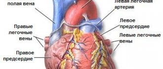
- the pulmonary artery emerging from the right ventricle and its branches in the lungs.
Artery through which venous blood flows
Venous blood flows through the pulmonary artery. This name is due to the fact that all the vessels leaving the heart are called arteries, and those coming to it are called veins. Therefore, in the veins of the lungs there is an arterial vein, rich in oxygen, and in the arterial network - carbon dioxide.
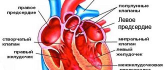
Which parts of the heart contain venous blood?
Venous blood is found in the right atrium and right ventricle of the heart. If there is a defect in the septum between the right and left parts, then a mixture of arterial and venous occurs.
Why are tests taken from a vein?
All metabolic products, hormones and toxins accumulate in the veins. Therefore, by venous blood it is possible to determine diseases of internal organs and the brain, which are manifested by changes in the composition of cells or plasma (liquid part). A puncture of a vein causes slight bleeding, which can be stopped very quickly by squeezing the vessel. If there is even slight damage to the artery, a tourniquet will be needed.
How to stop venous bleeding
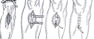
In order to stop venous bleeding, you need:
- lift the limb up;
- Apply a pressure bandage to the site of damage to the vessel; if the bleeding is severe, then below the wound you should not apply too much pressure to the soft tissue with a tourniquet.
After providing first aid, the patient must be examined by a doctor.
Expert opinion
Alena Ariko
Expert in Cardiology
You should not wash the wound yourself or remove traumatic objects from it or remove blood clots.
Features of the circulatory system: what kind of blood flows through the pulmonary arteries?
Anatomy
10.08.2016
18.1 thousand
12.1 thousand
7 min.
What kind of blood flows through the pulmonary arteries? Do arteries always contain arterial blood? If you remember school anatomy, you can easily navigate the principle of operation of the cardiovascular system.
The heart has a right and left side, each of which contains an atrium and a ventricle, which are separated by valves. These valves only allow blood to flow in one direction; it cannot flow in the opposite direction.
These parts are not connected to each other.
Venous blood always flows through the right atrium and the inferior vena cava; it does not contain much oxygen, but, on the contrary, is saturated with carbon dioxide. It flows into the right ventricle, it contracts and pushes it further.
It is divided into the right and left pulmonary arteries, which carry blood to the lungs. The artery is divided into lobar and segmental branches, and they diverge into arterioles and capillaries.
It is in the pulmonary space that venous blood is freed from carbon dioxide and enriched with oxygen, turning into arterial blood. The pulmonary vein carries blood to the left atrium and left ventricle.
It then has to overcome high pressure to be pushed into the aorta. After this, it spreads through the arteries and goes to the internal organs.
The artery branches into small capillaries, and towards the end of the path the pressure drops to a minimum. Oxygen and necessary substances penetrate into the tissue of the human body through a network of capillaries, and the liquid itself is saturated with water and carbon dioxide.
Dividing into a capillary network, the blood from arterial becomes venous. The network of capillaries merges into venules, which develop into larger veins and eventually enter the right atrium.
This is the circulatory cycle of a healthy person.
An artery refers to the type of vessel that carries blood away from the heart. The walls of the artery are thick, the fibers in the middle layer are elastic, and the muscles are smooth. These vessels can withstand a large flow of blood pushed out under pressure. They stretch, but do not tear, unlike other types of fabrics.
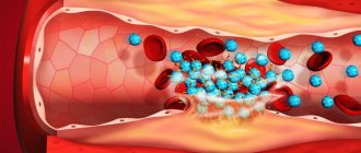
With thromboembolism, a blood clot, one or more, appears in the pulmonary arteries. They look like clots that float in liquid.
Typically, they begin in the main veins and separate from the vessel wall to continue their journey to another part of the system. This movement towards the pulmonary artery is especially dangerous.
Migrating blood clots are the most dangerous, since it is unknown in which part and how seriously they will block important gaps. They are called emboli, hence the name of the disease – embolism.
What kind of blood is called venous and how is it different from arterial? Venous in appearance is dark red in color, sometimes you can notice that it gives off a blue color, it is so dark. This effect is associated with the presence of carbon dioxide and metabolic products.
Venous blood has low acidity and is warmer in temperature than arterial blood. The mechanism of blood flow through the vein is associated with its close location to the upper layers of the skin. This is due to the structure of the venous network, due to valves that slow down the speed of fluid flow.
Venous blood does not contain many nutrients and has little sugar. For a number of reasons, it is precisely this that is taken for analysis during research.
The peculiarity of the pulmonary artery in anatomical terms is that it is presented as a paired blood vessel and belongs to the pulmonary circulation. It is connected to the pulmonary trunk, and, remarkably, it is the only vessel that carries venous blood to the respiratory organ.
The pulmonary artery has two branches, they do not exceed 3 cm in diameter in a healthy person, the pulmonary trunk extends from the right side of the heart. The main task of the pulmonary arteries is to transport venous blood to the lung. Thus, venous blood flows through the pulmonary artery, despite the name of this vessel.
If there are any disturbances in the human body, the transport of blood through the pulmonary artery is disrupted. The most dangerous diseases are: pulmonary embolism, embolism. It becomes impossible to transfer fluid due to the presence of blood clots and blockages.
If the pulmonary artery becomes blocked by fatty deposits, air bubbles, a foreign body, or a tumor, the natural flow of blood is disrupted.
Disturbed blood flow and problems with the walls of blood vessels slow down the resorption of the blood clot, so normal blood circulation is not restored.
If pulmonary artery stenosis occurs, the outflow tract of the right ventricle narrows in the area of the valves. The most unpleasant thing that happens because of this is that the pressure in the pulmonary arteries and the right side of the ventricle is disrupted. The problem is also associated with the development of defects in the interatrial region, the pressure of the right atrium increases, and insufficiency occurs.
The pulmonary artery has an extremely fragile structure; it has thin walls; compared to the large aorta, they are simply lost. The branches are short, the entire pulmonary arterial system has a larger diameter than the systemic part of the arteries.
This vessel is not only thin, but also elastic; it gives the arterial mesh the ability to stretch up to 7 ml/mm Hg. This characteristic is inherent in the entire systemic arterial bed. This property allows the pulmonary artery to adapt to the volumes of the right ventricle.
The pulmonary vein is as short as the pulmonary artery. It supplies fluid to the left side of the atrium, from where it exits into the circulation.
Venous blood flows through the pulmonary arteries - this is a normal process that is tied to the circulation. If the system is disrupted, the entire cardiovascular part of the body suffers. Vital arteries should remain elastic and free of blood clots for as long as possible.
The heart operates on an autonomous principle, generating electrical impulses that travel through the muscles and allow them to contract.
These impulse tremors appear with a given regularity, there are about 75 of them in 60 seconds. The conduction system of the heart has sinus nodes, from which nerve fibers come. The heart muscle needs oxygen.
It reaches it through arteries called coronary arteries.
The right and left pulmonary veins carry arterial blood that flows from the lungs. The movement of these veins begins from the gate of the lung, as a rule, two come out of each lobe.
It is normal for a person to have up to five pulmonary veins. Each pair is divided into superior and inferior pulmonary veins. They go to the left side of the atrium and enter the posterolateral region.
The right pulmonary vein appears longer than the left and is located lower.
In the pulmonary veins, the origin is associated with a powerful capillary network, pulmonary acini. The capillaries merge and form a large venous network.
The pulmonary artery is located in the periarterial lymphatic space, the capsule and fissure that separates the walls of the arteries from the stretching tissue of the lungs. When there are changes in tension inside the lungs, pressure acts on these gaps.
When a person inhales air, space expands, and when he exhales, it contracts. When the arteries fill with venous blood, they pulsate, and a large amount of fluid stretches the walls of the vessel, creating high pressure.
Despite the pronounced effect, nearby structures do not experience discomfort.
The pulmonary arteriole has mural muscle tissue, and the precapillaries do not have the periarterial lymphatic space, the same gap as veins and venules. They are woven into the lung tissue. The lumen of blood vessels is associated with tension due to the growth of alveolar tissue.
Due to their attachment to the periphery, if the volume of air in the lungs increases, the vessels become longer with inhalation.
This process affects the flow of blood from the lung and affects the activity of the heart as a whole due to the fact that when the lumen narrows, the existing elongation increases resistance.
The pulmonary artery, or pulmonary trunk, is the main vessel in the pulmonary circulation. It is the only one through which venous blood flows, not enriched with oxygen.
With pulmonary hypertension, the pressure level increases, this is due to increased resistance of the vascular bed of the lung or increased blood flow. Such pathologies are usually secondary, and if the cause cannot be found, they are designated as primary. When suffering from pulmonary hypertension, the blood vessels narrow and hypertrophy significantly.
In the presence of the disease, the patient experiences an increase in blood pressure, which is associated with the artery. It grows gradually, progressing. It all ends with the person developing heart failure and ending his life in the hands of doctors.
Even if the symptoms of the disease are not clearly expressed, you need to be careful about possible pathology. In the treatment of pulmonary hypertension, a whole range of drugs is used, from oxygen-containing inhalations to diuretics.
Predicting the situation is related to the initial cause of pressure surges.
The pulmonary artery contains venous blood, despite the general belief that only arterial blood should flow through the arteries.
Pulmonary embolism does not always manifest itself actively, immediately leading to heart failure. Most often, embolism is expressed in slight tachycardia and chest pain. All this may not be noticed the first time.
When a patient experiences shortness of breath when walking short distances, the temperature rises, the person wheezes when breathing, then they run to the doctor.
A pulmonary embolism can lead to a collapsed lung, which is life-threatening.
If you send the blood to a specialist’s laboratory and do not tell him what it is, he will immediately determine by the chemical composition what kind of liquid is in front of him and where it came from. The chemistry of arterial and venous blood is very different.
It is considered a healthy indicator when oxygen in the artery contains up to 100 mm Hg.
If you take a drop of arterial blood, there will be carbon dioxide molecules in it, but to a lesser extent, it is rich in oxygen and nutrients.
The opposite is true with venous blood, which is predominantly filled with gas and has little oxygen. It transports breakdown products of cellular material. In laboratory tests, the acid-base balance level is 7.4, and the venous level is 7.35.
Since blood does not disappear anywhere from the human body, it turns from arterial to venous. This process is called gas exchange, since during the process the liquid gives up oxygen and receives carbon dioxide. Oxygen enters the blood from the air. Despite this, the pulmonary artery contains venous blood that is not rich in oxygen, but devoid of all nutrients.
In order to understand what processes occur in your body, you need to know the blood distribution system and blood circulation. Blood is directly related to pressure; if the walls of blood vessels are affected, the pressure increases.
It cannot be kept at a high level, since the network of arteries and veins throughout the body, if not functioning correctly, can seriously harm not only the heart, but also other internal organs.
To monitor how blood flows through vital arteries, such as the pulmonary arteries, you need to check your condition with a doctor, avoid high blood pressure, avoid stressful situations and get quality rest.
Source: https://vashflebolog.com/anatomy/kakaya-krov-techet-po-legochnym-arteriyam.html
Arterial and venous blood
Differences between arterial and venous blood: dark blood flows from the veins, it is thicker, it clots faster, the bleeding is less intense, the stream is smooth and not strained. Venous blood is more suitable for laboratory tests than capillary blood from a finger. It flows through the pulmonary arteries, and in the capillaries of the alveoli it turns into arterial artery.
Which one is darker
Venous blood is darker. Its color depends on the form of hemoglobin. In the arterial blood, it is combined with oxygen (oxyhemoglobin), which gives it a bright scarlet color. The venous contains both oxyhemoglobin and 2 other forms:
- reduced (gave oxygen to cells, but has not yet added carbon dioxide);
- carboxyhemoglobin (a compound with carbon dioxide).
Most of the latter pigment, so the color becomes dark cherry.
Capillary and venous blood: differences
The main differences between capillary blood (from a finger) and venous blood are the contents:
- cells, especially platelets (higher in the venous), leukocytes (higher in the capillary);
- glucose (higher in the veins).
Analysis from a vein is considered more accurate, since it does not contain impurities of tissue fluid or surface epithelium of the skin. Also, circulatory disorders, vascular spasm, and fever can affect the indicators of capillary blood tests. For many types of laboratory diagnostics, a sufficient volume of material is needed, and up to 0.5 ml can be taken from a finger.
Therefore, capillary blood can be used only at the first stage of the examination. It will give reliable results only when determining hemoglobin, red blood cells and ESR. For biochemical and immunological analysis, studies of hormonal levels, as well as, if necessary, an in-depth study of cellular composition, blood from a vein is needed.
Why does venous blood clot faster?
Venous blood clots faster due to high platelet levels. These blood platelets form the basis of the clot, they are connected to each other, and fibrin threads give strength to the formed blood clots.
How to determine the type of bleeding
To determine the type of bleeding, you need to pay attention to the signs that distinguish them:
| Sign | Venous | Arterial |
| Blood color | Dark | Light red |
| Jet type | Rovnaya | Pulsating |
| Speed | Slow | Fast |
| Pulsation below injury | Regular | Absent or thready pulse |
| Skin color and temperature | Pale with a bluish tint, cold | Normal color, some swelling, warm |
Through which veins does arterial blood flow?
Arterial blood flows through the veins that leave the lungs to the left atrium. Also, the entire internal venous network of the lungs is filled with hemoglobin, saturated with oxygen. Therefore, the contents in them are exactly the same in characteristics as in the arteries of all other organs (except the lungs).
Where venous blood turns into arterial blood
Venous blood is converted into arterial blood in the capillary network of the alveoli (vesicles) of the lungs. The pulmonary arteries branch into thin capillaries; they entwine the alveolar walls. Carbon dioxide enters the lumen of the pulmonary vesicle, and oxygen from it passes into the blood.
Oxygen molecules join the reduced hemoglobin, which has given up carbon dioxide, and it becomes oxyhemoglobin. Therefore, the blood changes its color and properties - from dark venous to light arterial. Through four pulmonary veins (two left and two right) it goes to the heart.
Treatment methods
Thromboembolism is a condition that requires urgent intervention. A patient with this diagnosis is placed in intensive care and placed on strict bed rest. Use an oxygen mask or another method of oxygen supply to avoid oxygen starvation. A venous catheter is installed. The faster these measures are carried out, the less likely the development of negative consequences (impaired blood supply to the lungs, development of chronic pulmonary hypertension and sepsis).
Vessel stenosis, if pronounced, is eliminated surgically. To maintain the general condition of the patient and prevent the development of complications, the doctor prescribes medications.
Valve insufficiency in severe cases is eliminated surgically. In other conditions, drug treatment is used.
Medications
In the case of pulmonary embolism, medications are prescribed: heparin intravenously, antibiotics if necessary, dopamine and more. They supply oxygen.
For stenosis, the following may additionally be prescribed:
- diuretics;
- drugs against acute and chronic heart failure (glycosides);
- calcium channel blockers and others.
Valve insufficiency requires prescription:
- antibiotics;
- nitric acid salts to dilate blood vessels;
- diuretics;
- ACE inhibitors (block the process of vasoconstriction).
Traditional methods
Traditional methods are used only after consultation with the attending physician and diagnosis. Self-prescribing treatment for yourself can lead to a worsening of the condition.
One of the recipes that helps reduce blood cholesterol levels and strengthen the walls of blood vessels includes:
- Dill seeds;
- honey;
- water.
For 2 liters of water, use dill and honey in a ratio of 1 glass to two. The mixture of seeds and honey is poured with boiling water and left for a day in a thermos. Next, the infusion is filtered and stored in the refrigerator. Take orally twenty minutes before meals, a tablespoon four times a day. After the infusion is over, you need to take a week break. If an allergic reaction occurs, stop taking it.
Other methods
These are methods that involve surgical intervention. Severe degrees of stenosis can only be eliminated using this method. Used: excision of the affected vessel wall, separation of the vessel walls, excision of the affected area of the myocardium.
Valve insufficiency in severe cases is corrected surgically:
- valve prosthetics (there are mechanical valves and biological valves);
- plastic surgery of your own valve;
- heart and lung transplant (performed in rare cases when the heart is significantly damaged).
It is possible to simultaneously correct several pathologies.
Incomplete separation of arterial and venous blood
Incomplete separation of arterial and venous blood occurs with heart defects and large vessels , as well as lung diseases:
- atrial septal defect;
- a hole in the septum between the ventricles;
- collapse of the walls (closing) of the alveoli of the lungs (atelectasis), filling with fluid (edema, pneumonia);
- fistula between a vein and an artery (congenital, less commonly acquired);
- transposition of the great vessels (aorta and pulmonary artery change places);
- underdevelopment of the heart chambers (two- and three-chamber);
- open arterial (ductus Botallus, connects the aorta and pulmonary artery).
Dark blood from a vein: what does it mean?
The dark color of blood from a vein means that the organ from which it flows is actively functioning. The higher the rate of metabolic processes, the more oxygen is absorbed from the blood, which means its color will be darker.
We recommend reading the article about venous congestion in the legs. From it you will learn about the causes and symptoms of the pathology, conservative and surgical treatment. And here is more information about vascular injury.
Venous blood flows from the organs, collects in the large vena cava and then into the right half of the heart. Through the pulmonary arteries it approaches the alveoli and turns into the arterial artery. The properties of blood from a vein are darker, thicker, and clot quickly. It is used for laboratory diagnostics, as it contains metabolic products, toxins, microbes, and hormones. Venous bleeding is accompanied by a slow and uniform release of blood; it is easier to stop than arterial bleeding.