What is hemostasis? What processes does it characterize in the human body?
Hemostasis is a complex of natural reactions of the body that are responsible for blood circulation, ensure its normal density, prevent pathological blood loss and stop bleeding in the event of a violation of the integrity of blood vessels.
The level of completeness of hemostasis depends on parameters such as:
- functionality of vascular walls;
- platelet volume in the blood, their activity;
- the state of the fibrinolysis system, as well as the blood coagulation system.
Another meaning of the term “hemostasis” is a set of medical measures aimed at preventing and stopping bleeding during injuries and during surgical operations.
What is the hemostasis system and what are its functions?
The diagram shows two of the four components (in the list below these are items 1 and 4)
The hemostasis system is a biological structure that allows you to maintain a fluid state of blood, as well as prevent and inhibit bleeding. It includes four main elements:
- the blood coagulation system, which is presented in the form of 13 factors and constitutes the coagulation link;
- platelets forming the platelet unit;
- anticoagulant structure (anticoagulant link);
- the fibrinolysis system, including plasminogen (a special circulating proenzyme), as well as its endogenous activators.
The physiological meaning of the hemostasis system is to maintain the optimal blood volume in the vessels for complete blood supply to the organs.
Normally, there is a slight predominance of anticoagulant processes. When there is a danger of blood loss, the balance immediately shifts towards procoagulants that reduce coagulation. If for some reason this does not happen, abnormal bleeding develops. When procoagulant activity is too high, thrombosis and embolism form. Stopping blood loss is the result of physiological processes realized through the interaction of different functional and structural elements.
The normal operation of the hemostasis system depends on the functionality of:
- liver and bone marrow;
- spleen and other hematopoietic organs.
How does bleeding stop?
The primary role is played by directly violating the integrity of the walls. After all, it is precisely as a result of this that collagen, formed as a result of the exposure of subendothelial tissue structures, is thrown into the bloodstream.
Then platelet activation begins. This occurs due to the appearance of von Willebrand factor in the blood, which in turn is caused by a sharp jump in the amount of protein.
Under the influence of a certain number of factors, they begin to swell, become covered with numerous processes and cover the damaged area.
The release of the contents occurs with the help of the formed collagen.
The last stage occurs with the help of adrenal hormones such as serotonin, adrenaline and norepinephrine, which, when released into the bloodstream, cause a spasm, due to which the bleeding gradually slows down.
Besides:
- platelet aggregation increases significantly;
- spasm of the blood vessel occurs with damage.
All processes included in platelet hemostasis significantly reduce the amount of blood that is released from the wound, and also ensure the accumulation of hemostatic substances in the area of damage.
Then the newly formed plug gradually acquires a denser structure and becomes more firmly attached to the damaged area. This occurs due to actomyosin-like proteins - thrombostenins, which make cells more dense by squeezing it out.
All together creates platelet hemostasis itself. In the damaged area, the formation of a coagulation link does not begin, but an unstable soft thrombus is formed, which, if necessary, may well stop the bleeding that has begun.
However, it is worth remembering that if the veins and arteries are damaged, this method will not bring the desired results, since the blood flow there is many times faster and under higher pressure.
Types and mechanisms
Three types of hemostasis are classified, which essentially represent its stages:
- vascular platelet;
- coagulation,
- fibrinolysis.
Scheme of human hemostasis in normal conditions and with disorders
Depending on the intensity of bleeding, one of them prevails in the process of thrombus formation, despite the fact that all three types begin to work simultaneously. They exist in a state of continuous interaction, complementing each other from the very beginning of the formation of a blood clot until its dissolution.
- Primary (vascular-platelet) hemostasis develops gradually:
- damage to the vessel leads to contraction of its walls, due to which bleeding stops after 30 seconds;
- platelets are directed and attached to the damaged area of the endothelium;
- a reverse accumulation of platelets is formed. They change shape, due to which they “stick” to each other and stick to the vascular wall. A primary or “white” (colorless) thrombus forms. Under the influence of thrombin, adhesion is irreversible;
- a hemostatic plug is formed, which differs from a blood clot in that it does not contain fibrin elements. Plasma coagulation factors accumulate on its surface. They give rise to the internal processes of coagulation hemostasis, which ends with the formation of fibrin threads. A dense blood clot forms on the basis of the plug. In certain pathologies (myocardial infarction, stroke), the fact of thrombus formation can cause serious complications if it cuts off the nutrition of vital organs. In such cases, doctors use special medications.
- Secondary (coagulation) hemostasis stops blood loss in those vessels where the primary one is insufficient. It lasts two minutes and essentially represents a reaction between plasma proteins, resulting in the formation of fibrin strands. This leads to stopping bleeding from the damaged vessel and makes it possible not to fear a relapse in the near future. Secondary hemostasis can be activated in two ways:
- external, when tissue thromboplastin enters the blood as a result of the interaction of collagen with the coagulation factor at the site of damage to the vascular wall;
- internal. If thromboplastin does not come from outside, the process is triggered by tissue factors. The internal and external mechanisms are interconnected thanks to kinin-kallikrein proteins.
- Fibrinolysis. It is responsible for restructuring fibrin threads into soluble combinations, restoring vessel patency, and maintaining optimal blood viscosity. In addition, it uses the ability of leukocytes to destroy pathogens and eliminates thrombosis.
In addition to the listed varieties, they are classified:
- Hormonal hemostasis, which represents the basis for the treatment of dysfunctional uterine bleeding (DUB) in women of any age. It involves the use of hormonal drugs, such as pills used for oral contraception.
- Endoscopic hemostasis is a set of special measures to stop ongoing bleeding. Among its methods: mechanical, physical and medicinal. Each of them, in turn, consists of a set of different procedures, depending on the nature of blood loss. In this case, they distinguish:
- temporary hemostasis is a preliminary stop of bleeding, which doctors perform urgently at the scene of the incident;
- final hemostasis, performed in the hospital.
- Plasma hemostasis is a sequence of transformations that occur in the blood plasma with the participation of 13 coagulation factors.
The main ultimate goal of all types and methods of hemostasis is to stop the bleeding process.
Mechanisms to eliminate bleeding
The vascular-platelet link of hemostasis consists of three primary structures that work in an orderly manner and simultaneously.
They are divided into three types, according to the type of working conditions:
- Vascular-platelet (primary).
- Coagulation (minor).
- Dissolution of the thrombus.
The main task of this system is that, through the action of thrombin, a protein called fibrinogen is converted into fibrin, which is insoluble in liquid. Each blood clot in the body is a unique combination of platelets and fibrin. They play a major role in renewing damaged blood vessel walls, and their growth factor helps in stimulating tissue regeneration.
Consists of several inseparable systems. Their stable functioning ensures constant neurohumoral regulation. The micro-mechanisms of negative and positive communication within the system function flawlessly, allowing the rapid creation of clots to prevent blood loss. And then just as quickly dissolve them as unnecessary.
Hemostasis disorders: causes and factors
Problems with one of the elements of hemostasis can pathologically affect the process itself. Among the reasons for the violation:
- DIC syndrome (disseminated intravascular coagulation). An anomaly in which blood clotting is impaired due to the large volume of thromboplastic substances released from the tissues. Often this occurs without any symptoms.
- Coagulopathy is a dysfunction of the coagulating and opposite blood systems. This pathology can be either congenital or acquired, including due to immune disorders.
- Thrombophilia is a disorder of the processes of coagulation and thrombus formation, leading to ischemia of tissues and organs.
- Hypocoagulative-hemorrhagic disorder. Characterized by deterioration of blood clotting.
Summarizing the causes of hemostasis disorders, we can state that they are:
- congenital or acquired;
- hypocoagulative or hypercoagulative;
- local (like thrombosis) or global (like DIC).
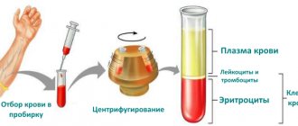
Table of blood clotting factors
| Clotting factors | |
| Factors | Properties |
| I – fibrinogen | Thrombin initiates the conversion of the first factor to fibrin |
| II – prothrombin | Synthesis in the liver only together with vitamin K |
| III – thromboplastin | With its participation, prothrombin is converted into thrombin |
| IV – calcium ions | Needed to activate clotting factors |
| V – proaccelerin | Stimulates the transition of prothrombin to thrombin |
| VI – whey accelerator | Initiates the transition of prothrombin to thrombin |
| VII – proconvertin | Acts on the third factor (activation) |
| VIII - antihemophilic factor A | X factor cofactor |
| IX - antihemophilic factor B (Christmas) | Activates factors VIII and IV |
| X – Stewart-Prower factor | Prothrombinase stimulation |
| XI – thromboplastin precursor | Activates factors VIII and IX |
| XII – Hageman factor | Takes part in the conversion of prekallikrein to kallikrein |
| XIII – fibrin-stabilizing factor | Stabilization of the formed fibrin mass |
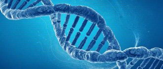
Polymorphism of hemostasis genes
These are mutational changes in genes responsible for hemostasis, which lead to various pathologies. Polymorphism happens:
- Congenital and representing hereditary transmission in a descending line, disorders of the properties of blood and the structure of blood vessels. The child runs the risk of inheriting such problems from one or both parents. The abnormality may affect one or more genes. Symptoms of carriage appear:
- in childhood;
- during pregnancy;
- during therapy with hormone replacement drugs, the use of oral contraceptives;
- never. Blood clotting disorders in close relatives are a reason to consult a doctor to check the functionality of your own hemostatic system.
- Acquired, which is the result of exposure to various factors. For example, antiphospholipid syndrome - an autoimmune pathology - leads to the fact that the body begins to synthesize antibodies to “native” phospholipids, which leads to blood clotting disorders. This is rare, but mutations in hemostasis genes also occur for reasons such as:
- prolonged exposure to chronic infections, stress;
- oncological or endocrine pathologies;
- injuries;
- taking certain medications.
Doctors are increasingly adding tobacco smoking to the list of factors, since nicotine affects blood clotting, which means it may be one of the acquired causes of mutation of hemostasis genes.
The consequences of congenital defects of hemostasis genes are such conditions as:
- thrombophilia is a pathology caused by increased blood clotting, resulting in an increased risk of thrombosis (the development of vascular blood clots);
- fetoplacental insufficiency during pregnancy;
- miscarriage;
- the birth of underdeveloped or dead children.
The polymorphism factor of hemostasis genes can be identified by undergoing a special hemotest.
Clinical picture of the disease
The main symptoms are:
Mutation analysis
This is a diagnostic method that allows you to accurately identify predispositions to blood diseases, which is especially important before and during pregnancy. The analysis identifies genetic abnormalities that carry the risk of late spontaneous abortion, intrauterine developmental defects, fetal blood loss, and maternal blood cancer pathologies.
It was said above that congenital abnormalities of hemostasis genes may not manifest themselves in any way, but in critical circumstances for the body, for example, during pregnancy, childbirth or during surgery, dysfunctions associated with the process of blood clotting are sometimes detected, which can lead to dangerous consequences .
There is a list of hemostasis genes that are more often susceptible to mutation:
- G20210A - prothrombin gene. Its changes are manifested by hereditary thrombophilias. The likelihood of thrombosis significantly increases the risk of developing heart attacks and strokes, and the use of contraceptive pills greatly increases the risk of blood clots. During pregnancy, a mutating gene causes miscarriage, premature placental abruption, and developmental delays in the unborn child.
- G1691A is a factor 5 gene subject to Leiden mutation. The consequence is the likelihood of fetal death in the second and third trimesters.
- FGB G455A - fibrinogen genes. Their changes lead to the formation of blood clots in the deep veins, thromboembolism, as well as problems with pregnancy.
- MTRR and MTHFR are folate metabolism genes. Due to their anomalies, the fetus develops defects of the nervous system, heart and blood vessels, and urogenital organs.
- MTHFR C677T is another gene that is responsible for folic acid metabolism. Due to its changes, the risk of developing atherosclerosis doubles, and the threat of the development of nervous system abnormalities in the fetus increases.
- GPIa C807T is a glycoprotein gene. The consequences of mutations are thrombosis and thromboembolism, the risk of heart attacks and strokes in youth
- PAI-1 4G/5G is the gene responsible for plasminogen activator inhibitor. Due to its mutational changes, pregnant women experience early and late miscarriages, gestosis, and premature expulsion of the placenta.
Considering that all gene abnormalities in one way or another affect the outcome of pregnancy, based on the results of the study, the couple will be able to decide not to plan it. This happens when there is too high a chance that pregnancy will have serious complications. This is not easy for many, but in such cases it is better to push emotions into the background, take a sober look at the situation and make a thoughtful decision.
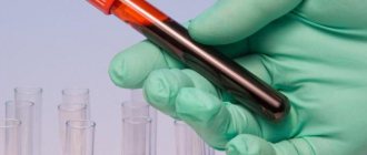
How to take blood for hemostasis
This test is practically no different from others that pregnant women regularly have to take. You can't eat for eight hours before the test, so it's best to plan it early in the morning and bring sandwiches and a thermos with you to have a snack after the test.
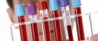
Expert commentary
Taking blood from a vein for hemostasis is not much different from regular blood taking. Blood is taken in the morning, on an empty stomach, from the antecubital vein with a wide-bore needle; only short-term application of a tourniquet is allowed; it is important not to take fatty foods the day before. Collection into vacutainers is preferable. There is nothing more useless than processing blood after 3 hours from the moment of collection, without indicating what medications the patient is receiving and without taking into account the symptoms and clinical picture of the disease (the latter gives the right direction to further diagnostic searches).
How it is carried out and why. Indications for use
Any doctor can refer you for testing, and the reasons for diagnosis will be such conditions as:
- phlebeurysm;
- vein thrombosis;
- pregnancy planning;
- fetal death at the intrauterine stage of development;
- prescription of hormone replacement drugs and oral contraceptives;
- placental abruption;
- intrauterine anomalies of fetal development;
- fetoplacental insufficiency and toxicosis;
- early miscarriages;
- stroke;
- myocardial infarction;
- thromboembolism at a young age, as well as its recurring episodes;
- excess body weight;
- upcoming surgery;
- cardiovascular pathologies in blood relatives;
In addition, analysis for gene mutations of the hemostatic system is recommended in cases such as:
- infertility;
- upcoming in vitro fertilization (IVF) procedure;
- the presence of signs of bleeding (including from the gums, from small cuts received during shaving), small bruises on the body;
- menstruation accompanied by abnormally high blood loss;
- the need to assess the condition of the liver.
For the analysis, you need to donate blood from a finger and a vein on an empty stomach. In some clinics, a cheek swab is taken, through which a sample of the epithelium of the inside of the cheek is obtained. It is painless and quick, which makes it possible to examine those who are pathologically afraid of injections, for example, children. After the results are ready, a consultation with a hematologist is indicated, who will professionally describe them.
The study is carried out using the PCR method, which stands for polymerase chain reaction. The obtained data are compared with control samples. It is taken into account that when carrying a child, the level of blood clotting always increases slightly. This is not considered a pathology, but if there are mutations, the process is aggravated.
To find out how high the risk is that the mutation will be inherited, doctors advise taking a genetic test. This is expensive, but this type of study is highly recommended for those who have already had cases of thrombosis in their family. It is important to understand that mutations in hemostasis genes that are not detected in a timely manner can cause fetal death or lead to severe developmental defects.
Heparin tolerance
If the introduction of heparin sharply increases the time of clot formation, then they speak of reduced tolerance (stability), but if the introduction of heparin does not slow down (or slightly changes), they speak of increased plasma tolerance to heparin.
Standards of tolerance to heparin in the hemostasis system:
- Tolerance to heparin of nitrate plasma = 10-16 minutes, (in 75% of people - 11-14 minutes, in 90% - 10-16 minutes)
- Plasma oxalate heparin tolerance = 10-15 minutes
Prolongation of the onset of coagulation (hypocoagulation) occurs in coagulopathies (hemophilic conditions, thrombopenia), and reduction of the period (hypercoagulation) occurs in thromboembolic conditions, after surgical interventions, and in cardiac decompensation.
Interpretation of results
In addition to genetic abnormalities, the study makes it possible to determine the cellular structure of hemostasis, which is extremely important for pregnancy management.
There are two types of gene disorders:
- the most dangerous polymorphism is homozygous, when the risk of thrombosis is extremely high;
- Heterozygous is considered less dangerous.
This is the basis for the principles of deciphering data obtained during a study on the probability of polymorphism:
- No mutations were found, that is, the genes responsible for the elements of the hemostatic system did not undergo changes.
- A mutation in heterozygous form means that a person is a carrier of a trait that can cause pathology.
- Mutation in homozygous form. This means that two genes with anomalies were discovered, that is, the likelihood of characteristic pathologies is high.
It is important to understand that only a geneticist or hematologist can correctly interpret the results, so you should not try to do this yourself, especially if you lack special education and experience.
Specialists will fully assess the degree of risk from pathologies associated with impaired blood clotting. They will also suggest the optimal prevention plan.
So what is the blood coagulation system?
The blood coagulation system or hemostasis is a unique protective system of the body, the tasks of which are, on the one hand, protection from bleeding, i.e. closing a vessel defect with a thrombus, on the other hand, preventing the formation of blood clots inside the vessel, where it is not needed.
Why is this so important?
In the process of life, a person is constantly faced with various microtraumas, and if not for hemostasis, then any small cut or injection would turn into a serious, even life-threatening problem. Everyone knows the disease of the heir to the Russian throne, Tsarevich Alexei - hemophilia, this is a condition of non-clotting of blood that is inherited.
The hemostatic system must be in balance. The coagulation system is balanced by the anticoagulation system. If a striking example of a blood coagulation disorder is the disease hemophilia, then an equally striking example of a violation of the anticoagulation system is thrombosis. This is truly “enemy number one”, hidden, asymptomatic, sudden. Moreover, it hits the most important thing in our body - blood circulation.
Blood in our body performs a very important function - it supplies cells with oxygen and other substances necessary for life. Blood clots seal vessels, capillaries, arteries, which complicates or completely prevents blood flow, in which case sad consequences for the body occur, sometimes irreversible. Thrombosis underlies such common diseases as myocardial infarction and ischemic stroke (according to WHO, they rank first in the world).
Treatment of disorders: methods and their effectiveness
If the results of the study reveal structures with disturbed morphology, it is worth consulting with a doctor about the prospects for the development of pregnancy and the possible state of health of the unborn child.
Unfortunately, gene abnormalities cannot be corrected or cured. However, when the examination reveals their consequences - blood clotting pathologies - the doctor prescribes medical measures.
Drug treatment of thrombophilia
For example, in the initial stages of thrombophilia, a course of conservative treatment is indicated, which includes the use of:
- disaggregants - drugs that improve blood flow and prevent the formation of microthrombi;
- anticoagulants, whose action is also aimed at preventing thrombosis;
- fibrinolytics, which are injected directly into the thrombosed area of the vessel;
- medications to strengthen vascular walls;
- folic acid preparations (for hereditary thrombophilia).
An excellent therapeutic effect is observed after:
- donor blood or plasma transfusions;
- thrombocytopheresis. This is the name of a special procedure for obtaining platelets. They enrich donor plasma, which is then administered to a person suffering from thrombophilia. When tissues are damaged, this allows them to speed up their regeneration.
As for treatment during pregnancy, it has a number of features, since not all drugs are approved for use. In cases where thrombosis occurred before conception, anticoagulants are prescribed. One of the popular drugs in this class is Heparin. It does not cross the placenta and does not affect the fetus. After childbirth, the course is supplemented with Warfarin, which is approved for use during breastfeeding. Treatment is carried out only for severe forms of thrombophilia, and women with milder forms are simply observed. They are prescribed tests more often than healthy pregnant women, so that prompt measures can be taken if the blood condition changes.
Diet for thrombophilia
Compliance with a special diet prescribed by your doctor is important for the successful fight against thrombophilia. To reduce the risk of thrombosis, it involves avoiding consumption of:
- fatty meat and offal;
- lard;
- fried food;
- rich broths;
- chocolate;
- coffee and other drinks containing it;
- legumes;
- hard cheeses.
In this case, the diet should have enough:
- fresh vegetables and fruits, berries (especially dried apricots, cranberries and grapes);
- seafood;
- cereals
Don't forget about the drinking regime. To normalize blood clotting processes, those suffering from thrombophilia are recommended to consume at least 2.0–2.5 liters of fluid per day. At the same time, it is better to drink clean water, herbal teas and compotes.
Traditional treatment methods
They are not an independent method to overcome pathology, but successfully complement the treatment prescribed by the doctor. The permission of a hematologist to use methods of traditional therapy, as well as agreement with him on the dosages and duration of the course, is mandatory. This is due to the fact that, in combination with pharmaceutical drugs of similar action, such treatment can lead to excessive blood thinning.
So, the recipes:
- Garlic. It dissolves fibrin, which promotes the formation of blood clots, and also strengthens vascular walls. A couple of fresh cloves daily will be an excellent prevention of thrombosis;
- Tincture of Japanese Sophora. 100 g of plant seeds are poured with half a liter of vodka and left for half a month in the dark and cool. After 15 drops of tincture, dilute in half a glass of water and take before meals. The recommended course duration is 1 month.
- Horse chestnut peel tincture. Take 50 g of the peel, pour in 0.5 liters of vodka and leave for 14 days, after which you take 20 drops of the tincture before meals, having previously dissolved them in 100 ml of water.
- Sweet clover infusion. Two teaspoons of the herb are poured into half a liter of boiling water and left for at least 6 hours, after which it is filtered and taken half a glass three times a day.
When the body has concomitant pathologies, the treatment of which also does not exclude the use of traditional therapy, it is important not to use plants that thicken the blood.
Hemostasis before pregnancy
In order to avoid complications associated with increased blood clotting during pregnancy, it is worthwhile in advance, at the stage of preparation for pregnancy, to do an analysis for mutations in the genes of the hemostatic system (if necessary, consult with a geneticist) and a hemostasiogram. In some cases, doctors simply give recommendations to carefully monitor the state of the hemostatic system during pregnancy, and sometimes prescribe preventive treatment.
A hemostasiogram at the planning stage is necessary if there were:
- thrombotic complications under the age of 50 in close relatives (heart attack, stroke, deep vein thrombosis, etc.);
- varicose veins in a woman and/or her close female relatives;
- recurrent miscarriage (two or more missed pregnancies or a history of miscarriage).
Expert commentary
Sometimes there is a need to monitor the state of the hemostatic system even before pregnancy - if there is a history of a tendency to thrombus formation. The most accessible marker of thrombus formation is D-dimer. In the case of hereditary disorders, it is wiser to anticipate the appearance of signs of intravascular thrombin deposition (“high D-dimer”). If necessary, low molecular weight heparins (LMWH) are used during preparation for pregnancy. This happens in the following cases:
- aggravated personal or family history of thromboembolism;
- persistent migraine during hormone replacement therapy while taking combined oral contraceptives;
- verified thrombophilia;
- APS syndrome (VAC positive);
- hyperhomocysteinemia (>15 µmol/l).
It is advisable to at least once assess the complete hemostasis, indicating the relationships in this unique system; in the future, it is enough to monitor individual key positions in which violations were identified.