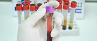
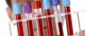
GENERAL URINE ANALYSIS
It shows the quality of functioning of the excretory system. Before collecting urine, it is necessary to toilet the genitals. An average portion of urine is used for analysis. Urine must be delivered to the laboratory no later than 2 hours after collection.
COLOR
from straw to yellow. The saturation of the yellow color of urine depends on the concentration of substances dissolved in it. The color changes when taking medications (salicylates, etc.) or eating certain foods (beets, blueberries). Turbid urine means the presence of impurities of salts (phosphates, urates, calcium oxalates), bacteria, red blood cells, which may indicate inflammatory kidney diseases.
ACIDITY
urine (RN) depends on the nature of the diet. If you like meat foods, then an acidic urine reaction will be observed during a urine test; if you are a vegetarian or follow a dairy diet, then the urine reaction will be alkaline. With a mixed diet, mainly acidic metabolic products are formed, so it is believed that the urine reaction is normally acidic. An alkaline reaction of urine is characteristic of chronic urinary tract infection, and is also observed with diarrhea and vomiting. The acidity of urine increases in feverish conditions, diabetes mellitus, tuberculosis of the kidneys or bladder, and renal failure.
SPECIFIC GRAVITY
(specific density) characterizes the filtering function of the kidneys and depends on the amount of excreted organic compounds (urea, uric acid, salts), chlorine, sodium, potassium, as well as the amount of urine excreted. Normally, the specific gravity is 1010-1030. Changes in the specific gravity of urine towards a decrease may indicate chronic renal failure. An increase in specific gravity indicates inflammatory kidney diseases (glomerulonephritis), possible diabetes mellitus, large fluid losses or low fluid intake.
PROTEIN
is absent in the urine of a healthy person. Its appearance usually indicates kidney disease, an exacerbation of chronic kidney disease.
GLUCOSE
Normally, it is absent in a general urinalysis.
LEUCOCYTES
Normally they can be present in urine in an amount of 0-5 per field of view. An increase in the number of leukocytes in the urine (leukocyturia, pyuria) in combination with bacteriuria and necessarily in the presence of any symptoms (for example, frequent painful urination, or increased body temperature, or pain in the lumbar region) indicates inflammation of an infectious nature in the kidneys or urinary tract ways.
erythrocytes and bacteria
. Red blood cells can normally be present in urine in an amount of 0-3 per field of view. Bacteria are normally absent in a general urine test. The presence of bacteria is a sign of chronic or acute diseases of the kidneys and urinary tract. A particularly dangerous phenomenon is asymptomatic bacteriuria, that is, the presence of changes in tests in the absence of patient complaints. It is dangerous because it can last as long as desired without appropriate treatment and observation; during pregnancy, inflammatory diseases of the urinary system develop, which has a negative impact on the course of pregnancy and the condition of the fetus.
CYLINDERS
in
a general urinalysis. Cylindruria is a symptom of kidney damage, so it is always accompanied by the presence of protein and renal epithelium in the urine.
Once detected changes in urine are not a diagnosis.
To clarify the situation, the doctor will prescribe additional examinations.
Why are veins blue and not red?
Veins carry burgundy blood. They appear blue due to many factors. Primarily due to the color perception of the human eye.
Color is the wavelength of light emitted by an object or reflected by an object from another light source. Red light has the longest wavelength (700 nm). This means that it passes through objects and is not reflected from them. It also passes through the skin and, reaching the veins, is absorbed by hemoglobin. If you shine a red light on your hand, it will reflect everywhere except where the veins are. There it will turn black as it is absorbed. With this trick, doctors can find hard-to-reach veins.
Violet light is the shortest wavelength (400 nm). Blue has approximately the same indicators - 475 nm. It disperses easily and does not penetrate deep into the skin, but is reflected from it. If you look at your hand under a blue light, you won't be able to find any veins.
Attention!
This trick is often used against those who like to go to clubs or other places. Violet or blue light in the toilet leaves no chance of revealing your veins.
Place your hand under regular white light. There are other colors in it, not just white. The veins will be blue, as it will be reflected from it, and the red will go deep into the skin and be absorbed there.
Why don't we see other vessels through which blood flows?
A person does not see the vessels because they are too deep under the skin. The light doesn't reach there. If the blood vessel is closer than 0.5 mm, it already absorbs all the blue light. Red is partially reflected, so people see this part of the skin as ruddy and pinkish. Veins that are clearly visible are located at a distance of no more than 0.5 mm from the surface of the skin.
Why can't we see arteries under the skin?
Most of the blood is in the veins, so they are much larger than arteries and vessels. Because blood puts more pressure on the arteries, they have thicker walls. Because of this, they are not as transparent and cannot be seen through the skin. If they were visible, arteries would likely look exactly like veins. Although the liquid in them is bright red.
What color are veins really?
Empty blood vessels have a red-brown tint. The veins are about the same color, since there are few differences between them. Veins have thin walls, while arteries have thicker and more muscular walls. This difference can only be seen in a cross section.
BLOOD CHEMISTRY
This analysis allows the doctor to judge the condition of the internal organs and their enzymatic function. The test is taken on an empty stomach (in the morning), blood is taken from a vein.
GLUCOSE
is a source of energy for cells. To absorb glucose, cells need normal levels of insulin, a pancreatic hormone. Normal glucose levels are from 3.3 to 5.5 mmol/l. A decrease in glucose indicates starvation, with poorly chosen treatment for diabetes. An increase in glucose levels indicates diabetes. However, it can also be physiological - after eating.
TOTAL BILIRUBIN
- component of bile. Normally no more than 20.5 mmol/l. High numbers may appear after 24-48 hours of fasting, during a long-term diet, or with liver disease.
UREA
- a product of protein metabolism that is removed by the kidneys. The norm is 4.2 - 8.3 mmol/l or 2.1-7.1 mmol/l (G). Its increase indicates a violation of the excretory function of the kidneys.
URIC ACID
– a product of nucleic acid metabolism, excreted by the kidneys. The norm is from 179 to 476 µmol/l. In healthy people, its level in the blood and urine can increase with a high content of chemical purines in food (they are found in meat, wine) and decrease with diet. An increase in uric acid occurs with gout, leukemia, acute infections, liver diseases, chronic eczema, psoriasis, and kidney diseases.
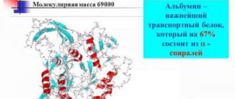
TOTAL PROTEIN
– is part of all anatomical structures, transports substances through the blood and into cells, accelerates the course of biochemical reactions, regulates metabolism and much more. The norm is 65-85 g/l. Total protein consists of two fractions: albumin and globulin. Albumin – no less than 54%. A decrease in the level of total protein occurs with kidney disease, fasting, and long-term inflammatory diseases. An increase in level may occur with some blood diseases, with systemic connective tissue diseases, with cirrhosis of the liver.
CREATININE
– a product of protein metabolism, excreted by the kidneys. Its increase also indicates a violation of the excretory function of the kidneys. The norm is 44-150 µmol/l.
AMYLASE
-an enzyme produced by the cells of the pancreas and parotid salivary glands. The norm is from 0.8 to 3.2 IU/l. An increase in its level indicates diseases of the pancreas. Decreased blood levels may indicate hepatitis.
TOTAL CHOLESTEROL
- a substance that comes from outside and forms in the body. With its participation, sex and some other hormones, vitamins, and bile acids are formed. The norm is from 3.6 to 6.7 mmol/l. The level increases with diabetes mellitus, atherosclerosis, chronic kidney disease, and decreased thyroid function. Cholesterol levels decrease with increased thyroid function, chronic heart failure, and some types of anemia.
CALCIUM
– an element involved in the conduction of nerve impulses, blood clotting and is part of bone tissue and tooth enamel. The norm is 2.15-2.5 mmol/l. An increase in calcium levels may be associated with increased function of the parathyroid gland, excess vitamin D, while a decrease may be associated with vitamin D deficiency, or impaired renal function.
POTASSIUM, SODIUM, CHLORIDE
provide the electrical properties of cell membranes and are part of the internal fluid of the body (extracellular fluid in tissues, blood, gastric juice). A change in their quantity is possible with fasting, dehydration, impaired renal function and adrenal cortex.
The norm is sodium – 135-145 mmol/l, potassium – 2.23-2.57 mmol/l, chlorides – 97-110 mmol/l.
MAGNESIUM
– an element that is part of a number of enzymes necessary for the functioning of the heart, nervous and muscle tissue. An increase in its level is possible if the function of the kidneys or adrenal glands is impaired, and a decrease is possible if the function of the parathyroid glands is impaired.
The norm is 0.65-1.05 mmol/l.
PHOSPHORUS UNLIMITED
– an element that is part of nucleic acids, bone tissue and the main energy supply systems of the cell. Regulated in parallel with calcium levels.
The norm is 0.87-1.45 mmol/l.
PHOSPHOTASE ALKALINE
– an enzyme produced in bone tissue, liver, intestines, placenta, and lungs. Serves for a general assessment of these organs.
The norm is 38-126 IU/l.
IRON
– a substance that is part of hemoglobin and is involved in the transfer of oxygen in the blood. A decrease in level indicates anemia.
The norm is 9-31.1 µmol/l.
TRIGLYCIRIDES
– The level of triglycerides can be used to judge dietary patterns. It can increase with consumption of large amounts of animal fats and decrease with a vegetarian diet.
The norm is from 0.43 to 1.81 mmol/l.
ALANINE AMINOTRANSFERASE (ALT)
- a liver enzyme involved in amino acid metabolism. An increase in the enzyme is possible in case of dysfunction of the liver or organs where ALT normally accumulates (heart, skeletal muscles, nervous tissue, kidneys).
The norm is up to 31 U/l.
ASPARATE AMINE TRANSFERASE (AST) –
liver enzyme involved in amino acid metabolism.
The norm is up to 31 U/l.
Blood in children
The composition of the blood in children is characterized by its instability - during the process of growth, the ratio of the main components constantly changes. In addition, the indicators strongly depend on external factors: diet, daily routine, physical activity. The level of leukocytes in children's blood is increased, since it is during this period that the active formation of immunity occurs - blood cells constantly encounter new antigens and antibodies are produced.
As a percentage, the amount of blood in a newborn is much higher than in an adult - it is about 14% of body weight, it turns out that there are about 150 ml per 1 kg of weight. In the first 12 hours, children's blood is characterized by an increased level of immature red blood cells and hemoglobin. However, within the first day these indicators drop significantly. The fact is that red blood cells in the blood of newborns live much shorter than in an adult body - they are destroyed on average in 12 days.
Premature babies often experience anemia in the first months of life. If, with such a decrease in hemoglobin, the general state of health does not cause concern and no additional symptoms appear, then early anemia of prematurity is not considered dangerous and is a normal reaction to adaptation to new conditions.
After the birth of a child, up to 150 ml of blood with specific characteristics is stored in the placenta and umbilical vein. Previously, it was not given much importance, but today umbilical cord blood is increasingly preserved. It contains a large number of stem cells, which can be used in the treatment of various diseases. They are unique in their characteristics, since they are not differentiated and can produce offspring of any specialized cell types.
COAGULOGRAM. HEMOSTASIOGRAM
COAGULOGRAM
(blood test for hemostasis) is a necessary stage in the study of blood clotting during pregnancy, before operations, in the postoperative period, i.e. in situations where the patient expects some blood loss, as well as with varicose veins of the lower extremities, autoimmune diseases and liver diseases. Violation of blood clotting, especially its increase or hypercoagulation, can lead to dangerous consequences for the body, causing a heart attack, stroke, thrombosis.
During pregnancy, a coagulogram always shows increased blood clotting. If coagulation values are higher than normal, then blood clots can form in the vessels of the placenta, as a result of which the baby does not receive enough oxygen, which can lead to miscarriage, premature birth or the birth of a child with severe brain damage.
Blood hemostasis is maintained due to the balance of three systems:
A coagulation system that activates platelets, their adhesion to the vascular wall and gluing (main components: fibrinogen, platelets, calcium, vascular wall).
Anticoagulant system that controls blood clotting and prevents spontaneous thrombus formation (antithrombin III)
Fibrinolytic system that dissolves clots (plasmin).
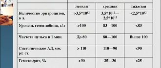
Red blood counts are normal
- Hemoglobin (Hb)
- Men = 132 – 164 g/l
- Women = 115 –145 g/l
- Hemoglobin fractions
- A1 = 96 – 98%, A2 = 3%, A3 – traces, F = 2%
- Red blood cells
- Men =4.0 – 5.1 million in 1 mm3 (x10 12/l)
- Women = 3.7 – 4.7 million per 1 mm3
- Red blood cell diameter
- 5.0 µm – 0.5%;
- 6.0 µm – 4%;
- 7.0 µm – 39%;
- 8.0 µm – 54%
;
- 9.0 microns – 2.5%.
- Reticulocytes = 0.5 – 1.5% of the number of red blood cells
- Hematocrit
- Men = 40 – 52%
- Women = 36 – 48%
- Color index
= 0,85 – 1,05
(Hb x 3 : first 2 digits number of red blood cells)
- Mean erythrocyte volume ( MCV)
= 80 – 100 fL - Average
Hb in erythrocyte ( MCH ) = 24 – 35 pg - Mean
erythrocyte Hb MCHC ) = 30 – 38 g/dL - Osmotic resistance of red blood cells
- onset of hemolysis = 0.5 – 0.45% NaCl
- end of hemolysis = 0.4 – 0.35% NaCl
- Iron content in blood plasma (50 – 150 mg/dl):
— men: 13-80 µmol/l
— women: 11-25 µmol/l
- Total iron-binding capacity of blood serum (TIBC) 250 – 400 mg/dl or 48-71 µmol/l
- CST = 16 – 54%
- Plasma ferritin = 20 – 350 mg / dl
- Iron – containing normoblasts in the bone marrow = 20 – 40%
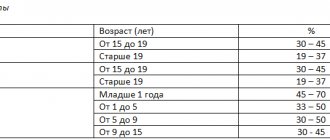
- Requirements for the collection and preparation of biological material for hematological studies.
- Requirements for transportation and storage of biological material for hematological studies.
Correctly filling out a referral for a study of the hemostasis system:
- Full name, age, gender of the patient
- Time of blood sampling for research
- Clinical diagnosis (briefly)
- Hematocrit
- Hemorrhagic syndrome (nasal, uterine bleeding, etc.), thrombosis (specify location), shock, multiple organ failure, trauma, prolonged compression syndrome, burn, etc.)
- Indicate medications taken that affect hemostasis parameters, indicating the dosage, method and date of last administration
- The time interval for sampling (blood), dietary and exercise restrictions are observed:
- Routine blood sampling is carried out from 7 to 9 am, with the patient lying or sitting. When studying hemostasis in dynamics, it is advisable to take blood sampling in the same body position as the previous sampling.
- Blood is taken on an empty stomach, 12-14 hours after the last meal.
- Before drawing blood, it is advisable for the patient to rest for 15 minutes.
- Exceptions:
hemostasis studies by cito, APTT assessment.
- On the eve of the planned examination (within 24 hours), the patient abstains from significant physical activity and drinking alcohol. It is advisable that the patient does not eat fatty foods on the eve of blood sampling.
Surgical intervention leads to changes in hemostasis for a period of several days to several weeks.
Factors affecting hemostasis include injections, infusions, transfusions, punctures, biopsies, massage, dialysis, administration of radiocontrast agents, immunoscintiography, ionizing radiation, endoscopic examination, and special diets.
RULES FOR BLOOD COLLECTION
- Blood is taken from a peripheral vein (usually the ulnar vein).
- Blood is collected only using a vacuum collection system into tubes with special color markings depending on the anticoagulant used (sodium citrate 3.2%)
in the ratio: 1 volume of sodium citrate to 9 volumes of blood.
- For research platelet level
blood is taken into a tube
with EDTA
(lilac color marking). A platelet level test is prescribed in the absence of a clinical blood test or when pathological platelet levels are obtained in a clinical blood test - Short-term application of a tourniquet is acceptable, no more than 60 seconds. Remove the tourniquet immediately after the needle enters the vein. To collect blood for the study of the coagulation system, it is important that you cannot massage the veins or knock on them.
- Use a blood collection needle with an internal diameter of 0.7-1 mm (size 19-22).
- The use of a syringe is unacceptable due to the activation of platelets and blood clotting factors by the turbulent movement of blood and its mixing with air (foaming). This is eliminated when using vacuum containers.
- After inserting the needle into the vein, attach a vacuum container, the blood will begin to flow into the container by gravity.
- Take blood for coagulation testing at second
test tube, use the first one for other studies, for example, biochemical ones.
If the coagulation test tube is to be drawn first, draw the first 5 mL of blood into an empty tube and discard it to prevent tissue thromboplastin from contaminating the sample. - Immediately after collection, carefully mix the blood without foaming with the anticoagulant (invert the tube 3-4 times).
- After collecting biological material, check the blood sample. Careful checking of blood samples avoids the following errors: incorrect blood/citrate volume ratio; partially coagulated blood.
The blood is delivered to the laboratory within 45 minutes after it is collected. During transportation, blood should not be shaken. Blood samples should not be transported at temperatures below 4°C or above 30°C.
Medical laboratory assistant (medical technologist) carries out:
- incoming control of delivered blood, including: 1)checking that the referral form is filled out correctly. 2)
checking the sufficiency of the delivered blood volume using the mark on the tube, 3) checking the sample for the absence of clots by slowly tilting the tube.
- centrifugation of delivered blood to obtain:
platelet rich plasma (
centrifugation parameters: rpm = 1000 rpm (about 150-200g), centrifugation time 5-7 minutes.
To select optimal centrifugation conditions, they are guided by centrifugal force (g). You can use the formula:
g = 1.118 x 0.00001r x n2,
where r is the radius of the centrifuge - the distance in centimeters between the axis of the rotor and the center of the test tube in the centrifuge nest; n is the number of revolutions per minute.
platelet-poor plasma (
centrifugation parameters: rpm = ~2500–3000 rpm (about 1500–2000g), centrifugation time 10–20 minutes. To completely remove platelets, repeated centrifugation is performed, the centrifugation parameters remain the same.
3. After centrifugation, the plasma is checked for color and transparency: a hemolyzed sample, icteric or lipemic (chylous) plasma cannot be examined.
- After centrifugation, the supernatant is collected.
Preferably
study hemostasis indicators within
2 hours after blood collection
, but
it is allowed
to study coagulological indicators within
4 hours
if the plasma was separated from the sediment of erythrocytes and leukocytes.
If long-term storage
“fresh” samples (no later than 2 hours after blood collection)
of platelet-free plasma
be frozen
once and stored for up to 2 weeks at a temperature of -20°C or up to 6 months at a temperature of -70°C. Plasma must be thawed quickly in warm water (+36°C), mixed thoroughly and examined immediately. After freezing, changes in aPTT may occur.
- Methods for preparing microslides for hematological studies, methods of fixation and staining.
- Methods for preparing blood smears for hematological studies.
Preparation and processing of glass. New clean glasses can be used after degreasing in Nikiforov's mixture (equal parts of 96% ethyl alcohol and ether). Used glasses are soaked for 24 hours in a warm soap solution or washing powder solution (1 tablespoon of powder per 5 liters of water). After a day, the solution is drained and the glass is washed with running water. Then they are again poured with a warm soap solution or a solution of washing powder and brought to a boil, boiled in the same solution for 5-10 minutes (no more, to avoid clouding of the glass). After the solution has cooled, it is drained again, the glass is rinsed with running water, and each glass is washed with a brush under running water. Glasses treated in this way are laid out on a clean sheet to dry. Clean glasses for degreasing are placed in Nikiforov’s mixture or 96% ethyl alcohol for 60 minutes, then wiped dry and stored in a clean container with a wide neck. It is recommended to grasp clean glass either with tweezers or by hand by the side edges.
Preparation of smears. A small drop of blood is applied to a dry prepared slide closer to the short side with a glass rod (or directly from the finger prick site). Leave the glass in a horizontal position and smear the blood on the glass using dry, clean, ground glass, holding it at an angle of 45°. With a short edge, after waiting until all the blood has spread over it, quickly draw it across the slide. You should not press hard on the slide, as this can damage the blood cells. The smears are dried in air and marked (preferably with a simple pencil). The dried smear should be uniformly thin, yellowish in color, of sufficient size, located at a distance of 1.0-1.5 cm from the edges of the glass, occupy almost the entire length of the glass and end in a “whisk”. Thick (dense pink) smears should not be used, since the cell morphology in them is difficult to distinguish.
Standard high-quality smears are obtained using automatic devices designed for preparing smears and produced by various companies.
Fixation and staining of blood smears. Before staining, blood smears are usually fixed for 5–10 minutes in methyl alcohol to prevent hemolysis, which can occur upon contact with water during staining with water-soluble dye or upon subsequent contact with water. Fixation techniques are described below along with staining techniques. Fixation is not required for Wright and Leishman staining, since these paints contain methyl alcohol.
Dry smears can be stored for 2 days in a dry, warm place; in a hot, humid atmosphere without fixation they last much less.
Blood cells contain basophilic and acidophilic structures that differ in reaction (pH). The nuclei are basophilic and stain blue. Highly basophilic (acidic) basophil granules are also blue. Hemoglobin (being basic) is stained acidophilic, i.e. red. Staining using a combination of acidic and basic dyes is called Romanovsky staining and has various modifications (Pappenheim, Wright, Nocht, Leishman, Giemsa, Janer, etc.). Methylene blue is usually used as the main dye, but toluidine blue is also used. The acid dyes used are eosin, azure I and azure II.
Criteria for good staining: pink color of erythrocytes, violet staining of granularity of neutrophils on a pink background, delicate azurophilic granularity of monocytes.
To dilute paint, it is best to use neutral, fresh distilled water. Stale distilled water becomes acidic due to the capture of carbon dioxide from the atmosphere. If distilled water is alkaline, the red blood cells turn a dirty bluish-green color; that part of the leukocytes that should be stained blue acquires a violet tint, the eosinophil granules become bluish and greenish instead of pink, and the neutrophil granules are recolored. In acidic water, red blood cells turn bright orange, and the nuclei of white blood cells are very pale. The ideal is distilled water with a pH of 7.0, which is preserved by buffering. You can use ready-to-use buffer tablets dissolved in distilled water.
For staining bone marrow smears, a pH of 7.4–7.5 is ideal.
The staining methods according to Nocht and Pappenheim were adopted as unified.
Reagents for fixation: 1) methyl alcohol or 2) eosin-methylene blue solution according to May-Grunwald.
Reagents for coloring smears according to Nokht: 1) basic solution of azure I: 1 g of paint is dissolved in 1 liter of distilled water, left in a dark glass container for 12-14 days at room temperature, after which it is used; 2) basic solution of potassium eosin: 1 g of paint is dissolved in 1 liter of distilled water, left in a dark glass container for 12-14 days at room temperature, after which it is used; 3) phosphate buffer (Weise mixture), pH 7.4-7.5: mix 0.49 g of potassium dihydrogen phosphate (KH2PO4) and 0.909 g of sodium hydrogen phosphate (Na2HPO4) and dissolve in 1 liter of distilled water; 4) working solution of azure-eosin: before use, mix 25 ml of azur II stock solution, 20 ml of potassium eosin stock solution and 55 ml of a buffer solution (the proportions of dyes can vary, they are established experimentally when preparing fresh batches of stock solutions).
Reagents for staining smears according to Pappenheim: 1) eosin-methylene blue solution according to May-Grunwald. In the absence of a ready-made dye solution, it is prepared by dissolving 1 g of dry paint in 1 liter of methyl alcohol; 2) working solution of azure-eosin according to Nokht.
Fixation of strokes. The fixing solution is poured into a cuvette or into a wide-necked container with a ground-in stopper. The smears are placed in a container, which is immersed in a cuvette or placed one at a time in a dish for 5–10 minutes. The container with the glasses is removed from the fixing solution (or the glasses are removed with tweezers and placed on a tripod) and left in the air until completely dry.
Staining according to Nokht. Dried fixed strokes, without removing them from the container, are placed in a cuvette with a working paint solution for a strictly defined time (20–45 minutes), which is selected experimentally for each batch of paint. If there is no cuvette with a container, the glass is placed horizontally on two glass rods placed parallel (“rails”) with a stroke upward and 3–4 ml of the working paint solution is poured onto them. Remove the container with glasses from the cuvette with the dye and place it in a cuvette with tap water (if there are no containers, wash off the paint from the glasses with water without removing them from the glass rods). The smears are air dried.
Pappenheim staining. Dry, unfixed blood smears are placed in a container and lowered into a cuvette with May-Grunwald solution for 3-5 minutes (or 3-4 ml of dye is pipetted onto an unfixed smear). The container with smears is rinsed in a cuvette with distilled water, and then placed in a cuvette with azure-eosin paint according to Nocht for 8–15 minutes. On glass placed on the “rails”, without draining the dye, add distilled water for 1 minute, and then pour paint onto the smear for 8–15 minutes, then wash off the paint with water. The smears are air dried.
Romanovsky-Giemsa staining. It is produced in the same way as according to Nokht. A ready-made Romanovsky-Giemsa solution is used as a dye, which is diluted before use at the rate of 1 drop of dye per 1 ml of distilled water. The coloring time is determined experimentally for each batch of dye (25–40 min).
Wright staining. Reagents. 0.2 g of Wright's paint (dry powder, BDH/E. Merck) is dissolved in 100 ml of methyl alcohol and allowed to stand for several days. If the red blood cells are not stained clearly enough, prepare a 0.25% or 0.3% solution.
Progress of painting. Apply a few drops of paint to the smear , leave for 2-3 minutes (make sure the paint does not dry out), then pour an equal amount of buffered water onto the smear. If the paint is mature, foam or a film with a metallic sheen will appear on the surface of the diluted paint. Diluted paint leave on the smear for 2-3 minutes, and then wash off with a buffer solution or water. Do not allow the paint to settle on the surface of the smear. If this happens, fill the smear with undiluted paint for 15-20 s and then again with a buffer solution or water.
Preparation of phosphate buffer.
Solution A (0.2 M KH2PO4): dissolve 27.2 g of salt in 1 liter of distilled water.
Solution B (0.2 M Na2HPO4): dissolve 35.6 g of Na2HPO4 × 2H20 in 1 liter of distilled water.
To obtain 100 ml of a buffer solution with the required pH, solutions A and B should be drained in the quantities indicated in the table. The pH value is checked using a pH meter.
smear on flora
smear on flora
- microscopy of scrapings from the urethra, the contents of the posterior wall of the vagina and the cervix.
Decoding the smear:
- squamous epithelium
is a layer of cells lining the vagina and cervix. In a normal smear, epithelium should be present. If the smear does not contain epithelium, then the gynecologist has reason to assume a lack of estrogen, an excess of male sex hormones. The absence of squamous epithelium in the smear indicates atrophy of epithelial cells. - leukocytes
- the norm is up to 15 units in the field of view. A small number of leukocytes will be considered normal, since leukocytes perform a protective function and prevent infection from entering the woman’s genitals. Elevated leukocytes in the smear are observed during inflammation of the vagina (colpitis, vaginitis). The more leukocytes in the smear, the more acute the disease. - rods
make up the normal microflora of the vagina. Apart from rods, there should be no other microorganisms in the smear. - small sticks
are most often gardnerella - the causative agents of gardnerellosis or vaginal dysbacteriosis. - “key” cells
(atypical cells) are squamous epithelial cells glued to a small rod. As with Gardnerella, if the smears contain atypical cells, the doctor can diagnose vaginal dysbiosis. - fungus
is a sign of candidiasis (thrush). In latent (asymptomatic) stages of thrush, the fungus in the smear can be detected in the form of spores.
Even if the smear results show the presence of cocci, small rods and “clue” cells indicating bacterial vaginosis, the smear results alone may not be enough to make a diagnosis. It is necessary to conduct an additional examination: bacteriological culture and DNA diagnostics (PCR smear).
BAKPOSEV
The bacteriological method of examining a smear taken from the vagina or urethra is that this material is placed in a special nutrient medium that promotes the proliferation of certain bacteria. Bakposev allows you to differentiate nonspecific bacterial flora, determine the species and quantity of the pathogen. In addition, which is very important for subsequent treatment, bacterial culture makes it possible to determine sensitivity to antibacterial drugs
PCR DIAGNOSTICS
PCR – polymerase chain reaction. The main advantage of the DNA method is that it allows you to determine small quantities of the pathogen, as well as persistent forms of pathogens that are encountered in the treatment of latent and chronic infections. The sensitivity and specificity of the PCR method is high – 95%.
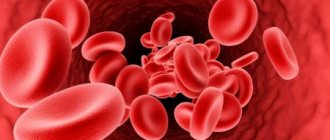
Hemoglobin
The blood, enriched with oxygen in the lungs, disperses to the vital organs of the body. At this moment it has a bright scarlet color. This occurs due to bonding with oxygen, resulting in oxyhemoglobin. As it passes through the body, it distributes oxygen and becomes hemoglobin again. Next, hemoglobin absorbs carbon dioxide from tissues and is transformed into carbohemoglobin. At this moment, the color of the blood changes to dark red. Immature red blood cells also have a bluish tint, and as they grow they turn gray and then turn red.
The color of the blood may vary. Answers to the questions why blood is dark red or bright red. A person’s blood takes on a different shade depending on whether it moves towards the heart or away from it.
Very often people wonder why veins are blue and blood is red? The fact is that venous blood is blood that flows through the veins to the heart. This blood is saturated with carbon dioxide and deprived of oxygen, has lower acidity, contains less glucose and significantly more final metabolic products. In addition to being dark red, venous blood also has a bluish, blue tint. However, not so strong as to “stain” the veins blue.
Why is blood red? It's all about the process of passing light rays and the ability of bodies to reflect or absorb solar rays. In order to reach venous blood, the beam must pass through the skin, the fat layer, and the vein itself. The sun's ray consists of 7 colors, three of which the blood reflects (red, blue, yellow), the remaining colors are absorbed. Reflected rays pass through tissues a second time to enter the eye. At this moment, red rays and low-frequency light will be absorbed by the body, and blue light will be transmitted. We hope that we have answered why a person has dark red and bright red blood.
Science knows that different living organisms on the planet have different blood colors.
However, in humans it is red. Why is blood red? This question is asked by both children and adults.
What makes blood red is hemoglobin, which consists of:
- From a protein called globin;
- The non-protein element heme, which contains the ferrous ion.
It was possible to find out what gives the red color, but its elements turn out to be no less interesting. What elements give it this color is an equally interesting aspect.
Blood contains:
- Plasma.
The liquid is light yellow in color, with its help the cells in its composition can move. It is composed of 90 percent water, with the remaining 10 percent made up of organic and inorganic components. Plasma also contains vitamins and microelements. The light yellow liquid contains many useful substances. - The formed elements are blood cells.
There are three types of cells: white blood cells, platelets and red blood cells. Each type of cell has certain functions and characteristics.
These are white cells that protect the human body. They protect it from internal diseases and foreign microorganisms penetrating from the outside.
This is a white element in color. Its white tint cannot be ignored during laboratory tests, so such cells are identified quite simply.
These are very small colored plates whose main function is coagulation.
These cells are responsible for ensuring that the blood:
- It coagulated and did not flow out of the body;
- Coagulates quite quickly on the surface of the wound.
More than 90 percent of these cells are in the blood. It is also red because red blood cells have this hue.
They carry oxygen from the lungs to peripheral tissues and are continuously produced in the bone marrow. They live for about four months, then are destroyed in the liver and spleen.
Few people know that immature red blood cells are blue, then acquire a gray tint and only then become red.
There are quite a lot of human red blood cells, which is why oxygen reaches peripheral tissues so quickly.
Children often ask questions regarding the components of the human body. Blood is one of the most popular topics of discussion.
Explanations for children should be extremely simple, but at the same time informative. Blood contains many substances that differ in function.
Consists of plasma and special cells:
- Plasma is a liquid that contains useful substances. It has a light yellow tint.
- The formed elements are erythrocytes, leukocytes and platelets.
The presence of red cells - erythrocytes - explains its color. Red blood cells are red by nature, and their accumulation leads to the fact that a person’s blood is exactly this color.
smear for cytology
Cytology smear is a cytological examination of smears taken from the surface of the cervix and from the cervical canal. This test is performed annually on all women over 18 years of age who are sexually active. The procedure is absolutely painless. The examination is not carried out during menstruation and in the presence of an inflammatory process.
Normally, the smear reveals squamous and columnar epithelial cells without any features. The appearance of atypical cells in a smear is a signal of trouble. The cause may be inflammatory processes caused by urogenital infections (mycoplasma, gonococci, trichomonas, chlamydia, etc.), background diseases (erosion, ectopia, leukoplakia, polyps, etc.), as well as precancerous conditions (dysplasia) and malignant degeneration of cells.
Each pathology has its own cytological features, which will be described in the cytogram.
Further examinations depend on the results of cytology: colposcopy (examination of the cervix under magnification using a special device - a colposcope), PCR examination, PAP test, bacteriological examinations (cultures), biopsy followed by histology (taking a piece of tissue from suspicious areas and examination under a microscope).