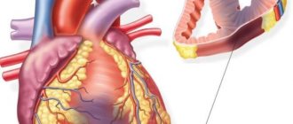
What is an enlarged heart
In medicine, an increase in the volume of the heart is called cardiomegaly or bovine heart. The change in the size of the main organ is due to the expansion of the myocardium and one of the chambers (left or right). This pathological phenomenon is called hypertrophy. Mostly, in pathology, the left ventricle is enlarged, and less often, the right ventricle is enlarged.
Normally, in an adult, the width of the heart is 9.5 cm, length – 11 cm, wall thickness – 7 cm, and weight – 250 g. The heart is often enlarged in size in professional athletes, pregnant women and adolescents. Often the anomaly appears due to prolonged stress.
Attention! Cardiomegaly is not an independent pathology, i.e.
it develops against the background of other diseases. The disease is mainly detected in older people. Diagnosing cardiomegaly is easy. As a rule, difficulties arise when determining a treatment regimen.
Congenital heart defect
The presence of this disease can be detected by a pediatrician immediately after the baby is born. Doctors can often make a diagnosis based on ultrasound data performed during pregnancy.
Pathology occurs very early, in the first weeks of pregnancy, when the heart is forming. It happens that heart defects appear in babies only on the 5th day of life. When listening to it, you can detect murmurs in the heart area. The sooner the problem is discovered, the better for the baby.
If the disease begins to progress, the person may die.
One of the most common types of defect is the absence of the third wall of the valve. A person can live a long time and feel great. But two walls will wear out much faster than the required three.
And as a result, the patient’s health will begin to deteriorate sharply. The stage of the disease can be detected by ultrasound.
Some types of defects do not have symptoms; doctors can detect the presence of the disease already in adulthood if other symptoms and abnormalities are present.
Causes
Sometimes the heart enlarges to the left on its own for unknown reasons. But more often the organ becomes larger under the influence of unfavorable factors that force it to work more intensely. Large heart syndrome can occur due to many reasons. The most common include:
- Congenital heart defects (CHD). The disorder develops in the child while still in the womb. If the defect is minor, then the heart enlarges gradually or never changes at all. Signs of cardiomegaly may appear immediately after birth, when extensive defects are observed.
- High blood pressure value. At normal blood pressure, the myocardium easily copes with the pressure created by the vascular system. At high levels, the organ experiences increased stress. It begins to gain mass, enlarging the left ventricle.
- Diseases of the cardiovascular system. For example, after a myocardial infarction, some area of the heart muscle dies. A scar forms on the exhausted, affected area. To compensate for this violation, other (healthy) parts of the myocardium begin to work intensively. Gradually the heart expands.
- Valve disease. Initially, organ enlargement due to valve disease was considered normal. After all, the large organ temporarily compensated for the resulting valvular changes. If the heart significantly increases in volume, then there is a high probability of weakening of the myocardium and the development of heart failure.
The following diseases and conditions can also lead to heart enlargement:
- pericarditis – accumulation of fluid near the heart;
- cardiomyopathy – damage to the heart muscle;
- rheumatic heart disease, which appears due to frequent sore throats and tonsillitis;
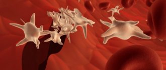
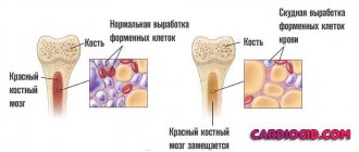
- anemia – a decrease in the number of red blood cells in the blood;
- hypothyroidism, hyperthyroidism – diseases of the thyroid gland;
- hemochromatosis is a genetic disease associated with impaired iron metabolism in the body;
- amyloidosis is a systemic disease that is caused by pathological deposition of amyloid in tissues;
- chronic lung pathologies (bronchial asthma);
- renal and liver failure;
- diabetes mellitus, obesity;
- severe stress and anxiety.
Attention! The left side of the heart is often enlarged when a person smokes and drinks alcohol.
The risk of cardiomegaly doubles when several causes of development are combined. For example, if a person has diabetes and hypertension at the same time. Pulmonary embolism, heart attack (right-sided), pulmonary hypertension, tricuspid valve insufficiency are the reasons that lead to enlargement of the right ventricle.
First aid for low heart rate
For normal and low blood pressure it will help:
- taking tinctures with a tonic effect (Leuzea, Eleutherococcus, ginseng);
- strong tea, coffee with sugar, honey;
- cold and hot shower;
- dark chocolate 2-3 slices;
- vigorous rubbing of the ears with palms, massage of the lobes;
- mustard plaster on the chest in the center of the sternum under the collarbones for 2 minutes.
If you have high blood pressure, you can take a diuretic (Hypothiazide, Furosemide). These remedies will raise the pulse rate, but if the cause is a disease, the effect will be short-lived.
Watch the video about helping with a low heart rate:
Pathogenesis
The mechanism of development of cardiomegaly depends on the reasons that led to the disorder. Left ventricular hypertrophy mainly occurs in people with coronary artery disease, hypertension, or metabolic syndrome (obesity). The myocardium begins to work more intensively with insufficient oxygen supply, gradually increasing in size.
A slightly different development mechanism is observed with valve disease and stenosis. With these disorders, blood does not completely flow to the adjacent part of the heart, which leads to stretching of one of the chambers. In the absence of treatment, there is a significant increase in not only the left ventricle, but also the atrium. In more severe cases, hypertrophy of the entire organ develops.
Low heart rate in an elderly person
In an elderly person, a low pulse can be a symptom of age-related changes in the heart and blood vessels, atherosclerosis, and cardiosclerosis. A full examination is required before increasing its frequency.
What should be the normal heart rate?
Heart rate depends on age. The average normal values for women and men are shown in the table.
| Age | Minimum | Norm | Maximum |
| 60–70 years | 50–55 | 60–70 | 160–170 |
| 70–80 years | 50–55 | 60–70 | 150–160 |
| 80–90 years | 45–50 | 55–65 | 140–150 |
| 90 years and older | 40–45 | 55–65 | 130–140 |
Reasons for changing frequency
Patients at risk of slowing the heart rate include:
- those who have had a stroke, heart attack, or suffer from angina pectoris;
- with low physical activity and on bed rest;
- in the presence of chronic inflammation of the digestive organs - stomach ulcer, cholecystitis, pancreatitis;
- with diabetes mellitus;
- taking medications from the group - beta blockers, cardiac glycosides, to lower blood pressure.
How to increase in women and men
To begin with, an examination is carried out - an ECG, including daily recording, ultrasound of the heart, stress tests to establish what the pulse should be in accordance with age and concomitant diseases
Depending on the diagnosis and manifestations of bradycardia, the following is used to increase heart rate in men and women:
- medications or discontinuation of medications that cause a slower heart rate;
- hormone replacement therapy for thyroid and adrenal insufficiency;
- thiazide diuretics for excess potassium;
- herbal remedies with a tonic effect;
- installation of a pacemaker.
Watch the video about increasing your heart rate:
Why does a low pulse occur at normal blood pressure?
Normal blood pressure and low pulse most often occur with neurocirculatory dystonia, as well as reactions to climate change, atmospheric pressure, and cold. The reasons are also:
- taking medications to lower blood pressure;
- metabolic disorders in the myocardium - cardiomyopathy during menopause, diabetes mellitus, myocardial dystrophy;
- peptic ulcer, urolithiasis, cholelithiasis (during a painful attack);
- recovery after a long infection.
This condition is not dangerous, especially if there is no severe weakness or dizziness. A rare pulse is the norm with a hereditary predisposition and sufficient fitness of the body. A reduced contraction rhythm and good health, adequate tolerance to physical activity mean a low risk of heart disease.
Myocardial dystrophy is one of the causes of low heart rate with normal blood pressure
Changes in heart rate with high blood pressure
Bradycardia and high blood pressure are characteristic of diseases of the thyroid gland and kidneys, hypertrophic and dilated cardiomyopathy (thickening of the myocardium and dilation of the ventricular cavities). This combination also occurs when using medications to treat hypertension. With a slow rhythm and hypertension, the nutrition of the heart muscle deteriorates, and circulatory failure occurs.
Why does it fall when blood pressure is low?
Slow heart rate and low blood pressure occur when:
- playing sports,
- late pregnancy,
- hypothermia.
Diseases associated with hypotension and bradycardia include:
- lack of blood flow to the heart - atherosclerosis of the coronary arteries, coronary artery disease with angina attacks;
- heart attack, post-infarction cardiosclerosis (scars in the myocardium);
- congenital anomalies of the structure of the sinus node (produces impulses for contractions), syndrome of its weakness.
Coronary heart disease causes low pulse with low pressure.
Pulse and pressure drop sharply in conditions that are often life-threatening:
- complex heart rhythm disorder;
- blockage of the branches of the pulmonary artery (thromboembolism);
- vascular collapse (fainting);
- acute poisoning;
- development of extensive myocardial infarction;
- severe infection.
Symptoms
Some people have no signs of an enlarged heart. Others, on the contrary, complain of the following symptoms:
- changes in heart rhythm (often arrhythmia);
- shortness of breath unrelated to physical activity;
- swelling of the limbs;
- chest pain;
- increased fatigue;
- noise in ears;
- headache;
- dizziness;
- fainting (rare);
- cough (with pulmonary pathology);
- sleep disturbance.
Clinical manifestations are more pronounced with left ventricular hypertrophy.
3Why do the right sections grow?
Arterial hypertension
The left ventricle and atrium are connected to each other through the bicuspid valve. Accordingly, damage to this valve can cause an increase in the left parts. Valvular mitral pathology can be congenital; narrowing or insufficiency can develop secondary. Also, enlargement of the left ventricle often occurs with heart defects such as aortic stenosis, most often this is a congenital defect. Recall that the aorta is the main efferent vessel from the left ventricle.
But the number 1 reason for the frequency of occurrence of left ventricular enlargement is “her majesty” arterial hypertension! High blood pressure in experienced hypertensive patients deforms the cardiac sections: first the left ventricle, then the atrium. Obesity and excess weight also negatively affect the above cardiac chambers. It is worth noting the hereditary factor in the development of left ventricular hypertrophy - hereditary forms of hypertrophic, dilated cardiomyopathy.
Tricuspid insufficiency
The tricuspid valve is located between the right ventricle and the atrium, and the right sections are in close proximity to the lungs. Lung diseases, such as obstructive disease, severe, frequent bronchitis, respiratory failure, increase pressure in the pulmonary circulation, thereby creating an increased load on the right sections, which can cause them to increase.
Congenital or acquired insufficiency of the 3-leaflet valve, tricuspid insufficiency also causes isolated enlargement of the right chambers. There is a separate arrhythmogenic right ventricular cardiomyopathy, in the origin of which the hereditary nature of transmission of the disease plays an important role.
Diagnostics
First of all, the cardiologist collects the patient's medical history. Then he palpates and taps the organ. Clinical symptoms of this pathological condition are characteristic of many cardiovascular diseases. Therefore, to confirm the diagnosis, the doctor prescribes the following studies:
- Radiography. A chest x-ray can show blood congestion and the shadow of an enlarged heart. If such signs are detected on x-ray, additional tests are performed.
- Electrocardiography (ECG). The study helps evaluate the physiological state of the heart muscle and its function.
- Ultrasound examination (ultrasound). Echocardiography is a study aimed at identifying changes in the organ, assessing the size of the heart and its valve apparatus.
- Magnetic resonance imaging (MRI), computed tomography (CT). The most modern and informative study that allows you to accurately determine heart disease.
In addition, a general blood and urine test, biochemical and immunological blood test will be required.
Attention! Sometimes the problem is discovered by chance, using fluorography. Although this does not always mean the development of cardiac pathology.
Acquired type
An adult may experience changes in hemodynamics after suffering from an acute respiratory viral infection or tonsillitis, if timely treatment was not carried out or the patient interrupted it without fully treating the disease. As a result, rheumatic heart disease develops. It affects people with reduced immunity, whose body simply cannot cope with streptococcal or staphylococcal infection. Therefore, you should not ignore the treatment of colds, so as not to give impetus to the development of heart defects.
The pathology can develop according to the compensated type, when the symptoms of circulatory disorders are not expressed, since the functioning of the heart is not impaired. The subcompensated form may mean that the patient does not feel unwell at rest. Discomfort occurs during physical work. The main indicator of the decompensated degree is the manifestation of heart failure without load.
When performing an ultrasound, the stage of hemodynamic disturbance inside the heart is determined. It can be weak, moderate or strongly expressed. Depending on the location of the flaw - left or right. In addition, there is damage to one valve, several (multi-valve), as well as a combination of valve defects and vasoconstriction.
The most commonly diagnosed type is rheumatic heart disease in adults. This is a consequence of rheumatism, which is a complication of infectious diseases. It mainly affects connective tissues and the human cardiac system. It is manifested by insufficient function of the valves, which do not close tightly, contribute to the disruption of blood outflow, and provoke stagnant processes, first in the pulmonary circulation, and then in the large circulation.
Not all patients develop rheumatism, leading to a defect, against the background of an infectious disease. The predominance of the hereditary factor plays a significant role.
Aortic form
The pathology consists of changes in the structure and dysfunction of the heart valve system. This leads to:
- the aortic valve does not close tightly;
- the mouth of the aorta narrows;
- combination of these two disorders.
Such defects are detected at birth and develop with age, leading to serious disruptions in the functioning of organs and systems, subsequently causing disability. In addition to general factors contributing to the disease, pathology can be provoked by:
- hypertension;
- injury to the heart;
- age-related enlargement of the aorta;
- atherosclerosis and valve calcification.
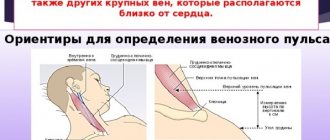
Visually, the cardiologist observes the patient:
- pale skin;
- strong pulsation of the cervical arteries;
- increased heart rate;
- constriction and dilation of the pupil (reaction to the phases of the heartbeat).
The doctor also determines the enlargement of the muscle organ and hears noises during contraction. In the initial stage of the disease, medical assistance is not provided. Such patients are given recommendations for correcting their work and rest regime. If the patient’s profession involves carrying heavy loads, long working hours, or constant physical labor, then it is advised to change jobs. In addition, emotional stress should be avoided.
As the disease progresses and moves to stage 3-4, the patient is given drug therapy.
- Calcium channel blockers (Anipamil, Falipamil) - the medicine normalizes heart rhythm and reduces the force of the stroke.
- Diuretic (“Furosemide”, “Lasix”) - the drug increases urination and removes excess fluid from the body.
- Vascular medicine (“Hydralazine”, “Diazoxide”) – relieves vascular spasm.
- Beta blockers (Bisoprolol, Metoprolol) - regulate heart rate.
If the therapeutic method of therapy is unsuccessful and the disease continues to progress, then surgery is indicated.
Treatment for heart enlargement
The treatment tactics are determined by the attending physician after all diagnostic measures have been carried out. Depending on the root cause of the problem and the severity of the condition, treatment can be either conservative or radical.
Drug therapy
If cardiovascular pathologies are to blame for the appearance of cardiomegaly, then the doctor prescribes medications from the following groups:
- diuretics;
- beta blockers;
- anticoagulants;
- antiarrhythmic drugs;
- angiotensin blockers;
- antihypertensive drugs;
- bronchodilators.
The goal of drug therapy is to normalize myocardial function, prevent the formation of blood clots, lower blood pressure, and reduce the risk of complications and heart attack.
Attention! Typically, a patient with an enlarged heart is prescribed lifelong medication.
Surgical intervention
If a large organ poses a danger to the patient, i.e. can lead to serious heart failure or arrhythmia, the doctor recommends surgery. Considering the degree of enlargement, the cardiologist may use one of the following surgical methods:
- coronary artery bypass surgery;
- implantation of a special device that stimulates the functioning of the ventricles;
- reconstruction or replacement of the valve with an artificial device;
- heart transplant surgery.
The transplant method is used only in the most severe cases, when the risk of developing severe complications increases and cardiomegaly threatens the patient’s life.
Traditional medicine methods
As a supplement to the main treatment, you can use the following recipes for herbal decoctions:
- For preparation you will need (50 g each) St. John's wort, birch buds, chamomile flowers, hawthorn fruits, immortelle flowers. Take a tablespoon of crushed collection per glass of boiling water. The composition is kept in a water bath for 15 minutes. The resulting product is filtered. Take 3 times a day, ¼ cup before meals. The course of treatment is 1-2 months.
- Take valerian rhizome and fennel fruits (200 g each), hawthorn fruits and motherwort (100 g each). The crushed components (1 tablespoon) are poured with a glass of boiling water and kept in a water bath for 15 minutes. Then infuse for two hours. The finished product is drunk after meals, ¼ cup 3 times a day. Duration of treatment – 1 month. The course can be repeated after 3 months.
Lifestyle changes
People with an enlarged heart should reconsider their lifestyle. First of all, you need to change your eating habits. You can reduce symptoms and reduce the load on your heart by following a diet. Any unhealthy food (fatty, smoked, salty, pickled, fried, etc.) is excluded from the diet.
The menu should include more fruits and vegetables. Dishes are steamed, baked or boiled. It is better to give preference to lean meats, seafood, and legumes.
You can improve your condition using the following methods:
- Sports activities. If the heart is weak, then cardiac exercises (walking, swimming) are indicated. The type of physical activity should be determined by the attending physician. By combining diet and exercise, you can quickly lose weight, which will have a positive effect on your heart function.
- Rejection of bad habits. Excessive alcohol consumption and smoking negatively affect the cardiovascular system, increasing the load on the heart. Therefore, if you have cardiomegaly, you should get rid of bad habits forever.
- Visit your doctor regularly. Patients with a large heart need to see a specialist more often to prevent the development of complications.
Cardiomegaly or enlarged heart?
Every year, hundreds of thousands of citizens die from cardiovascular pathologies around the world. In most cases, the reason for this is untimely consultation with a doctor and deterioration of cardiac function.
Enlargement of the organ is associated with the development of ventricular hypertrophy, accumulation of metabolic products and neoplastic processes. Cardiomegaly often occurs in healthy people, this includes athletes and pregnant women.
The volume of the heart varies within different limits for each person. If we talk about gender differences, then in men this organ is larger than in women. So for the age category from 20 to 30 years, the approximate heart volume will be the following values:
- women – 580 cm3;
- men - 760 cm3.
This figure also depends on body weight. A diagnosis of cardiomegaly should be made only after a thorough examination, because in some cases a slightly enlarged heart is the norm, which is strictly individual for each person.
Complications, prognosis
If cardiomegaly is not treated, complications may occur. The most dangerous of them include:
- heart failure;
- valvular insufficiency;
- heart attack;
- stroke;
- thromboembolism.
All of these conditions can be fatal. The prognosis of cardiomegaly depends on the causes of development, nature and duration of the disease. The prognosis will be extremely unfavorable for heart failure complicated by heart enlargement. Without treatment, death can occur within 2-3 years.
A more successful prognosis in the absence of heart failure and therapy (after surgery). A more accurate prognostic conclusion can only be given by the attending physician who monitors the patient’s condition over time.
Why is low heart rate dangerous?
When the rhythm slows down, the minute volume of blood circulation decreases. This means that little blood flows to the internal organs and brain. The result is an attack:
- the skin becomes cold, pale and covered with sweat;
- the vision darkens and the patient loses consciousness;
- in the absence of help, acute heart failure is possible.
Most often, consciousness is restored on its own, but in case of a prolonged attack, emergency medical care is needed. With constant bradycardia, brain nutrition decreases and discirculatory encephalopathy develops. Its consequences:
- memory loss, blackouts;
- dizziness, unsteadiness when walking;
- unclear speech, “swallowing” of syllables and words;
- deterioration of movements and sensitivity in the limbs;
- insomnia.
Prevention
Maintaining a healthy lifestyle is the best way to prevent ventricular hypertrophy. To avoid the appearance of a large heart, you must:
- adhere to proper nutrition;
- monitor body weight, avoid obesity;
- limit salt intake;
- give up coffee, alcohol, smoking;
- engage in a feasible sport;
- avoid stressful situations;
- follow the daily routine.
A large heart is a dangerous disease that requires therapy. Cardiomegaly can lead to serious complications and decreased quality of life. Therefore, you need to strictly follow all the specialist’s recommendations, avoiding the problem from worsening.
Prognosis for children and adults
Cardiomegaly is rare in children. As a rule, it is congenital with a disappointing prognosis:
- 30% of children die before 3 months of age;
- Only 25% survive with signs of heart failure;
- 45% of children recover.
In children, the causes of a large heart are congenital heart abnormalities, infections or maternal intoxication with alcohol during pregnancy.
Attention! Alcoholic cardiomegaly is especially dangerous in adults. After diagnosis, only 40% of people live 3 years. Patients die from heart failure, or thromboembolism.
If fluorography reveals an enlarged heart, consult a doctor. Based on which section and how enlarged it is, specialists determine the cause of the pathology. To clarify the diagnosis, additional laboratory or instrumental research will be needed.