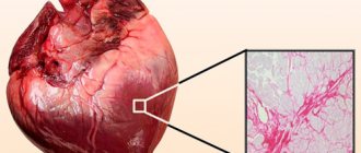
Cardiac ischemia and its treatment
The term “cardiac ischemia” does not imply a diagnosis, but a pathological condition in which the blood supply to part of the myocardium is disrupted.
The heart muscle works non-stop throughout your life, ensuring the movement of blood throughout the body. But the myocardium itself must also receive oxygen and nutrients through the coronary arteries to support performance and restore energy balance. Violation of vascular patency from a short-term spasm to complete blocking of the diameter causes coronary heart disease (CHD). This is the diagnosis according to the modern cardiological classification.
Diagnosis of myocardial infarction
When diagnosing a heart attack, several basic methods of examining the patient are used:
- Blood analysis. Myoglobin (a protein that transports oxygen to cells) can be detected in it approximately 5–6 hours after the onset of a heart attack. In addition, after 8–10 hours, the patient’s blood shows an increased activity of creatine phosphokinase by more than 50%. It later decreases to normal levels (after about two days).
Important : to exclude the possibility of a heart attack, creatine phosphokinase activity is monitored three times every 5–8 hours. And only with three consecutive negative results, the diagnosis of “heart attack” is not confirmed.
- Electrocardiogram (echocardiogram). This type of examination gives the doctor a complete picture of the patient's heart health. The transcripts show all possible pathological waves/segments, etc., arising against the background of a heart attack.
- Coronary angiography. It involves the introduction of a radiopaque substance into the blood and further monitoring of the work of all vessels and arteries of the heart using an X-ray machine. The image will show all areas of stenosis or thrombosis of blood vessels, which will enable the doctor to choose the method of surgical intervention (if necessary).
In addition to the listed methods, the doctor can also use differential diagnosis. More precisely, to track the patient’s condition based on such important signs:
- Intensity and frequency of pain. In case of a heart attack, it cannot be stopped even with nitroglycerin.
- Changes in pain when changing body position.
Only a comprehensive and quick diagnosis allows the doctor to adequately assess the patient’s condition and prescribe competent therapy.
Why is myocardial ischemia dangerous?
Cardiac ischemia is the main cause of sudden mortality in the working age population. More often it develops in men from 40 to 50 years old, after 50 years it equally affects both men and women.
Exclusion of a large area of the myocardium from movement leads to a decrease in the force of contractions. Neighboring muscle fibers are not able to take on the overload. This contributes to the gradual development of heart failure.
The weak scar of the heart wall is thinned by constant tremors. The development of an aneurysm and myocardial rupture is possible.
Treatment
Subsequently, infarction of the lower myocardial wall is treated in a hospital setting. Most often, the patient is prescribed conservative treatment. In this case, drugs such as thrombolytics are used, which dissolve blood clots blocking the arteries. Ticlopidine is considered the most effective drug in this group, but Aspirin is sometimes prescribed to prevent relapses.
In addition, doctors recommend anticoagulants that reduce blood clotting activity (for example, Dicoumarin) and beta-blockers to avoid arrhythmia and reduce the area of necrosis (Atenolol). Analgesics may be prescribed to relieve pain, and Amiodarone is used to restore heart rhythm.
If conservative treatment does not produce results, then surgical methods are used, including coronary artery bypass grafting. This decision can only be made by the attending physician.
People after myocardial infarction are prescribed therapeutic nutrition. More potassium is introduced into the diet, the amount of fat and bad cholesterol is reduced.
Why does coronary disease develop?
Work on studying coronary artery disease, preventing severe forms of ischemia and new treatment methods is being carried out in all countries. The main causes of the disease have been identified.
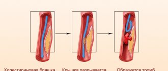
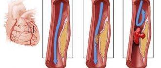
- Narrowing of the coronary arteries is possible with a pronounced atherosclerotic process. Cardiac vessels are the type of arteries in which atherosclerotic plaques are deposited between the inner and middle layers. Their increase leads to a gradual narrowing of the lumen of the coronary arteries, slows down blood flow and helps platelet deposition, which further causes thrombosis and blocks the vessel. Among the factors contributing to atherosclerosis are hypertension, diabetes mellitus and endocrine disorders, obesity, and lack of physical activity.
- Not everyone knows that before the deposition of low-density lipoproteins in the plaque, the intima of the artery is damaged by viruses (influenza, cytomegaloviruses, adenoviruses). This is confirmed by the simultaneous increase in the manifestations of coronary heart disease and the spread of influenza infection among the population. Prevention of influenza prevents the cause of ischemia.
- Stressful conditions affect the coronary vessels, causing spasm under the influence of adrenaline. With good stability, the spasm is quickly relieved, the blood supply is not disrupted. But in people with frequent overstrain of nervous regulation, this does not happen. Prolonged spasm causes malnutrition.
- Nicotine and alcohol act as provoking factors for vasospasm, disruption of nervous control function. Cigarette smoke contains substances that directly paralyze blood vessels.
- Hereditary predisposition plays a role in families with these factors.
Changes in the myocardium on the ECG - what does it mean and is it dangerous?
An ECG can diagnose most heart pathologies. The reasons for their appearance are due to concomitant diseases and lifestyle characteristics of the patient.
What does this mean if changes in the myocardium are detected on the ECG? In most cases, the patient requires conservative treatment and lifestyle modification.
Description of the procedure
An electrocardiogram (ECG) is one of the most informative, simple and accessible cardiological studies. It analyzes the characteristics of the electrical charge that causes the heart muscle to contract.
Dynamic recording of charge characteristics is carried out in several areas of the muscle. The electrocardiograph reads information from electrodes placed on the ankles, wrists and chest skin in the area where the heart is projected, and converts them into graphs.
Normal and deviations - possible reasons
Normally, the electrical activity of the myocardial areas, which is recorded by the ECG, should be uniform. This means that intracellular biochemical exchange in heart cells occurs without pathologies and allows the heart muscle to produce mechanical energy for contractions.
If the balance in the internal environment of the body is disturbed for various reasons, the following characteristics are recorded on the ECG :
- diffuse changes in the myocardium;
- focal changes in the myocardium.
The reasons for such changes in the myocardium on the ECG can be either harmless conditions that do not threaten the life and health of the subject, or serious dystrophic pathologies requiring emergency medical care.
One of these serious pathologies is myocarditis, or inflammation of the heart muscle. Regardless of its etiology, areas of inflammation can be located either in the form of foci or diffusely throughout the heart tissue.
Causes of myocarditis:
- rheumatism as a consequence of scarlet fever, tonsillitis, chronic tonsillitis;
- complications of typhus, scarlet fever;
- consequences of viral diseases: influenza, rubella, measles;
- autoimmune diseases: rheumatoid arthritis, systemic lupus erythematosus.
One of the reasons for changes in muscle tissue may be cardiodystrophy - a metabolic disorder in heart cells without damage to the coronary arteries. Lack of cell nutrition leads to changes in their normal functioning and impaired contractility.
Causes of cardiac dystrophy:
- Ingress of toxic metabolic products into the blood due to severe impairment of kidney and liver function;
- Endocrine diseases: hyperthyroidism, diabetes mellitus, adrenal tumor, and, as a result, excess hormones or metabolic disorders;
- Constant psycho-emotional stress, stress, chronic fatigue, starvation, unbalanced diet with nutritional deficiencies;
- In children, a combination of increased stress with a sedentary lifestyle, vegetative-vascular dystonia;
- Lack of hemoglobin (anemia) and its consequences - oxygen starvation of myocardial cells;
- Severe infectious diseases in acute and chronic form: influenza, tuberculosis, malaria;
- Dehydration of the body;
- Avitaminosis;
- Alcohol intoxication, occupational hazards.
Determination by cardiogram
With diffuse lesions of the heart, deviations from the normal pattern are observed in all leads. They look like numerous areas with impaired conduction of electrical impulses.
This is expressed on the cardiogram as a decrease in T waves, which are responsible for ventricular repolarization. With focal lesions, such deviations are recorded in one or two leads. These deviations are expressed on the graph as negative T waves in the leads.
If focal changes are represented, for example, by scars remaining in the connective tissue after a heart attack, they appear on the cardiogram as electrically inert areas.
What diseases do they accompany?
Pathological changes in the myocardium detected on the ECG may be accompanied by impaired blood supply to the heart muscle, reprolarization processes, inflammatory processes and other metabolic changes.
A patient with diffuse changes may exhibit the following symptoms:
- dyspnea,
- chest pain,
- increased fatigue,
- cyanosis (blanching) of the skin,
- rapid heartbeat (tachycardia).
Such manifestations most often become the reason for an electrocardiogram. In medical practice, there are many examples when myocardial pathologies did not cause noticeable changes in the well-being of patients and were discovered during preventive examinations.
Diseases accompanied by changes in the heart muscle:
- Myocardial dystrophy is a violation of biochemical metabolic processes occurring in the heart;
- Allergic, toxic, infectious myocarditis - inflammation of the myocardium of various etiologies;
- Myocardiosclerosis – replacement of cardiac muscle cells with connective tissue as a consequence of inflammation or metabolic diseases;
- Disturbances of water-salt metabolism ;
- Hypertrophy of parts of the heart muscle.
Additional examinations are needed to differentiate them.
Additional diagnostic tests
These cardiograms, despite their information content, cannot be the basis for making an accurate diagnosis. In order to fully assess the degree of changes in the myocardium, the cardiologist prescribes additional diagnostic measures:
- General clinical blood test - assesses the level of hemoglobin and such indicators of the inflammatory process as the level of leukocytes in the blood and ESR (erythrocyte sedimentation rate);
- Analysis of blood biochemistry - indicators of protein, cholesterol, glucose levels are assessed to analyze the functioning of the kidneys and liver;
- General clinical urine analysis - kidney function indicators are assessed;
- Ultrasound for suspected pathology of internal organs - according to indications;
- Daily monitoring of ECG indicators;
- Carrying out an ECG with stress ;
- Ultrasound of the heart (echocardiography) – the condition of the parts of the heart is assessed to determine the cause of myocardial pathology: expansion (dilatation), hypertrophy of the heart muscle, signs of decreased contractility of the myocardium, disruption of its motor activity.
After analyzing the medical history and laboratory and instrumental examination data, the cardiologist determines the method of treating the changes.
Treatment for focal and diffuse disorders
In the treatment of myocardial pathologies, various groups of drugs are used:
- Corticosteroid hormones – as an antiallergic agent;
- Cardiac glycosides - for the treatment of diffuse changes in the myocardium, manifestations of heart failure (ATP, Cocarboxylase);
- Diuretics - to prevent edema;
- Means for improving metabolism (Panangin, Magnerot, Asparkam);
- Antioxidants (Mexidol, Actovegin) - to eliminate the negative effects of lipid oxidation products;
- Antibiotics – for anti-inflammatory therapy;
- Drugs for the treatment of concomitant diseases;
- Vitamin preparations.
If conservative treatment does not lead to significant improvements in the condition of a patient with myocardial diseases, he undergoes surgery to implant a myocardial pacemaker.
In addition to medications, the patient is recommended to change his lifestyle and establish a balanced diet. For a patient with such pathological manifestations, physical activity, drinking alcohol and smoking are unacceptable. He is prescribed physical therapy and feasible labor.
Basic principles of dietary nutrition:
- The consumption of salt and excess liquid is limited to a minimum;
- Spicy and fatty foods are not recommended;
- The menu should include vegetables, fruits, lean fish and meat, and dairy products.
Myocardial changes detected on the ECG require additional laboratory and instrumental examination . If necessary, the cardiologist will prescribe treatment in a hospital or on an outpatient basis. Timely measures taken will help avoid serious complications.
Source: https://oserdce.com/diagnostika/ekg/izmeneniya-miokarda.html
Main clinical forms of IHD
The international classification has approved the clinical forms of the disease with characteristic manifestations and symptoms. This is necessary to develop treatment standards at all stages of medical care.
"Pectoris pectoris" or angina pectoris
A typical symptom of angina is pressing pain behind the sternum radiating to the left into the shoulder, fingers, under the shoulder blade, and into the jaw. Other features:
- Attacks are associated with physical activity, overeating, anxiety, and inhalation of cold air.
- The duration of pain is from 15 to 30 minutes. They go away on their own after stopping the exercise and taking nitroglycerin.
- The stable course of the disease is favorable for selecting treatment. Patients carry medications with them that quickly relieve pain, stop smoking, and control physical activity.
- An unstable form is more difficult to deal with. Seizures occur unexpectedly, for no apparent reason. Increased frequency indicates a pre-infarction state. A similar appearance may occur for the first time in life or develop after an acute heart attack.
- For a progressive form of angina, prolonged attacks at night, accompanied by suffocation, are typical. The dose of nitroglycerin has to be increased.
Myocardial infarction
The pain is localized in the anterior parts of the chest (irradiation as in angina pectoris), is intense and persistent, and lasts up to several days. In specialized departments, anesthesia is used to completely remove them. It usually starts in the morning. Nausea, vomiting, dizziness, increasing weakness, and shortness of breath are observed. Sometimes a burning sensation appears in the epigastrium and bloating occurs.
Depending on the prevalence, varying degrees of damage to the activity of the left ventricle of the heart are observed, the rhythm is disturbed, and acute heart failure develops. A state of shock with a sharp drop in blood pressure requires intensive care and does not always have a successful outcome.
Myocardial infarction must be treated in a specialized department.
Cardiosclerosis
This form of ischemia is characterized by the development of multiple scar foci. The cause of cardiosclerosis is the overgrowing of the lumen of the heart vessels with atherosclerotic plaques.
If the affected area is small, the course of the disease progresses gradually, but inevitably leads to heart failure with shortness of breath, tachycardia, and swelling in the legs.
IHD without pain
This form was identified in the classification to characterize symptoms in people with a high pain threshold, those whom we call “patient.”
Often such a clinic is observed in the elderly, alcoholics, and people with diabetes. Instead of pain, patients feel weakness, nausea, palpitations, increased shortness of breath, and heartburn.
Sudden death
With primary cardiac arrest, which occurs within a few hours after the onset of an attack of pain, complications quickly develop. Resuscitation can be successful if specialized care is provided immediately. Most often, death occurs in people with multiple risk factors.
Ischemia of the posterior wall of the heart - what is it?
The strongest muscle in the human body is the heart. It is capable of pumping up to 7000 liters of blood per day at a speed of 1.6 km/h, which is comparable to the operation of a powerful pump. At the same time, the human heart is characterized by increased sensitivity to hypoxia, which often leads to damage to cardiac tissue.
The basic method in the study of heart disease is the ECG. Registration of electrical impulses carried out in all leads will help to detect even outdated signs of myocardial ischemia.
People who have previously experienced oxygen deprivation should be especially vigilant and regularly resort to medical examination in order to prevent repeated spasm of the coronary arteries.
Signs of ischemia on the ECG
What is ischemia of the heart muscle
Coronary heart disease is an acute or chronic condition that occurs as a result of impaired arterial blood flow to the myocardium due to spasm or blockage of the coronary vessels.
When the heart does not receive the amount of oxygen it needs, areas of connective tissue form in the lumen of the muscle fibers, which have lost the ability to function at full strength..
The process of myocardial damage always begins with minor ischemia, which, without proper treatment, eventually leads to a true infarction.
The pathogenesis of coronary heart disease is approximately as follows:
- Stable angina. It is characterized by paroxysmal pressing pain behind the sternum, which occurs under the influence of physical activity and gradually disappears when stressful conditions are excluded.
- Unstable angina. It represents an intermediate period between stable myocardial ischemia and the development of complications. Its clinical sign is chest pain, which develops even at rest and can lead to cell damage.
- Small focal myocardial infarction. This is a very insidious variant of IHD, characterized by the absence of a pathological Q wave on the ECG and microscopic foci of necrosis; it often goes unnoticed, as it is disguised as an acute attack of angina.
- Q-myocardial infarction. One of the dangerous complications of cardiac muscle ischemia is a large-focal infarction, characterized by transmural myocardial damage with ST segment elevation and the formation of an additional Q wave, which does not disappear even after complete replacement of areas of necrosis with connective tissue.
ECG signs of myocardial ischemia
Since the signs of the ischemic process are the same in some subtypes of the disease, there are a number of additional studies to identify a heart attack. Early markers of cardiac necrosis include myoglobin and creatine phosphokinase.
For a more accurate diagnosis, it is useful to examine the level of lactate dehydrogenase, aspartate aminotransferase and troponins after 6–8 hours.
ST segment elevation can be present not only during a heart attack; it often occurs with unstable angina, and therefore all visible changes in the waves on the ECG must be taken into account.
How does ischemia manifest on an electrocardiogram?
It is quite difficult to give an unambiguous answer to what the result of recording electrical impulses in coronary heart disease on film will look like.
With the development of myocardial hypoxia, the movement of electrical potentials slows down somewhat, Potassium ions leave the cells, negatively affecting resting potentials.
Compensatory processes are activated, the heart is overstrained, a pressing pain appears behind the sternum, and the patient is disturbed by a pronounced feeling of lack of air.
Characteristic signs of oxygen starvation of cardiac tissue:
- ST segment depression, horizontal or oblique;
- reduction of the T wave, as well as a shift below the horizontal line;
- widening of the T wave due to slower ventricular repolarization;
- the appearance of a pathological Q wave with large-focal necrosis;
- dynamics of changes in the ECG (indicates the freshness of the process).
ECG signs of myocardial ischemia
Additionally, the figure may show signs of blockades and arrhythmias that arose as complications of the ischemic process. In most cases, with myocardial ischemia on the ECG, the QRS complex retains its normal shape, since oxygen deficiency primarily affects the repolarization (restoration) of the ventricles, which normally completes the cardiac cycle.
Treatment
First of all, the treatment of ischemic diseases involves:
- Moderate physical activity (exercise, walking) and avoiding excessive overload.
- Compliance with a special diet (the same as that prescribed for atherosclerosis) aimed at improving metabolism. If body weight is significantly higher than normal, it must be reduced by reducing the amount of food and reducing its calorie content.
- Drug therapy, medications for which are prescribed by a doctor individually.
All patients, without exception, are prescribed antiplatelet agents - acetylsalicylic acid, on the basis of which they are created, shows excellent results in the fight against pathology. If necessary, the doctor additionally prescribes the use of anticoagulants. In case of a heart attack, heparin is required.
Beta-blockers are considered very important drugs, which help regulate the heart rate and reduce the need for oxygen, thereby increasing the life expectancy of patients.
Fibrates and statins help reduce the content of atherogenic cholesterol fractions, while increasing the amount of antiatherogenic ones.
Nitroglycerin is very effective in relieving pain. It is used both in the form of tablets and injections
However, this drug should be used with extreme caution by hypotensive patients, since its side effects can include a sharp decrease in blood pressure, fainting and dizziness.
To remove excess fluid that creates a load on the myocardium, diuretics are used - thiazide, diuretics and loop drugs.
Almost all treatment regimens for ischemic diseases include ACE inhibitors, since they relieve vascular spasms and normalize blood pressure, stabilizing its values.
If a patient has cardiac arrhythmia, he is prescribed antiarrhythmic drugs. For tachycardia, beta blockers will be required, for other forms - cordarone or amiodarone.
In cases where the arteries are very severely affected and drug therapy does not have the desired effect, surgical correction is performed. Both more gentle techniques (stenting or balloon angioplasty) and radical ones (coronary artery bypass grafting) are used.
Ischemic heart diseases always have an extremely serious prognosis.
The vast majority of people become disabled as a result of this pathology, and the risk of complications and even death is extremely high.
Due to the widespread prevalence of the disease, specialists are making every possible effort to find the best way to treat the pathology and its successful prevention.
https://.com/watch?v=MNpzKJilcbk
ECG depending on the location of the ischemic area
The inner layer (endocardium) is more susceptible to lack of oxygen, since it is supplied with blood much worse than the epicardium, and receives much greater pressure from the blood that fills the ventricles.
ECG results may vary greatly depending on the location and volume of cardiomyocytes affected.
Myocardial hypoxia is often indicated by a change in the ST segment, which consists of depression greater than 0.5 mm in depth in at least two adjacent leads. Depression can be either horizontal or downward.
Deviations on the ECG will be directly related to the ischemic area:
- damage to the anterior wall of the left ventricle near the endocardium - characterized by a high positive T wave with a sharp end, distinguished by visible symmetry;
- oxygen starvation of the anterior wall of the left ventricle with transmural damage to myocardial tissue is one of the most dangerous types of hypoxia, characterized by a biphasic or drooping flattened T wave;
- subendocardial ischemia, localized near the endocardium of the posterior wall of the left ventricle - the T wave on this version of the ECG will be reduced and almost flat;
- subepicardial ischemia at the anterior wall of the left ventricle will be indicated on the ECG by a negative T wave with a sharp apex;
Possible ECG changes in cardiac ischemia - transmural lesion of the posterior wall of the left ventricle - characterized by an elevated positive T wave with a sharp, symmetrically placed apex.
When an obliquely ascending placement of the ST segment is present in the figure, it can be compared with the presence of severe tachycardia in the patient. In this case, after eliminating the stress factor and stopping the tachycardia, the result of the electrocardiogram will show normal.
If the patient was able to undergo an electrocardiographic study during the acute stage of a heart attack, then on the ECG it will be possible to see depression of the ST segment of an obliquely ascending nature, which turns into “coronary waves” of T, characterized by an impressive amplitude.
Diagnostic signs
Diagnosis of coronary artery disease begins with an examination by a doctor and questioning of the patient. A clear description of the complex of symptoms indicates signs of one of the forms of myocardial ischemia. Auscultation can reveal rhythm disturbances. The disease does not produce typical noises.
In the ambulance
At the ambulance level, an ECG study is carried out. Doctors of various specialties are familiar with the signs of acute coronary pathology. The ECG tape shows the rhythm of contractions, an increase in the load on individual parts of the heart, the zone and size of ischemia, necrosis (in case of a heart attack), focal or diffuse cardiosclerosis are determined.
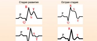
Stages of development of a heart attack with ST segment elevation
The most acute stage of STEMI
First, a high coronary T appears on the cardiogram. The high coronary T before ST elevation is present for a very short time, so it is not always possible to register it. Then there is an increase in the ST segment, which merges with a high T.
Scheme 2. ECG fragments in the most acute stage of STEMI
Acute stage STEMI
Lasts up to 2-3 days (sometimes up to 2 weeks). The beginning of the acute phase is considered to be the appearance of the Q wave, which reflects the formation of a zone of myocardial necrosis. As the acute phase develops, the Q wave deepens, and the ST segment begins to decline towards the isoline. The T wave becomes negative.
Scheme 3. ECG fragment in the acute stage of STEMI
Subacute stage of STEMI.
Lasts for several weeks. The ECG records a decrease in the ST segment to the level of the isoline. At the same time, a deep negative T wave is formed.
Scheme 4. ECG fragment in the subacute stage
Scar stage STEMI
Dead myocardial cells are replaced by connective tissue, which leads to the formation of a scar. The areas of the myocardium surrounding the scar undergo compensatory hypertrophy. The number of leads on the ECG in which changes associated with a heart attack are recorded is reduced. The Q wave may become less deep, and the T wave returns to the baseline. The formed picture of post-infarction cardiosclerosis on the ECG persists throughout the patient’s life.
Sometimes the ST segment does not reach the isoline and remains elevated almost throughout life. As a rule, the QRS complex takes on the QS form, which may indicate the formation of a cardiac aneurysm.
Scheme 5. ECG fragments with post-infarction changes
Diagram 5 on the left shows a fragment of a cardiogram indicating a myocardial infarction. On the right is a fragment of a cardiogram in which the ST segment has not reached isolia and a QS complex has formed, turning into increased ST. This picture is called a “frozen ECG” and indicates a post-infarction cardiac aneurysm.
How is ischemia treated?
Treatment of coronary heart disease is carried out on an outpatient basis or in a hospital, depending on the form and severity of the patient’s condition.
If angina attacks become more frequent, bed rest is required. Acute myocardial infarction will require a longer gentle regimen. This is the only way to reduce the load on the heart.
Exercises for arms and legs are recommended to be done in bed without serious effort and only in the complete absence of pain.
Nutrition of the patient
The diet for ischemic heart disease is based on the principle of preventing risk factors from increasing: animal fats (cholesterol) and sweets (glucose) are limited as much as possible.
The diet excludes: fatty meat, lard, butter, smoked foods, all fried foods, sweet confectionery and candies. To relieve bloating, vegetable dishes with cabbage, legumes, and spicy seasonings are not recommended.
Coffee, strong tea, chocolate, cocoa are not recommended.
Dairy products (kefir, cottage cheese, sour cream) can be consumed with reduced fat content.
Boiled fish, poultry dishes, vegetable oil, cereals, salads from fresh and boiled vegetables are shown. Not limited to fruits.
You can drink green tea, juices, still mineral water.
Drug therapy
In a serious condition, treatment is determined by symptomatic needs: anti-shock measures, painkillers, drugs that affect fibrinolysis (if possible, dissolve a blood clot in the coronaries).
In the future, patients must take:
- nitrates (reduce the need for oxygen in the heart muscle);
- statins (lower cholesterol);
- anticoagulants (deprive platelets of the ability to stick together and resist thrombus formation).
For initial symptoms of heart failure, diuretics are prescribed. Patients with hypertension complicated by ischemia will have to reconsider previously taken medications and their dosage.
Patients are asked to carry nitroglycerin preparations or a small spray can (Isoket) with them at all times. The occurrence of pain in the heart area cannot be tolerated. You should immediately take the recommended products under the tongue.
For treatment, anxiety and stress, hard work, and physical activity are strictly contraindicated for patients.
Folk remedies
Treatment with folk remedies helps lower blood cholesterol, adds vitamins and protective forces during the recovery period. It is not recommended to try to use traditional methods to treat pain in the heart. This is a good addition to your diet.
For ischemic disease it is recommended:
- Eat viburnum berries in the pits;
- wash down nutmeg with milk;
- take oatmeal jelly, decoction before meals;
- add cardamom to cereals and drinks;
- drink tincture of young pine cones;
- the famous recipe “for all diseases” - honey with garlic and lemon;
- a decoction of woodlice, nettle, and bluehead herbs;
- tinctures of valerian, hawthorn, motherwort are sold in the pharmacy in ready-made form, relieve unnecessary stress, improve the rhythm of contractions.
All treatment methods are good if the patient has an internal intention to heal. Little things like quitting smoking and drinking are unimportant when it comes to life or early death.
source
What is myocardial ischemia of the inferior wall of the left ventricle
When a specialist indicates myocardial ischemia, he means one of several pathological conditions (heart attack, angina, heart failure or cardiosclerosis).
The development of IHD is provoked by physical inactivity, abuse of fatty foods and smoking. Ischemia is best detected by ECG.
This inexpensive and informative research method with a high probability determines not only the degree of myocardial damage, but also the localization of the process.
The first signs of coronary heart disease
The signs characterizing IHD directly depend on the type of disease. In addition, in some cases, ischemia is asymptomatic. This can complicate the diagnostic process.
The following signs of ischemic disease are distinguished:
- pressing pain that intensifies due to stress or physical activity;
- the appearance of shortness of breath even after minor exertion;
- heart rhythm disturbances;
- general weakness, fatigue;
- swelling of the legs;
- sudden fear of death.
An ECG for coronary heart disease allows you to assess the location, distribution and depth of disorders in the myocardium.
An ECG for ischemic heart disease, depending on the form, contains the following data:
- The appearance on a graphical reflection of coronary teeth with sharp ends, characterized by symmetry and significant amplitude. This is due to insufficient blood supply and hypoxia of cardiac tissue. As a result, the rate of repolarization of organ cells decreases. Depending on the location of the area affected by coronary artery disease, coronary waves can be either positive or negative.
- Signs of ischemia on the ECG, expressed in T-waves with further displacement of the ST segment for 15–30 minutes, occur with acute myocardial infarction. However, in some cases they indicate the development of other diseases (alcoholic cardiomyopathy, vagotonia, etc.).
- Signs of ischemic heart disease on the ECG, reflected in the form of a shift of the ST segment above or below the isoline, are recorded during ischemic damage. In this case, a deviation of less than or equal to 0.5 millimeters is within the normal range.
- In case of ischemic damage, a characteristic sign on the ECG is the occurrence of the phenomenon of reverse changes. The first signs of coronary heart disease with subepicardial damage, according to the readings of the electrodes located above the affected area, are ST segment elevation. Electrodes recording readings from the opposite side of the heart muscle will determine the depression of this segment.
- The ECG conclusion for coronary artery disease, indicating myocardial infarction, is based on identifying Q waves that have values above normal. A gradual increase in the amplitude of the R waves is also detected.
These are not all the signs that can be read from an electrocardiogram. However, a detailed assessment of the research data must be entrusted to a specialist.
Electrocardiogram during pacing
In pacing, the heart is activated by artificial, rhythmically delivered electrical impulses from a pacemaker. The ventricle to which the stimulating electrode is attached is the first to be excited, then the excitation passes to the other ventricle.
The course of the impulse resembles its distribution during bundle branch block. Each electrical impulse from the pacemaker creates a mark on the ECG - an artifact, which is a plumb line located in front of the QRS complex.
1) constant pulse frequency;
2) artifact before the ventricular QRS complex;
3) widened and deformed ventricular QRS complex, reminiscent of the shape of the ventricular complex with complete blockade of one of the bundle branches;
ECG at rest
An electrocardiogram performed in a patient with CAD at rest is the simplest method of assessment. The procedure is carried out without preparatory measures, regardless of the time of day.
In this case, electrodes are installed on the body. They are located on the limbs and sternum. The average duration of a resting ECG is 5–7 minutes.
The study has no side effects and can be repeated, if necessary, an unlimited number of times.
The study allows us to identify the following signs of IHD:
- rhythm disturbance;
- hypertrophic change in the myocardium;
- symptoms of previous myocardial infarction;
- cardiac cycle disorders.
Topical diagnosis of myocardial infarction localization
Topical diagnosis (localization) of myocardial infarction is determined by those leads in which changes characteristic of myocardial infarction are detected.
Table. Typical locations of myocardial infarction
Accelerated ectopic rhythms are non-attackable normoor tachycardias emanating from the atria, AV junction or ventricles. The cause of such arrhythmias is an increased frequency of impulses from underlying centers of automatism, exceeding the sinus frequency and the interception of the function of the main pacemaker of the heart by a more active ectopic focus.
1) correct non-sinus rhythm with a frequency of more than 60 beats per minute,
2) gradual onset and end of an episode of non-sinus rhythm. If the frequency of the accelerated ectopic rhythm exceeds 90
beats per minute, this rhythm is called non-paroxysmal tachycardia from the corresponding part of the heart (supraventricular or ventricular).
ECG during an attack or immediately after it
The procedure allows you to determine the affected area in case of coronary artery disease. Recommended if symptoms are detected only during an attack and then completely stop. The following are the signs of IHD:
- Amplitude and polarity of T-waves, deviation of the indicator from the norm. With ischemic heart disease, the teeth can be symmetrical negative and have a height of more than 6–8 millimeters due to muscle relaxation due to tissue hypoxia.
- In addition, tall, positive, symmetrical T-waves may be recorded in heart disease. They are detected during the diagnosis of subepicardial ischemic heart disease. The indicator is recorded below the active electrode.
- T-waves may also have a flattened, lowered, biphasic pattern. The indicator is detected when diagnosing coronary heart disease when an active electrode is placed in the peripheral zone of coronary artery disease.
- Despite the detected signs of coronary heart disease, the ST segment does not have deviations from normal values.
- The QRS complex does not differ from the usual appearance in coronary heart disease.
Interpretation of electrocardiogram sections for ischemic heart disease
When and how often to do it
An ECG is included in the list of mandatory tests when passing a medical commission. Therefore, once a year, a healthy adult should have a cardiogram. This is important because heart problems do not always show symptoms.
If a person has already been diagnosed with myocardial ischemia, he needs to undergo an ECG at least once every 1-3 months, and if his condition worsens, immediately during an attack. Only in this case is it possible to avoid the development of a heart attack. With the same frequency, it is necessary to check the condition of the heart in diseases that are provoking factors of ischemia:
- diabetes;
- hypertension;
- severe toxicosis in pregnant women;
- systemic pathologies;
- disorders of the thyroid gland.
Cardiography should be done a little more often in elderly people and people whose profession is associated with a high risk of developing coronary artery disease.
ECG monitoring
ECG for ischemic heart disease is mainly based on the Holter method. Wherein:
- a small device is attached to the patient’s body;
- data is recorded throughout the day;
- the information is stored in the device’s memory and evaluated at the end of the procedure.
This technique allows you to assess the state of the patient’s heart function over a 24-hour period in everyday life. Based on the data, it is possible to determine the prerequisites and signs of angina attacks.
Left ventricular hypertrophy
Left atrial hypertrophy leads to an increase in the total time of impulse propagation through the atria.
1) a significant increase in the duration of the P wave - more than 0.12 s;
Source: https://tepcontrol.ru/chto-takoe-ishemiya-miocarda-nizhney-stenki-levogo-zheludochka/
Myocardial ischemia: causes, symptoms, diagnosis, treatment
© Author: A. Olesya Valerievna, candidate of medical sciences, practicing physician, teacher at a medical university, especially for SosudInfo.ru (about the authors)
Myocardial ischemia is a pathological condition consisting of a lack of oxygen to the heart, which is manifested by angina pectoris, heart attack, and various changes in the rhythm of contractions. Ischemia is based on atherosclerosis, thrombus formation or spasm of the heart arteries.
Myocardial ischemia forms the basis of coronary heart disease (CHD), the most common pathology of the cardiovascular system in humans. According to statistics, at least half of elderly men and a third of women suffer from it, and mortality from various forms of ischemia reaches 30%.
The disease has no geographical boundaries and is common in both developing and developed countries with a high level of medicine. For a long time, IHD can be asymptomatic, only occasionally making itself felt by unpleasant sensations in the heart area.
Painless myocardial ischemia is of great importance. The disease does not manifest itself in any way for many years, but can cause a massive heart attack and sudden death. According to some data, this form of pathology affects up to 20% of practically healthy people, but with risk factors.
Dystrophic changes in the myocardium
The muscles of the human heart wall, thanks to extremely complex heterogeneous, autogenous and neurohumoral processes in the body, are capable of generating a bioelectric impulse from biochemical processes into mechanical energy responsible for creating rhythmic movements of the heart. And the consequences of changes in the balance of the body, first of all, result in changes in the myocardium of the posterior and lower walls of the ventricle of the heart. They may be cardiac pathologies, hormonal imbalances, or the consequences of a viral infection.
What it is
The pathology of any area related to cardiology is called diffuse changes in the myocardium. This condition may be a consequence of a disease or be caused by a malfunction of the mechanism of physical and chemical reactions of the body.
The main task of myocardial muscle tissue is to conduct transmitted electrical voltage along neuromuscular fibers.
Under the influence of diffuse changes, the heart cannot function normally - the frequency of contractions of the heart muscle regularly decreases and the heart rhythm is disturbed.
A concentration of cells is formed that have undergone degenerative changes and are unable to transmit nerve impulses through themselves, electrophysiologically manifesting themselves as a necrotic zone.
Damage to the functionality of the myocardial cellular system can be diffuse - evenly distributed throughout all tissues of the heart and focal - isolated, for example, during the formation of connective tissue in place of irreversibly damaged cells. Scars are overgrown connective tissue that manifests itself as electrically inert, incapable of conducting nerve impulses and leading to cardiac hypertrophy.
Causes
The causes of degenerative changes in the myocardium of the ventricles of the heart are caused by various processes.
Myocarditis
This pathology develops due to pathogenic microorganisms that have entered the cells of the heart muscle. There are isolated and diffuse cardiac myocarditis.
Healthy heart and myocarditis
It is a consequence of heart diseases such as:
- Rheumatic disease caused by group A B-hemolytic streptococcus.
- Diphtheria, rickettsiosis,
- Respiratory viral diseases,
- Various disorders of the immune system.
Dystrophic
They arise due to disruption of physical and chemical processes in myocardial cells. It has a non-inflammatory nature. It is characterized by the appearance of damaged muscle fibers that do not participate in the contractile activity of the myocardium, and are also unable to receive sufficient amounts of nutrients.
This leads to the death and decomposition of such cells and the appearance of necrotic foci in the muscular layer of the myocardium. Dystrophic changes on the ECG of the heart do not manifest themselves in any way.
The causes of myocardial dystrophy are:
- Intoxication with breakdown products due to electrolyte diseases of the renal system.
- Endocrine metabolic disorders.
- Physiological reasons.
- Decreased hemoglobin and insufficient oxygen supply to myocardial cells.
- Acute and chronic respiratory viral diseases.
- Diseases accompanied by disruption of the thermoregulation process and dehydration of the body.
- Avitaminosis.
- Intoxication caused by chemicals (alcohol, nicotine, pesticides).
- Increased intellectual load with a lack of physical activity can occur in children and provokes vegetative-vascular dystonia.
Metabolic changes occur due to changes in the balanced composition of ions in myocardial cells and a decrease in the electrolyte composition of the blood,
The pathology causes a decrease in the contractility of heart cells and a failure of the entire energy metabolism. Metabolic changes in the heart appear on the ECG as deviations of segments and complexes from the isoline.
- The causes are conditions caused by a lack of products involved in metabolic processes
- Conditions that provoke dystrophic changes in the myocardium.
- Formation of cholesterol plaques on the walls of the coronary artery.
- Impaired blood flow in the coronary arteries of the heart.
- Arterial essential hypertension.
- Heart rhythm disturbances.
- Enlargement of the heart muscle.
Failure of metabolic processes in cardiomyocytes and nonspecific changes in the myocardium are a manifestation of insufficient blood supply to the heart muscle, which can lead to the development of various pathologies.
Impaired blood flow in the coronary arteries
Mild myocardial changes in childhood and old age are considered normal.
Scarring
They cause the growth of connective tissue on the heart muscle. Scar changes on the heart can be diffuse and focal.
Occurs in conditions such as:
- cardiac ischemia. Causes necrosis of individual areas of the myocardium and forms a focal scar.
- transferred inflammatory process. Causes the death of muscle fibers throughout the heart muscle area.
Symptoms
With diffuse changes in the myocardium, symptoms are not observed. You can observe clinical manifestations of the underlying cardiac disease, which has caused changes in the heart muscle. Then the symptoms of changes in the myocardium of the left ventricle of the heart appear as secondary.
These include:
- painful sensation or feeling of discomfort behind the sternum due to ischemia of the heart muscle;
- violation of the frequency and depth of breathing and excessive accumulation of fluid in cardiosclerosis;
- consequences of myocardial infarction;
- involuntary contraction and relaxation of muscles (tremor);
- sudden weight loss and eye pathologies due to hyperthyroidism;
- pallor of the skin, disorientation in space and a feeling of fatigue with anemia.
When diffuse changes appear on the ECG, you need to pay attention to the manifestation of symptoms. If signs of myocardial changes are present, further examination should be performed.
Diagnostics
In the absence of a concomitant disease associated with disruption of the cardiovascular system, specific clinical signs of diffuse or focal changes do not appear.
Therefore, myocardial changes can be diagnosed using a cardiogram. The basis for such changes can be a variety of factors, from harmless to those causing significant harm to health.
The decision on the advisability of further examination is made by the cardiologist.
In the absence of clinical signs of serious pathology and minor changes in the myocardium in adults, no further examination is required. It is enough to take preventive measures aimed at normalizing blood pressure levels, adhering to the principles of diet, and additional intake of vitamins and macroelements. Taking health promotion measures is also recommended.
If there is reason to assume the presence of significant degenerative changes, a thorough diagnosis of myocardial changes is carried out, including:
- General blood and urine analysis.
- Analysis of kidney parameters, number of red blood cells and white blood cells.
- Blood chemistry.
- Reflects the state of all body systems. Also provides information about metabolism.
- Ultrasound examination of the heart, liver and kidneys.
- Electrocardiography.
- Analysis of the manifestations of deviations from normal heart rhythm - the presence of pathological Q waves, decreased T waves and other symptoms.
- Holter monitoring and functional tests,
- Shows morphological and functional changes of the heart and its valve apparatus.
Myocardial changes are well diagnosed on an ECG
Treatment
Treatment of changes in the myocardium of the ventricles of the heart is based on complex therapy. The initial stage is based on changing the usual lifestyle and creating a diet taking into account the specific nature of the disease.
Diet
With diffuse changes of a non-inflammatory nature in the initial stages, a properly selected diet, good sleep and adequate rest are of utmost importance.
To fully nourish the heart muscle with the necessary energy resources, you should follow a diet that excludes foods with high cholesterol levels, flour and sweets, alcohol and coffee, spices that irritate the walls of the stomach.
Foods and vitamins to strengthen your heart
It is recommended to divide the meal into six approaches. Must be used:
- Lean meat and chicken.
- Sea fish.
- A sufficient amount of vegetables and fruits rich in iron and calcium (apricots, bananas, carrots, potatoes, spinach, nuts).
- Whole milk products.
- Whole grain cereals are rich in complex carbohydrates.
Drugs
Drug therapy consists of prescribing drugs that improve cellular metabolism.
Typically, when there is a change in the myocardium, drugs such as:
- Panangin, asparkam avexima, magnesium orotate dihydrate, magne B6 - are responsible for the conduction of nerve impulses, serve as the prevention of cardiac arrhythmia, and stimulate metabolic processes in the myocardium.
- Actovegin and Mexidol are antioxidants that help activate energy processes in the cell and affect its functional metabolism.
- Retinol, ascorbic acid, tocopherol, thiamine and pyrodoxine.
If the cause of diffuse changes is a cardiac disease, then therapy is aimed at treating it. For example, the prescription of drugs that replenish iron deficiency in case of disturbances in hemoglobin levels, hormonal therapy for disorders of the endocrine system, drugs aimed at stabilizing blood pressure in case of essential hypertension.
Do not be upset if the ECG results reveal diffuse changes in the myocardium. The presence of minor changes in the absence of symptoms of serious cardiovascular diseases can occur with metabolic disorders. But you shouldn’t put off visiting a doctor. If necessary, only a cardiologist can prescribe examination and treatment.
Source: https://SostavKrovi.ru/sosudy/serdca/distroficheskie-izmeneniya-miokarda.html
What happens in the heart during ischemia?
The main symptom of IHD is pain, which occurs both in the chronic course of the disease and in its acute forms. The occurrence of pain is based on irritation of nerve receptors by metabolic products that are formed under hypoxic conditions. The heart continuously works, pumping enormous volumes of blood, so the cost of oxygen and nutrients is very high.
Blood flows to the heart muscle through the coronary vessels, and collateral blood flow in the heart is limited, so when the arteries are damaged, the myocardium always suffers. An atherosclerotic plaque, a blood clot, or a sudden spasm of blood vessels creates an obstacle to blood flow, as a result of which muscle cells do not receive enough blood, pain and characteristic structural changes in the myocardium appear.
In cases of chronic myocardial ischemia, usually with atherosclerosis, the heart muscle is constantly “starving”; against this background, stimulation of fibroblast cells that form connective tissue fibers occurs, and cardiosclerosis develops. Involvement of nerve bundles contributes to arrhythmia.
Vascular accidents due to thrombosis, plaque rupture, spasm are accompanied by a complete and sudden cessation of blood flow through the vessels, the blood does not reach the heart muscle, and acute myocardial ischemia “results” in a heart attack - necrosis of the heart muscle. Often, acute forms of the disease occur against the background of long-term chronic ischemia.
Ischemic changes are usually recorded in the left half of the heart, since it experiences a significantly greater load than the right parts. The thickness of the myocardium here is greater, and good blood flow is needed to provide it with oxygen. Ischemia of the wall of the left ventricle usually forms the basis of ischemic heart disease, and this is where the main events in necrosis of the heart muscle “unfold.”
Clinical case
A 58-year-old man was brought to me with complaints of sudden shortness of breath, severe sweating, and no typical chest pain.
On auscultation, moist fine bubbling rales were heard in the lower parts of the lungs. A general blood test and electrocardiography did not yield any results. EchoCG revealed an area of akinesia in the basal parts of the left ventricle. The first troponin test was negative, the second one became positive 1 hour after hospitalization. As a result, “Acute myocardial infarction of the inferior wall of the left ventricle” was diagnosed. AHF 1" The patient received treatment, which consisted of prescribing antiplatelet agents (Aspeter), anticoagulants (Enoxaparin), beta blockers (Metoprolol) and nitrates (Nitroglycerin). The general condition stabilized after 10 days, there were no complications.
Knowledge of the specific symptoms of acute myocardial infarction is necessary not only for doctors, but for all people, at least in order to promptly seek medical help.
Manifestations of myocardial ischemia
Clinical signs of cardiac ischemia depend on the degree of arterial damage and the course of the pathology. The most common type of ischemia is angina pectoris, when pain appears during physical effort. For example, a patient climbed the stairs, went for a run, and the result was chest pain.
Symptoms of angina include:
- Pain in the heart area, behind the sternum, spreading to the left arm, interscapular area, intensifying or appearing with physical stress;
- Shortness of breath when walking fast, emotional overload.
First aid
If there is a suspicion that this is a posterobasal myocardial infarction, then you should immediately call an ambulance. Before doctors arrive, the patient should be positioned so that his head is elevated. If a person suffers from shortness of breath, it is better to sit him down.
It is recommended to place a Nitroglycerin tablet under the tongue (the drug is also available in the form of a spray, and this option is considered more convenient when providing first aid).
Nitroglycerin most often cannot completely relieve pain. Therefore, many try to immediately give the second tablet. This is incorrect, since the interval between doses of the drug should be at least 15 minutes. Moreover, Nitroglycerin must be given, especially for shortness of breath, since it eliminates precisely this symptom.
Important information: Algorithm of actions for providing first emergency aid to a person with myocardial infarction at home before the ambulance arrives
The mechanism of pathology development
People over 40 years old have a history of a disease such as atherosclerosis. It provokes a narrowing of the lumen of blood vessels, as a result the elasticity of the membranes changes, and deposits form on them.
These disorders cause oxygen deficiency in organs, tissues, and most importantly, the heart, which leads to necrosis of certain areas. The lesion can be located in different places, one of which is the posterior wall of the myocardium.
It is divided into the diaphragmatic and basal sections, which is why the names of infarctions come from:
- Posterior phrenic - the side of the left ventricle adjacent to the diaphragm. The lower coronary blood canal is blocked by a thrombus, which leads to large-focal lesions.
- Posterobasal - occurs as a result of occlusion of the distal parts of the right coronary artery or the circumflex branch of the left coronary artery. This type of pathology is observed with an extensive heart attack.
All affected areas of cardiac muscle tissue cannot be resuscitated. They are covered with fibrous tissue, which cannot perform all the necessary functions. Therefore, to prevent recurrence of an acute attack, it is recommended to constantly take medications and lead a healthy lifestyle.
Diagnostics
When diagnosing myocardial infarction, the following methods are used:
- Physical. The examination begins with collecting anamnesis, examining the patient, and palpating tissue to detect the point of the heart that fits tightly to the chest. If the lymph nodes are enlarged, it means that an inflammatory process is developing. The pulse rate is also determined. During an attack, a complete cessation of heart contractions may occur. The chest is tapped to clarify the boundaries of the organ. They tap rhythms and tones using a stethoscope, and measure blood pressure and temperature.
- Hardware. The main method for determining disorders is electrocardiography. Inferior myocardial infarction appears very accurately on the ECG. An ultrasound examination is performed to assess the condition of the heart. During its course, they determine the speed of blood flow, the presence of blood clots, aneurysms, and find out the condition of the vessels. Foci of necrosis can be detected by scintigraphy. Vascular patency is checked using coronary angiography. If these manipulations do not provide accurate information, then magnetic resonance imaging is performed.
- Laboratory. The blood is examined for the presence of markers of necrosis. A heart attack is accompanied by the destruction of cardiomyocytes, and their destroyed components enter the bloodstream. If a blood test confirms their presence, then there is no doubt about the diagnosis.
Also read: Features of right ventricular myocardial infarction