Types and causes of pathology
What is a change in the myocardium? This is the name for any deviations that interfere with the normal functioning of an organ. They can have varying degrees of severity. Depending on the reasons that led to the failure, this pathology may be of the following nature:
- diffuse;
- dystrophic;
- metabolic;
- focal.
Any of the above conditions requires timely treatment. Otherwise, the development of coronary heart disease may begin due to lack of oxygen.
Diffuse changes in the myocardium
They are characterized by the incorrect functioning of all cardiac cells when the muscle is affected evenly. Most often they occur due to inflammation, which includes myocarditis. Sometimes the development of pathology occurs with significant physical exertion and as a result of taking certain medications.
Against the background of the disease, changes in metabolic processes in the heart muscle occur. As a result, the organ ceases to synthesize oxygen in the amount necessary for the normal functioning of the circulatory system. Experts identify several reasons for the development of this disease:
- excessive alcohol consumption;
- great physical activity;
- stress;
- frequent hypothermia;
- chronic infections.
If the disease is detected in a timely manner, then competent treatment will help return the heart to a normal state. In most cases, this will require the patient to reduce physical activity to a moderate level, review their diet and begin to lead a healthy lifestyle.
Dystrophic
Observed in the left ventricle and occur due to a lack of nutrients that the heart muscle should receive. In medicine, this condition is called cardiac dystrophy. The reasons for its occurrence are as follows:
- intoxication due to impaired kidney and liver function;
- anemia;
- nervous overstrain;
- diabetes mellitus and disruptions in the endocrine system;
- poor nutrition, which leads to the development of vitamin deficiency;
- dehydration due to poisoning;
- chronic diseases and infectious diseases;
- intoxication resulting from alcohol abuse or taking medications.
Such disturbances in the functioning of the atria often occur in students and schoolchildren experiencing serious emotional and mental overload. Since the child’s body cannot yet boast of mature metabolic processes, such changes are considered the norm. The same applies to older people whose metabolism slows down due to age-related characteristics.
Focal scar
These are cicatricial changes in the myocardium, which are characterized by a local location. Most often they arise as a result of a previous heart attack. With this pathology, areas of cardiosclerosis are clearly visible on the ECG. Their appearance can be observed on one or several heart walls, and the lesions themselves can be both small and large.
Most often, scar type pathology develops due to:
- diabetes mellitus;
- alcohol abuse;
- smoking;
- nervous breakdowns;
- excessive physical activity;
- high blood pressure.
The main factor in the formation of scar changes is cholesterol deposits on the vascular walls. This, in turn, leads to atherosclerosis.
Metabolic
These are mostly minor changes in the ventricles, revealed only as a result of an ECG examination. Most often, with such a pathology, the patient does not feel any changes in the functioning of his body. The disease is characterized by simple treatment, during which it is only necessary to exclude provoking factors, which may be the following:
- arterial hypertension;
- angina pectoris;
- pancreatitis;
- heart defects;
- infections;
- inflammation of the vascular walls.
Dysmetabolic disorders can occur due to alcohol abuse or smoking, obesity, or taking certain chemical drugs. When an illness is detected, the patient should first give up bad habits.
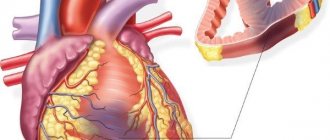
Focal cicatricial lesion of the myocardium of the lower localization
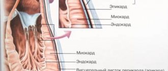
The myocardium is the heart muscle; some of its structural changes are often provoked by external and internal factors.
Transformations do not always indicate pathology or any negative disorder, but in any case, they need to be focused on. After all, the heart is an important organ of the human body; it is akin to a car engine: it converts biochemical reactions into mechanical energy.
The movements of the heart muscle must maintain rhythm; any disturbances in this process and changes in the myocardium are shown by an electrocardiogram (ECG).
Signs of a problem
Cardiac activity depends on many criteria that affect intracellular metabolism in the tissues of the heart muscle. The constancy of the internal environment can be periodically disrupted, which can lead to disruptions in the functioning of the heart cells.
Diffuse changes in the myocardium are not considered a disease; it is a syndrome that means an accumulation of changed cells with impaired conduction of electrical impulses in a given area, clearly displayed on the ECG.
It is important to determine the cause of such failures; it may be hormonal in nature, infectious in origin, or a consequence of heart disease of varying severity.
Changes are not always only diffuse, covering sectors in each department of the organ. They can be focal as a result of the formation of scars in the myocardium of any size. The scar is a connective tissue that does not conduct impulses; the electrical inertia of this area is visible on the cardiogram.
The variety of myocardial diseases is very large, but the general signs of problems with the cardiovascular system and symptoms of myocardial changes are as follows:
- burning and pressing pain behind the sternum;
- shortness of breath at the slightest physical exertion or even at rest;
- disturbances of heart rhythm and contraction frequency;
- increased fatigue, general weakness, chronic fatigue.
The primary change in the heart muscle provokes the development of certain processes:
- myocardial hypoxia;
- circulatory disorders;
- disruptions in the transport of oxygen to cells and tissues;
- irreversible necrotic consequences.
A critical case of the development of myocarditis is an acute infarction; its course also varies.
Causes of myocardial changes
The detected deviations have different origins. The reasons can be minor or significant. The latter provoke a fatal outcome. A thorough examination will reveal the problem to an experienced cardiologist.
Changes in the myocardium can form several groups of factors:
- Inflammatory. They cause myocarditis. Its nature can be infectious or aseptic, that is, pathogenic microorganisms do not take part in this process. Typically, such areas have a diffuse location, but sometimes there are foci of inflammation.
Manifestations of myocarditis, expressed with varying degrees of intensity, accompany the following pathologies:
- typhus, diphtheria;
- acute rheumatic fever or rheumatism of streptococcal origin, which is a consequence of tonsillitis, tonsillitis, scarlet fever;
- weakened immune system (systemic lupus erythematosus, rheumatoid arthritis affecting the heart, etc.);
- damage by viruses of rubella, measles, influenza, etc.
- Dystrophic.
Caused by a malfunction of heart cells and a disorder of metabolic processes. Here, the provocateurs are not inflammatory processes and diseases; they do not affect the coronary arteries. Changes in the myocardium are caused by a lack of necessary nutrition, so the heart muscle begins to contract unevenly. This problem is called cardiac dystrophy, its causes are as follows:
- diseases of the endocrine system: hyperfunction of the thyroid gland, diabetes mellitus, tumor of the adrenal glands, as a result, an excessive amount of hormones or a lack of glucose in the heart cells provoke disruptions in metabolic processes within these cells;
- liver and kidney failure lead to the accumulation of toxins in the blood resulting from metabolic processes;
- anemia - a decrease in hemoglobin levels - brings with it a lack of air for the heart muscle cells;
- dehydration, fever;
- severe physical conditions: frequent stress, hard work, constant overwork, malnutrition and starvation;
- mental stress combined with increased emotional stress leads to changes in the myocardium in children, especially if the child is not active enough; here among the consequences are vegetative-vascular dystonia and disruptions in the control of the nervous system of the heart;
- infections: tuberculosis, influenza, malaria;
- intoxication - acute or chronic, including alcoholism, work in hazardous industries, constant contact with chemicals;
- food unsaturated with vitamins.
- Metabolic – occur due to disruption of repolarization processes in muscles. Inside each cell, an exchange of sodium and potassium ions occurs, the output is energy, which, in the process of certain transformations, becomes the driving force for relaxation and contraction of the cell. These mechanisms are called repolarization and depolarization. The electrolyte composition of the blood is fixed, and when it changes, the metabolism in muscle cells also changes. Among the reasons here are myocardial hypertrophy, arterial hypertension, coronary heart disease, rhythm disturbances, atherosclerosis of the coronary arteries. In other words, in any case, the blood supply to the heart is disrupted, and there is a lack of nutrients and microelements.
- Scarring is evidence of a previously occurring inflammatory process; there could have been a heart attack, and the myocardial cells died. Myocarditis leaves behind scar changes - cardiosclerosis. They are usually diffuse in nature, and the infarction leaves behind focal scarring changes.
Source: https://lechimsosudy.com/ochagovo-rubcovoe-porazhenie-miocarda-nizhnej/
Symptoms
Diffuse minor changes in the myocardium are not accompanied by severe symptoms. But there may be a clinical manifestation of the underlying disease, which causes problems in the heart. Most patients with this disease complain of the following sensations:
- discomfort or pain behind the sternum observed during ischemia;
- with hyperthyroidism, sudden weight loss is possible;
- muscle tremors (involuntary tension followed by relaxation);
- with cardiosclerosis, disturbances in the depth and frequency of breathing may occur as a result of fluid accumulation;
- pale skin, problems with orientation in space, fatigue, which manifest themselves with anemia.
What diseases do they accompany?
Pathological changes in the myocardium detected on the ECG may be accompanied by impaired blood supply to the heart muscle, reprolarization processes, inflammatory processes and other metabolic changes.
A patient with diffuse changes may exhibit the following symptoms:
- dyspnea,
- chest pain,
- increased fatigue,
- cyanosis (blanching) of the skin,
- rapid heartbeat (tachycardia).
Diseases accompanied by changes in the heart muscle:
- Myocardial dystrophy is a violation of biochemical metabolic processes occurring in the heart;
- Allergic, toxic, infectious myocarditis - inflammation of the myocardium of various etiologies;
- Myocardiosclerosis – replacement of cardiac muscle cells with connective tissue as a consequence of inflammation or metabolic diseases;
- Disturbances of water-salt metabolism ;
- Hypertrophy of parts of the heart muscle.
Additional examinations are needed to differentiate them.
Diagnostic features
The main method for detecting myocardial changes is an electrocardiogram. This technique allows you to identify the smallest pathologies and begin timely treatment. To decipher ECG data, a specialist has to spend no more than 15 minutes.
What do myocardial changes on an ECG mean? This type of diagnosis allows you to identify the following disorders:
- thickening of the muscle walls and anterior septum;
- ischemic lesion, also determining its depth and size;
- location of the infarction;
- disturbances in heart rhythm;
- enlarged cardiac cavities;
- toxic damage to the myocardium;
- disruptions in electrolyte metabolism.
A cardiogram allows you to accurately determine pronounced changes in the myocardium of the heart:
- Myocarditis. A cardiogram shows heart rhythm disturbances and the presence of inflammation. On the ECG result, such pathologies are manifested by a decrease in waves in all leads.
- Ischemia. It manifests itself as a change in the shape, polarity and amplitude of the T waves, which are responsible for the ischemic zone.
- Myocardial dystrophy. ECG data indicate the development of myocarditis, therefore, to determine the pathology, it is necessary to conduct laboratory tests, in particular, blood biochemistry.
- Myocardial infarction. The cardiogram shows an upward shift of the ST segments.
- Transmural septal necrosis. With this disease, damage to the wall of the heart muscle occurs, which is irreversible. On the ECG result, this pathology is manifested by the absence of the R wave.
- Necrosis of the heart muscle. Characterized by irreversible death of organ cells. Displayed on the graph as a pathological Q wave.
Symptoms
Depending on the type of disorders in the heart muscle, the symptoms in patients are specific. In addition to them, changes in the myocardium are observed on the ECG, which means that the pathological process has affected a large number of cells.
Dystrophic
All patients have different symptoms of changes in the heart. They can be expressed already in the initial stage or be insignificant, and in some cases the process is asymptomatic. The addition of additional signs is evidence of a worsening prognosis for recovery.
Dystrophic changes are of the following types:
- ischemic;
- focal;
- dyshormonal.
Signs of disturbances in the tissues of the heart occur in cases of short-term circulatory disturbances and lack of oxygen. It is important to take into account that characteristic changes in the myocardium also appear on the ECG, which means that the pathological process has affected its normal function. The clinical picture observed in the ischemic form is characteristic of angina pectoris. Its main symptoms are the following:
- Paroxysmal pain in the chest, which is associated with physical stress or previous emotional stress. They quickly disappear after taking a Nitroglycerin tablet. Its duration most often is no more than 15 minutes. The pain is felt not only in the heart area, but also radiates to the left arm, collarbone, lower jaw or neck.
- Increased blood pressure.
- Interruptions in the heart area.
- Feeling of lack of air.
In some cases, patients do not feel discomfort in the chest, but only changes in the myocardium are visible on the ECG, which means that the pathological process can lead to the development of complications.
A vivid picture of an attack with severe pain may alternate with a feeling of numbness or tingling in the fingers of the left hand.
Focal changes in the myocardium are observed during a heart attack, which is characterized by intense and prolonged chest pain. Its duration reaches several hours or days. Patients describe it as tearing, burning, spreading to the left arm or lower jaw, neck. Usually, after taking Nitroglycerin, the condition does not improve. In addition to pain, there is a feeling of fear, abdominal pain, nausea, severe weakness and cold sweat.
With dishormonal disorders, the heart muscle is affected, and the pathological process can progress rapidly. This is due to the effects of hormones during thyroid dysfunction or after menopause. Patients with this type of change complain of irritability, dizziness, disturbed sleep and weight loss. Among the signs of heart damage, they note stabbing pain spreading to the left arm, rapid or slow heartbeat.
Diffuse
All heart cells are affected by the pathological process of myocarditis. It can occur with or without the participation of microorganisms (aseptic). The main signs of diffuse manifestations are:
- Severe weakness, fatigue.
- Shortness of breath on exertion.
- Interruptions in the heart.
- Increased sweating.
- Pale skin, sometimes with a bluish tint.
- Swelling of veins in the neck.
- Increased heart rate.
- Reduced blood pressure.
Some patients with myocarditis show no signs of the disease for a long time. In some cases, the pathology takes a malignant course, a severe rhythm disturbance occurs, and the severity of all the listed symptoms is present.
Metabolic
There are acute and chronic pathologies. The first case is life-threatening and can result in the death of the patient if there is no help.
Chronic changes in the myocardium have a blurred picture. The main symptoms are fatigue and pain in the heart area. Unpleasant sensations are most often noted in the apex, less often - behind the sternum.
If the process lasts a long time, then cardiosclerosis is formed (connective tissue replaces normal heart cells) of dystrophic origin.
The changes that occur in the myocardium have different origins and clinical presentations. Many of them begin with nonspecific manifestations, and in some patients they may be absent for a long time. Therefore, in order to preserve heart function, when unpleasant signs appear, it is necessary to be examined by a cardiologist and promptly treated.
Treatment
Therapy is based on an integrated approach. Treatment should begin with changing your lifestyle, as well as creating the right diet, taking into account the nature of the disease.
If changes are identified in a timely manner, their elimination is more or less easy. Most often, drugs are prescribed aimed at normalizing the functioning of the organ. This helps prevent the development of heart failure. The most popular medications are Trompangin and Panangin. The doctor may also prescribe certain traditional medicines.
In the most severe cases, surgery may be required. The patient’s future life largely depends on the timeliness and quality of the operation.
Pronounced changes in the myocardium can occur for a number of reasons. In the early stages of their development, they do not have serious symptoms, but later obvious signs of the disease begin to appear. When the first suspicion of the development of the disease appears, the patient must immediately consult a doctor to undergo an examination and receive qualified assistance.
Diagnosing and fixing the problem
Minor changes in the myocardium will not require drastic measures. The patient will be advised to adjust blood pressure, take a course of vitamins and adhere to a healthy lifestyle.
More serious changes in the myocardium already imply the presence of a disease; for diagnosis, the following measures are usually performed:
- Clinical blood test. Examines hemoglobin levels and inflammation criteria.
- Biochemistry of blood. Determines the condition of the liver, kidneys, the amount of glucose, protein, cholesterol.
- General urine analysis. Evaluates renal activity.
- Ultrasound. Visual examination of internal organs.
- ECG. Diffuse changes are indicated by a decrease in T waves, responsible for ventricular repolarization. Focal changes are indicated by negative T waves in 1–2 sectors.
- Echocardiogram. The most informative method that identifies the causes of changes in the heart muscle thanks to clear visualization of its parts.
Therapy must be combined with correction of diet and lifestyle. Changes in the myocardium of a dystrophic or metabolic nature by default require proper rest, adherence to sleep patterns and diet.
The heart responds well to those present in the diet:
- nuts;
- spinach;
- carrots and potatoes;
- apricots, peaches, bananas;
- lean poultry and meat;
- red fish and caviar;
- cereals, grains;
- dairy products.
Chocolate and confectionery products should be consumed to a minimum. Fatty meat and poultry are extremely rare. Soda, coffee and alcohol are excluded. You should also remove spicy, fatty, salty, spicy and fried foods.
The following drugs help improve metabolic processes in heart muscle cells:
- “Asparkam”, “Panangin”, “Magne B6”, “Magnerot” - potassium and magnesium stabilize the frequency of contractions.
- “Mexidol”, “Actovegin” are antioxidants that eliminate lipid oxidation products in myocardial cells.
- Vitamins A, B, C, E - without them, intracellular metabolism is impossible.
If the cause of myocardial changes is a disease, then appropriate therapy will correct the situation. The lack of hemoglobin is compensated for with iron-containing drugs; for myocardial inflammation, antibiotics and Prednisolone are prescribed; for cardiosclerosis, urinary agents and cardiac glycosides are indicated.
Focal changes in the myocardium mean ischemic damage to the heart muscle, ischemia and necrosis.
Main directions of diagnostics
Upon examination, as well as conversation with a patient with cardiosclerosis, certain cardiac symptoms are usually revealed.
But these symptoms, as a rule, are caused by an underlying disease that has led to the development of scarring changes in the heart.
These changes themselves do not have specific clinical manifestations and are diagnosed using instrumental research methods.
To detect cardiosclerosis, the following are most often used:
- electrocardiography (ECG);
- Ultrasound examination of the heart (Echo-CG).
ECG is a simple and widely available method.
In the presence of cicatricial changes in the heart, this method makes it possible to identify them, as well as assess their localization and extent. In addition, the ECG allows you to diagnose the underlying and concomitant cardiac pathology.
Echo-CG allows you to visualize the structures of the heart, assess their morphology and function. Using this method, it is possible to determine the presence of scar changes, their localization, and extent.
In addition, the method allows us to judge how these changes affect the contractility of the myocardium, as well as the functional activity of the heart as a whole.
Thus, Echo-CG is a slightly more informative research method.
However, ECG is a much more accessible and simpler method, both from an economic and practical point of view.
The economic aspect of the issue requires no comment. The practical aspect implies that ECG results can be interpreted by a doctor of any specialty.
But the results of Echo-CG can only be assessed by a functional diagnostics doctor who has undergone the appropriate specialization.
Diagnostics
In the absence of a concomitant disease associated with disruption of the cardiovascular system, specific clinical signs of diffuse or focal changes do not appear. Therefore, myocardial changes can be diagnosed using a cardiogram. The basis for such changes can be a variety of factors, from harmless to those causing significant harm to health. The decision on the advisability of further examination is made by the cardiologist.
In the absence of clinical signs of serious pathology and minor changes in the myocardium in adults, no further examination is required. It is enough to take preventive measures aimed at normalizing blood pressure levels, adhering to the principles of diet, and additional intake of vitamins and macroelements. Taking health promotion measures is also recommended.
If there is reason to assume the presence of significant degenerative changes, a thorough diagnosis of myocardial changes is carried out, including:
- General blood and urine analysis.
- Analysis of kidney parameters, number of red blood cells and white blood cells.
- Blood chemistry.
- Reflects the state of all body systems. Also provides information about metabolism.
- Ultrasound examination of the heart, liver and kidneys.
- Electrocardiography.
- Analysis of the manifestations of deviations from normal heart rhythm - the presence of pathological Q waves, decreased T waves and other symptoms.
- Holter monitoring and functional tests,
- Shows morphological and functional changes of the heart and its valve apparatus.
Myocardial changes are well diagnosed on an ECG
Electrocardiographic signs of pathology
Diffuse cardiosclerosis does not have specific ECG signs. With this variant of the pathological process, the following are most often observed:
- deviation of the electrical axis of the heart to the left;
- reduction in the voltage of the ventricular QRS complexes in all leads;
- other electrocardiographic changes characteristic of chronic coronary heart disease.
But focal cardiosclerosis has more specific ECG signs, allowing one to draw a conclusion about the presence of local cicatricial changes.
These signs include:
- reduction of the ST segment below the isoelectric line;
- a significant decrease in the T wave or even a negative T wave;
- pathological Q wave (in cases where the cause of scar formation was Q-myocardial infarction);
- other signs associated with disturbances in excitability and conduction processes (blockades, flickering, fluttering and others), which are based on cardiosclerotic changes in the heart muscle.
Thus, a number of electrocardiographic changes may lead a specialist to believe that the patient has cardiosclerosis. As mentioned above, a combination of various types of cardiosclerosis is usually observed.
In addition, in almost all cases there is a combination of several pathogenetic associated cardiac pathologies. For greater clarity, here are two classic examples.
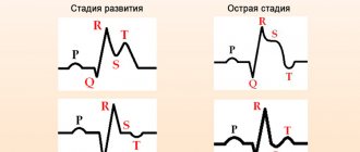
Figure No. 1. The ECG image shows changes characteristic of acute myocardial infarction of the posterior wall of the left ventricle, as well as focal cardiosclerosis.
Thus, we can conclude that most likely this patient had an acute recurrent myocardial infarction in combination with focal cardiosclerosis. Focal changes are most likely caused by a previous heart attack, that is, they are post-infarction in nature.
Figure No. 2. This ECG image shows changes characteristic of extensive focal cardiosclerosis in the form of a chronic aneurysm of the anterior wall of the left ventricle.
We can conclude that the identified disorders are a consequence of an extensive transmural myocardial infarction suffered in the past.
It should be noted that cardiosclerosis is a rather severe pathological process. Diffuse cardiosclerosis tends to progress, steadily reducing myocardial contractile activity.
Focal cardiosclerosis does not progress, but inevitably affects the functional activity of the heart muscle.
Thus, it is important to detect cardiosclerosis promptly and take appropriate therapy.
ECG, as mentioned above, is a simple and widely available method, so it is important for all cardiac patients to undergo this examination regularly.
This will not be particularly difficult and will allow you to monitor the condition of the heart, as well as timely identify developing pathological processes.
Myocardial changes: causes, symptoms, diagnosis, ECG, treatment
The muscles of the human heart wall, thanks to extremely complex heterogeneous, autogenous and neurohumoral processes in the body, are capable of generating a bioelectric impulse from biochemical processes into mechanical energy responsible for creating rhythmic movements of the heart. And the consequences of changes in the balance of the body, first of all, result in changes in the myocardium of the posterior and lower walls of the ventricle of the heart. They may be cardiac pathologies, hormonal imbalances, or the consequences of a viral infection.
What it is
The pathology of any area related to cardiology is called diffuse changes in the myocardium. This condition may be a consequence of a disease or be caused by a malfunction of the mechanism of physical and chemical reactions of the body.
The main task of myocardial muscle tissue is to conduct transmitted electrical voltage along neuromuscular fibers.
Under the influence of diffuse changes, the heart cannot function normally - the frequency of contractions of the heart muscle regularly decreases and the heart rhythm is disturbed.
A concentration of cells is formed that have undergone degenerative changes and are unable to transmit nerve impulses through themselves, electrophysiologically manifesting themselves as a necrotic zone.
Damage to the functionality of the myocardial cellular system can be diffuse - evenly distributed throughout all tissues of the heart and focal - isolated, for example, during the formation of connective tissue in place of irreversibly damaged cells. Scars are overgrown connective tissue that manifests itself as electrically inert, incapable of conducting nerve impulses and leading to cardiac hypertrophy.
Definition of pathology
Isolated changes in the LV most often manifest themselves as hypertrophy. Hypertrophy is the process of increasing the volume and mass of heart cells (cardiomyocytes) in response to increased stress.
The left ventricle can also be affected by infarction. A heart attack is the death of an area of tissue caused by a lack of blood supply. The heart is extremely sensitive to ischemia because it bears a huge load.
Norm and deviations
In a healthy body, the thickness of the wall of the left ventricle exceeds the thickness of the walls of other parts of the heart, since it bears the greatest functional load - providing blood supply to the entire body. Normally, this figure rarely exceeds 1 cm, and with hypertrophy it can reach 3 cm.
Causes
The causes of degenerative changes in the myocardium of the ventricles of the heart are caused by various processes.
Myocarditis
This pathology develops due to pathogenic microorganisms that have entered the cells of the heart muscle. There are isolated and diffuse cardiac myocarditis.
Healthy heart and myocarditis
It is a consequence of heart diseases such as:
- Rheumatic disease caused by group A B-hemolytic streptococcus.
- Diphtheria, rickettsiosis,
- Respiratory viral diseases,
- Various disorders of the immune system.
Dystrophic
They arise due to disruption of physical and chemical processes in myocardial cells. It has a non-inflammatory nature. It is characterized by the appearance of damaged muscle fibers that do not participate in the contractile activity of the myocardium, and are also unable to receive sufficient amounts of nutrients.
This leads to the death and decomposition of such cells and the appearance of necrotic foci in the muscular layer of the myocardium. Dystrophic changes on the ECG of the heart do not manifest themselves in any way.
The causes of myocardial dystrophy are:
- Intoxication with breakdown products due to electrolyte diseases of the renal system.
- Endocrine metabolic disorders.
- Physiological reasons.
- Decreased hemoglobin and insufficient oxygen supply to myocardial cells.
- Acute and chronic respiratory viral diseases.
- Diseases accompanied by disruption of the thermoregulation process and dehydration of the body.
- Avitaminosis.
- Intoxication caused by chemicals (alcohol, nicotine, pesticides).
- Increased intellectual load with a lack of physical activity can occur in children and provokes vegetative-vascular dystonia.
Metabolic
Metabolic changes occur due to changes in the balanced composition of ions in myocardial cells and a decrease in the electrolyte composition of the blood,
The pathology causes a decrease in the contractility of heart cells and a failure of the entire energy metabolism. Metabolic changes in the heart appear on the ECG as deviations of segments and complexes from the isoline.
- The causes are conditions caused by a lack of products involved in metabolic processes
- Conditions that provoke dystrophic changes in the myocardium.
- Formation of cholesterol plaques on the walls of the coronary artery.
- Impaired blood flow in the coronary arteries of the heart.
- Arterial essential hypertension.
- Heart rhythm disturbances.
- Enlargement of the heart muscle.
Failure of metabolic processes in cardiomyocytes and nonspecific changes in the myocardium are a manifestation of insufficient blood supply to the heart muscle, which can lead to the development of various pathologies.
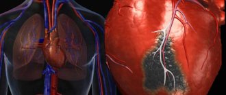
Impaired blood flow in the coronary arteries
Mild myocardial changes in childhood and old age are considered normal.
Scarring
They cause the growth of connective tissue on the heart muscle. Scar changes on the heart can be diffuse and focal.
Occurs in conditions such as:
- cardiac ischemia. Causes necrosis of individual areas of the myocardium and forms a focal scar.
- transferred inflammatory process. Causes the death of muscle fibers throughout the heart muscle area.
Source: https://ukb1.ru/drugoe/rubcovye-izmeneniya-peredne-peregorodochnoj-oblasti.html
Causes of cicatricial changes in the myocardium
The most common factors in the formation of coarse fibrous tissue in the heart muscle are inflammatory and atherosclerotic processes. At the same time, myocarditis occurs mainly in young people, in childhood and adolescence, and blockage of the coronary arteries due to cholesterol deposition is almost always detected in patients after 40 years of age.
We recommend reading the article about complications of myocardial infarction. From it you will learn about the stages of a heart attack, the classification of early and late complications, methods of treatment and prevention.
And here is more information about the ECG results for cardiac ischemia.
General information
Electrocardiography is the simplest diagnostic method for studying the functioning of the heart.
The essence of the examination is to record electrical impulses that accompany the contractile and restorative functions of the myocardium, called “depolarization” and “repolarization.” What do myocardial changes on an ECG mean?
Specific abnormalities in the electrocardiogram can be recorded during a routine medical examination and characterize the condition of the myocardium at the time of the examination. The function of the myocardium is to synthesize by cardiomyocytes , due to which the cavity contracts and ensures normal blood supply to the entire body. This process is carried out due to the cellular exchange of sodium and potassium ions in the cell. The functioning of the cardiac conduction system is recorded on an electrocardiogram using electrodes fixed on the limbs and chest.
Changes in the ECG are not a disease, but only a manifestation of some pathological processes occurring in the myocardium. With shifts in the biochemical activity in the heart cells, their contractility changes, which is reflected in the cardiogram when recording the conduction of impulses. Cardiomyocyte function can be impaired during inflammatory processes, for example, with myocarditis . Taking certain medications also affects the functioning of the heart muscle.
Long-term diabetes mellitus can gradually lead to atherosclerosis . Not only large vessels are affected, but also the coronary arteries that supply the myocardium. With inflammatory pathology in the gastrointestinal tract, the absorption of nutrients is impaired, which also negatively affects metabolic processes in cardiomyocytes.
What will the ECG show during changes?
For the first stage of diagnosing scar structures in the myocardium, an ECG is used; it can help in topical (location) diagnosis.
Left ventricle
Scar tissue leads to the formation of:
- abnormal Q in the first three standard leads, as well as V1 - 6;
- ST is located on the isoline;
- T is often positive, low and smooth.
In this case, the connective tissue fibers cannot generate signals, as well as the source of destruction. But the lesion becomes smaller due to the contraction of the remaining muscle fibers.
Bottom wall
Pathological Q is noted in the second standard lead, and a lower (negative) ventricular complex is also found there compared to the third standard lead.
Septal region
For a scarring infarction in the septal area, the Q waves in leads V1, V2 are of diagnostic value, and the R waves in V1,2,3 are low or cannot be determined.
EXAMPLE 2
Here is another Q in the same lead III. But here, after measurements, we see that its amplitude is not just 1/3 or 1/2 R, but almost equal to it - this is not the norm. In addition, the duration (width) of 0.04 s also goes beyond what is permitted. In the adjacent lead aVF, the Q wave is also pathological - it has a height of 1/3 R and a width of about 0.04 s. This gives us every right to talk about scar changes in the posterior wall (this is the area controlled by these leads)
Additional examinations
In addition to electrocardiographic examination, patients are prescribed:
- Ultrasound of the heart to assess the degree of myocardial hypertrophy and expansion of cavities;
- CT or MRI if there is a discrepancy between clinical signs and ECG data;
- myocardial scintigraphy to detect diffuse or focal defects in the accumulation of radioisotopes;
- blood tests - lipid profile, coagulogram, immunological complex, specific enzymes (troponin, myoglobin, creatine phosphokinase).
Classification
Depending on the size and location of the changed cardiomyocytes, the following are distinguished:
- Diffuse changes. Such a widespread lesion indicates the presence of multiple foci of altered cardiomyocytes. Characteristic signs are present in all leads on the ECG.
- Focal changes. Lesions are recorded only in certain leads that correspond to specific areas of the myocardium. Focal changes are a kind of clearly limited areas of connective, scar tissue, which are inert to conduct electricity.
When diagnosing changes on the ECG, a functional diagnostics doctor and cardiologist can state:
- early ventricular repolarization, which is manifested by a negative “T” wave;
- a decrease in the voltage of the r wave, which characterizes the contractility of the myocardium;
- rhythm disturbances;
- conduction disturbances.
Depending on these indicators, the causes of changes in the ECG are determined by nature:
- inflammatory;
- cicatricial;
- dystrophic;
- metabolic.
Dystrophic changes in the myocardium
Such changes in the ECG are formed due to insufficient nutrition of cardiomyocytes, which inevitably leads to a decrease in the contractility of the left ventricle. Diffuse-dystrophic changes in the myocardium are observed with:
- pathologies of the endocrine system: diabetes mellitus , adrenal dysfunction, disorders of the thyroid gland;
- pathologies of the renal system and liver: excessive amounts of toxic metabolic products negatively affect the functioning of the heart;
- chronic diseases of infectious origin: changes can be observed with tuberculosis , influenza , malaria , etc.;
- chronic iron deficiency anemia : constant oxygen starvation affects the functioning of cardiomyocytes;
- with an unbalanced diet, with vitamin deficiency in the diet;
- with excessive nervous and physical overload;
- with fever and concomitant dehydration;
- in case of poisoning with alcohol, medications or chemical components.
Metabolic changes in the myocardium
What it is? Characteristic nonspecific changes on the ECG are formed as a result of disturbances in intracellular metabolic processes associated with potassium and sodium ions.
Metabolic changes are associated with dystrophy of the heart muscle and appear when:
- ischemia , which is reflected on the cardiogram in the form of deviations of the T wave. Its polarity and shape changes in the leads corresponding to the damaged areas;
- myocardial infarction : the location of the ST segment changes on the ECG, which is located either above or below the isoline;
- death, necrosis of the myocardium, which is characterized by the appearance of an abnormal Q wave.
Scar changes
Areas of scar tissue form at the site of a former inflammatory process, necrosis, as a result of which normal, healthy cardiomyocytes lost their contractility and were replaced by connective tissue that does not have elasticity. Focal cicatricial changes on the ECG indicate a previous myocardial infarction .
- The lower wall of the left ventricle is characterized by changes in leads: II, III and a VF (indicates damage to the right, less often the left circumflex coronary artery).
- The anterior septal region is characterized by changes in leads: V1 and V2 (the left descending septal branch is damaged), or V2-V4 (the left descending coronary artery or its branches is involved).
- The anterior-lateral region is characterized by changes in leads: I, aVL, V4-V6 (the circumflex artery or the left descending artery is damaged).
- Anterior widespread infarction is characterized by changes in leads: I, aVL, V1-V6 (the left descending coronary branch is damaged).
How to treat deviations
It is not possible to influence already formed scars in the myocardium.
For this purpose, medications from various groups are prescribed:
- for angina pectoris - beta blockers (Bisoprol), nitrates (Cardiket), ACE inhibitors (Enap), diuretics (Trifas), anticoagulants (Aspirin, Clopidogrel);
- for myocarditis - antibiotics (Augmentin), anti-inflammatory (Nimid), antiviral and immunomodulators (Cycloferon), vitamin complexes (Milgamma);
- to improve myocardial nutrition - antioxidants (Kudesan, Cytochrome C), metabolism stimulants (Mexidol, Panangin, Riboxin);
- hypolipidemic - Tulip, Roxera;
- antiarrhythmic - Ritmonorm, Cordarone;
- cardiac glycosides - Korglykon, Digoxin.
If there is no result from drug therapy, and the threat of recurrent infarction remains, in case of severe rhythm disturbance, surgical treatment is performed: installation of a stent or shunt, pacemaker, suturing of the aneurysm.
We recommend reading the article about heart valve fibrosis. From it you will learn about the causes of the development of pathology, symptoms, methods of diagnosis and treatment, and prognosis for patients.
And here is more information about posterobasal infarction.
Scar formation in the heart muscle is the final stage after myocarditis or myocardial infarction; it is also considered the outcome of atherosclerotic lesions of the coronary vessels. An ECG is used to detect focal or diffuse myocardial scarring.
To clarify the diagnosis, an in-depth clinical and instrumental examination is recommended. Symptoms and prognosis of cardiosclerosis depend on the severity of the underlying pathology. There are no specific manifestations; complications can include various heart rhythm disturbances and circulatory failure. For treatment, drug therapy is used; in case of threatening conditions, surgery is prescribed.
Recognizing myocardial infarction on an ECG can be difficult due to the fact that different stages have different signs and variations of waveforms. For example, the acute and acute stage may not be noticeable in the first hours. Localization also has its own characteristics: the infarction on the ECG is transmural, q, anterior, posterior, transferred, large-focal, lateral, different.
A repeated myocardial infarction can occur within a month (then it is called recurrent), as well as 5 years or more. To prevent the consequences as much as possible, it is important to know the symptoms and carry out prevention. The prognosis is not the most optimistic for patients.
The T wave on the ECG is determined to identify pathologies of cardiac activity. It can be negative, high, biphasic, smoothed, flat, reduced, and depression of the coronary T wave can also be detected. Changes can also be in the ST, ST-T, QT segments. What is an alternation, discordant, absent, double-humped tooth.
Myocardial dystrophy, or dystrophic changes in the myocardium, can be associated with an incorrect lifestyle and work disorders. Diffuse, metabolic, and moderate changes can be detected during an ECG. To begin with, treatment involves taking vitamins.
Myocardial ischemia on the ECG shows the degree of heart damage. Anyone can figure out the meanings, but it’s better to leave the question to the experts.
Post-infarction cardiosclerosis occurs quite often. He may have an aneurysm or ischemic heart disease. Recognizing symptoms and timely diagnosis will help save lives, and ECG signs will help establish the correct diagnosis. Treatment is lengthy, rehabilitation is required, and there may be complications, including disability.
After suffering certain diseases, myocardial cardiosclerosis may develop. This pathology is characterized by rhythm disturbances and other unpleasant manifestations. Treatment needs to start the sooner the better.
Depending on the time of onset, as well as the complexity, the following complications of myocardial infarction are distinguished: early, late, acute, frequent. Their treatment is not easy. To avoid them, preventing complications will help.
It is quite difficult to diagnose, since subendocardial myocardial infarction quite often has an abnormal course. It is usually detected using ECG and laboratory examination methods. An acute heart attack threatens the patient's death.
Normal and deviations - possible reasons
Normally, the electrical activity of the myocardial areas, which is recorded by the ECG, should be uniform. This means that intracellular biochemical exchange in heart cells occurs without pathologies and allows the heart muscle to produce mechanical energy for contractions.
If the balance in the internal environment of the body is disturbed for various reasons,
the following characteristics are recorded on the ECG :
- diffuse changes in the myocardium;
- focal changes in the myocardium.
The reasons for such changes in the myocardium on the ECG can be either harmless conditions that do not threaten the life and health of the subject, or serious dystrophic pathologies requiring emergency medical care.
Causes of myocarditis:
- rheumatism as a consequence of scarlet fever, tonsillitis, chronic tonsillitis;
- complications of typhus, scarlet fever;
- consequences of viral diseases: influenza, rubella, measles;
- autoimmune diseases: rheumatoid arthritis, systemic lupus erythematosus.
One of the reasons for changes in muscle tissue may be cardiodystrophy - a metabolic disorder in heart cells without damage to the coronary arteries. Lack of cell nutrition leads to changes in their normal functioning and impaired contractility.
Causes of cardiac dystrophy:
- Ingress of toxic metabolic products into the blood due to severe impairment of kidney and liver function;
- Endocrine diseases: hyperthyroidism, diabetes mellitus, adrenal tumor, and, as a result, excess hormones or metabolic disorders;
- Constant psycho-emotional stress, stress, chronic fatigue, starvation, unbalanced diet with nutritional deficiencies;
- In children, a combination of increased stress with a sedentary lifestyle, vegetative-vascular dystonia;
- Lack of hemoglobin (anemia) and its consequences - oxygen starvation of myocardial cells;
- Severe infectious diseases in acute and chronic form: influenza, tuberculosis, malaria;
- Dehydration of the body;
- Avitaminosis;
- Alcohol intoxication, occupational hazards.
What are moderate changes in the myocardium?
Moderate changes in the myocardium are most often detected by ECG examination, ultrasound diagnostics or echocardiography. Changes in the myocardium are caused by inflammatory processes, diseases, hormonal imbalances and some external factors. Also, in most cases, age may be the cause. In old age, as a result of the aging of the body, and in adolescence and childhood, these are still incomplete processes of heart development.
Changes in the myocardium
As a result of cardiac activity, the human body receives the oxygen and nutrients it needs through the bloodstream. Therefore, if any disorder is diagnosed, it can affect the functional characteristics of other organs and lead to various troubles.
Moderate changes come in several types:
- Diffuse. In itself, such a disorder cannot be a separate disease, but is only a syndrome, the cause of which must be identified during examination by a doctor. This phenomenon can be explained as follows. Some of the cells, which are directly involved in biochemical processes, begin to work incorrectly and shrink. As a result, the electrical activity will be non-uniform. In simple terms, diffuse changes in the myocardium mean a part of the changed cells through which the conduction of electrical impulses is impaired.
- Focal. In this case, large or small scars form in the myocardium. The scars themselves are made of connective tissue, which is not inert and cannot conduct electrical impulses.
To find out what causes such changes, it is necessary to undergo a thorough examination. For a more detailed understanding of the etiology, a certain type of changes in the myocardium is identified.
Types of changes and reasons
As we have already understood, the etiology of such a disorder includes both harmless causes and quite serious diseases. Pathological changes are caused by various processes, for example, inflammatory changes. Here the root cause is myocarditis (inflammation of the heart muscle) of an infectious or aseptic nature. In the area of the left and right ventricles, as well as the left and right atria, myocarditis can occur in the following diseases:
- rheumatism;
- infectious diseases (rubella, measles, scarlet fever, typhus, diphtheria, etc.);
- systemic autoimmune diseases.
Preparation for an ECG during myocardial infarction and characteristics of the main zones
Electrocardiography is of great importance in diagnosing myocardial infarction today. With its help, the specialist makes a diagnosis and discovers exactly where the lesion is located. ECG changes during myocardial infarction depend both on the location of necrosis and on the location of the myocardium relative to the main electrode.
Necrotic zones
This disease is characterized by the presence of three zones. Each of them has its own electrocardiographic characteristics. So, experts highlight:
- Necrosis zone.
- Deformation zone.
- Ischemic zone.
During the study, all zones mutually influence each other, and therefore the range of changes can be very diverse.
The main signs of pathology on the ECG
The relevance of ECG for diagnosing myocardial infarction is not questioned. The changes observed during this study indicate the nature of the pathology, as well as the degree of progression and location.
Types of disease
ECG diagnosis of myocardial infarction helps to differentiate the main three types of this pathological condition. Thus, the ECG “speaks out” regarding:
- transmural infarction;
- subendocardial infarction;
- intramural infarction.
With the transmural type, the following ECG signs of myocardial infarction are observed:
- in the thickness of the left ventricular wall, about seventy percent of necrosis is observed;
- an abnormal Q wave is formed;
- the appearance of a pathological tooth with a small amplitude.
In the subendocardial type, electrocardiographic symptoms indicate the need for immediate medical intervention only if they persist within forty-eight hours.
The intramural type is quite rare.
This study also makes it possible to clarify in what form, complicated or uncomplicated, the anomaly develops.
There is also information regarding the stage of the disease. In particular, it is noted that in case of small-focal myocardial infarction, the ECG does not show the presence of an abnormal Q wave. At the same time, the presence of an abnormal R wave in the chest leads is noted.
Signs of a pathological process
The following ECG signs of myocardial infarction are observed:
- There is no abnormal R wave in the “supra-infarct” areas.
- An abnormal Q wave is present in the “supra-infarct” areas.
- In the “supra-infarct” areas, an elevation of the S and T segments is observed.
- In opposite areas there is a displacement of the S and T segments.
- In the “supra-infarct” areas, the presence of a negative T wave is noted.
Signs of an acute pathological process
Acute myocardial infarction on the ECG looks like this:
- Increasing the rate of contraction of the human heart.
- Clearly visible total elevation of the S and T segments.
- The presence of a pronounced depression of the S and T segments.
- Significant increase in the duration of the QRS complex.
- The presence of abnormal Q waves or the Q and S complex is noted.
Preparation and execution
Electrocardiography requires careful preparation of the patient. So, first you need to shave the hair surface where the electrodes will be placed. The next step will be to prepare the patient's skin. To do this, the specialist carefully wipes the skin with a swab soaked in an alcohol solution.
Then adhesive electrodes are installed on the patient's skin. Recording begins only after the exact time of its start has been established on a special device - a recorder.
The procedure assumes that a specialist monitors ECG curve complexes. This is possible by monitoring current complexes on the oscilloscope screen. At the same time, all available sounds are heard through the speaker.
Conclusion
It is important to remember that in the preliminary diagnosis of this pathological process, electrocardiography is an additional method of examination. Specific signs of this pathological process should be considered painful sensations localized behind the sternum. If a person has not consulted a doctor for a long time, stoically enduring pain, then electrocardiography should be replaced with an echocardiogram.
If the diagnosis is established correctly and in a timely manner, the treatment prognosis is favorable.
Changes in the myocardium on the ECG - what does it mean and is it dangerous?
An ECG can diagnose most heart pathologies. The reasons for their appearance are due to concomitant diseases and lifestyle characteristics of the patient.
What does this mean if changes in the myocardium are detected on the ECG? In most cases, the patient requires conservative treatment and lifestyle modification.
Description of the procedure
An electrocardiogram (ECG) is one of the most informative, simple and accessible cardiological studies. It analyzes the characteristics of the electrical charge that causes the heart muscle to contract.
Dynamic recording of charge characteristics is carried out in several areas of the muscle. The electrocardiograph reads information from electrodes placed on the ankles, wrists and chest skin in the area where the heart is projected, and converts them into graphs.
Normal and deviations - possible reasons
Normally, the electrical activity of the myocardial areas, which is recorded by the ECG, should be uniform. This means that intracellular biochemical exchange in heart cells occurs without pathologies and allows the heart muscle to produce mechanical energy for contractions.
If the balance in the internal environment of the body is disturbed for various reasons, the following characteristics are recorded on the ECG :
- diffuse changes in the myocardium;
- focal changes in the myocardium.
The reasons for such changes in the myocardium on the ECG can be either harmless conditions that do not threaten the life and health of the subject, or serious dystrophic pathologies requiring emergency medical care.
One of these serious pathologies is myocarditis, or inflammation of the heart muscle. Regardless of its etiology, areas of inflammation can be located either in the form of foci or diffusely throughout the heart tissue.
Causes of myocarditis:
- rheumatism as a consequence of scarlet fever, tonsillitis, chronic tonsillitis;
- complications of typhus, scarlet fever;
- consequences of viral diseases: influenza, rubella, measles;
- autoimmune diseases: rheumatoid arthritis, systemic lupus erythematosus.
One of the reasons for changes in muscle tissue may be cardiodystrophy - a metabolic disorder in heart cells without damage to the coronary arteries. Lack of cell nutrition leads to changes in their normal functioning and impaired contractility.
Causes of cardiac dystrophy:
- Ingress of toxic metabolic products into the blood due to severe impairment of kidney and liver function;
- Endocrine diseases: hyperthyroidism, diabetes mellitus, adrenal tumor, and, as a result, excess hormones or metabolic disorders;
- Constant psycho-emotional stress, stress, chronic fatigue, starvation, unbalanced diet with nutritional deficiencies;
- In children, a combination of increased stress with a sedentary lifestyle, vegetative-vascular dystonia;
- Lack of hemoglobin (anemia) and its consequences - oxygen starvation of myocardial cells;
- Severe infectious diseases in acute and chronic form: influenza, tuberculosis, malaria;
- Dehydration of the body;
- Avitaminosis;
- Alcohol intoxication, occupational hazards.
Determination by cardiogram
With diffuse lesions of the heart, deviations from the normal pattern are observed in all leads. They look like numerous areas with impaired conduction of electrical impulses.
This is expressed on the cardiogram as a decrease in T waves, which are responsible for ventricular repolarization. With focal lesions, such deviations are recorded in one or two leads. These deviations are expressed on the graph as negative T waves in the leads.
If focal changes are represented, for example, by scars remaining in the connective tissue after a heart attack, they appear on the cardiogram as electrically inert areas.
What diseases do they accompany?
Pathological changes in the myocardium detected on the ECG may be accompanied by impaired blood supply to the heart muscle, reprolarization processes, inflammatory processes and other metabolic changes.
A patient with diffuse changes may exhibit the following symptoms:
- dyspnea,
- chest pain,
- increased fatigue,
- cyanosis (blanching) of the skin,
- rapid heartbeat (tachycardia).
Such manifestations most often become the reason for an electrocardiogram. In medical practice, there are many examples when myocardial pathologies did not cause noticeable changes in the well-being of patients and were discovered during preventive examinations.
Diseases accompanied by changes in the heart muscle:
- Myocardial dystrophy is a violation of biochemical metabolic processes occurring in the heart;
- Allergic, toxic, infectious myocarditis - inflammation of the myocardium of various etiologies;
- Myocardiosclerosis – replacement of cardiac muscle cells with connective tissue as a consequence of inflammation or metabolic diseases;
- Disturbances of water-salt metabolism ;
- Hypertrophy of parts of the heart muscle.
Additional examinations are needed to differentiate them.
Additional diagnostic tests
These cardiograms, despite their information content, cannot be the basis for making an accurate diagnosis. In order to fully assess the degree of changes in the myocardium, the cardiologist prescribes additional diagnostic measures:
- General clinical blood test - assesses the level of hemoglobin and such indicators of the inflammatory process as the level of leukocytes in the blood and ESR (erythrocyte sedimentation rate);
- Analysis of blood biochemistry - indicators of protein, cholesterol, glucose levels are assessed to analyze the functioning of the kidneys and liver;
- General clinical urine analysis - kidney function indicators are assessed;
- Ultrasound for suspected pathology of internal organs - according to indications;
- Daily monitoring of ECG indicators;
- Carrying out an ECG with stress ;
- Ultrasound of the heart (echocardiography) – the condition of the parts of the heart is assessed to determine the cause of myocardial pathology: expansion (dilatation), hypertrophy of the heart muscle, signs of decreased contractility of the myocardium, disruption of its motor activity.
After analyzing the medical history and laboratory and instrumental examination data, the cardiologist determines the method of treating the changes.
Treatment for focal and diffuse disorders
In the treatment of myocardial pathologies, various groups of drugs are used:
- Corticosteroid hormones – as an antiallergic agent;
- Cardiac glycosides - for the treatment of diffuse changes in the myocardium, manifestations of heart failure (ATP, Cocarboxylase);
- Diuretics - to prevent edema;
- Means for improving metabolism (Panangin, Magnerot, Asparkam);
- Antioxidants (Mexidol, Actovegin) - to eliminate the negative effects of lipid oxidation products;
- Antibiotics – for anti-inflammatory therapy;
- Drugs for the treatment of concomitant diseases;
- Vitamin preparations.
If conservative treatment does not lead to significant improvements in the condition of a patient with myocardial diseases, he undergoes surgery to implant a myocardial pacemaker.
In addition to medications, the patient is recommended to change his lifestyle and establish a balanced diet. For a patient with such pathological manifestations, physical activity, drinking alcohol and smoking are unacceptable. He is prescribed physical therapy and feasible labor.
Basic principles of dietary nutrition:
- The consumption of salt and excess liquid is limited to a minimum;
- Spicy and fatty foods are not recommended;
- The menu should include vegetables, fruits, lean fish and meat, and dairy products.
Myocardial changes detected on the ECG require additional laboratory and instrumental examination . If necessary, the cardiologist will prescribe treatment in a hospital or on an outpatient basis. Timely measures taken will help avoid serious complications.
Source: https://oserdce.com/diagnostika/ekg/izmeneniya-miokarda.html