What is a venous catheter
The instrument is a thin hollow tube (cannula) equipped with a trocar (a hard pin with a sharp end) to facilitate insertion into the vessel. After administration, only the cannula is left, through which the medicinal solution enters the bloodstream, and the trocar is removed.
Before diagnosis, the doctor conducts an examination of the patient, which includes:
- Ultrasound of veins.
- Chest X-ray.
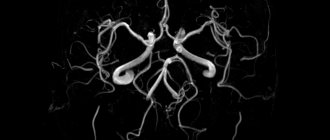
- MRI.
- Contrast phlebography.
How long does installation take? The procedure lasts on average about 40 minutes. Anesthesia at the insertion site may be required when inserting a tunneled catheter.
It takes about one hour to rehabilitate the patient after installing the instrument; the sutures are removed after seven days.
Classification
Intravenous catheters are classified according to many criteria.
By purpose
There are two types: central venous (CVC) and peripheral venous (PVC).
CVCs are designed for catheterization of large veins, such as the subclavian, internal jugular, and femoral. Using this instrument, medications and nutrients are administered and blood is drawn.
PVC is installed in peripheral vessels. As a rule, these are the veins of the extremities.
Comfortable butterfly catheters for peripheral veins are equipped with soft plastic wings with which they are attached to the skin
The “butterfly” is used for short-term infusions (up to 1 hour), since the needle is constantly in the vessel and can damage the vein if held longer. They are usually used in pediatrics and outpatient practice for puncturing small veins.
By size
The size of venous catheters is measured in gaits and is designated by the letter G. The thinner the instrument, the larger the gaits value. Each size has its own color, the same for all manufacturers. The size is selected depending on the application.
| Size | Color | Application area |
| 14G | Orange | Rapid infusion of large volumes of blood products or fluids |
| 16G | Grey | Transfusion of large volumes of blood or fluids |
| 17G | White | Transfusion of large volumes of blood or fluids |
| 18G | Green | Routine red blood cell transfusion |
| 20G | Pink | Long courses of intravenous therapy (two to three liters per day) |
| 22G | Blue | Long courses of intravenous therapy, oncology, pediatrics |
| 24G | Yellow | Sclerotic veins, pediatrics, oncology |
| 26G | Violet | Sclerotic veins, pediatrics, oncology |
By model
There are ported and non-ported catheters. Ported ones differ from non-ported ones in that they are equipped with an additional port for introducing liquid.
By design
Single-channel catheters have a single channel and end in one or more holes. They are used for periodic and continuous administration of medicinal solutions. They are used for both emergency care and long-term therapy.
Multichannel catheters have from 2 to 4 channels. Used for simultaneous infusion of incompatible drugs, blood collection and transfusion, hemodynamic monitoring, and visualization of the structure of blood vessels and the heart. They are often used for chemotherapy and long-term administration of antibacterial drugs.
By material
| Material | pros | Minuses |
| Teflon |
|
|
| Polyethylene |
|
|
| Silicone |
|
|
| Elastomeric hydrogel |
|
|
| Polyurethane |
| |
| PVC (polyvinyl chloride) |
|
|
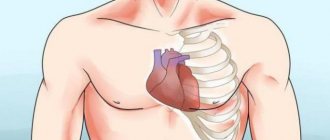
Central venous catheter
This is a long tube that is inserted into a large vessel to transport medications and nutrients. There are three access points for its installation: internal jugular, subclavian and femoral vein. The first option is most often used.
When installing a catheter in the internal jugular vein, there are fewer complications, pneumothorax occurs less often, and it is easier to stop bleeding if it occurs.
With subclavian access there is a high risk of pneumothorax and arterial damage.
When accessing through the femoral vein after catheterization, the patient will remain motionless, in addition, there is a risk of infection of the catheter. The advantages include easy entry into a large vein, which is important in case of emergency care, as well as the possibility of installing a temporary pacemaker
Kinds
There are several types of central catheters:
- Peripheral central. It is passed through a vein in the upper limb until it reaches a large vein near the heart.
- Tunnel. It is injected into a large jugular vein, through which blood returns to the heart, and is removed at a distance of 12 cm from the injection site through the skin.
- Non-tunnel. Installed in a large vein of the lower limb or neck.
- Port catheter. Inserted into a vein in the neck or shoulder. The titanium port is installed under the skin. It is equipped with a membrane that is pierced with a special needle through which fluids can be injected for a week.
Indications for use
A central venous catheter is installed in the following cases:
Also read: Technique for collecting blood from a vein
- To introduce nutrition if its entry through the gastrointestinal tract is impossible.
- During the behavior of chemotherapy.
- For rapid administration of large volumes of solution.
- With prolonged administration of fluids or drugs.
- During hemodialysis.
- In case of inaccessibility of veins in the arms.
- When administering substances that irritate peripheral veins.
- During blood transfusion.
- With periodic blood sampling.
Contraindications
There are several contraindications to central venous catheterization, which are relative, therefore, according to vital indications, a CVC will be installed in any case.
The main contraindications include:
- Inflammatory processes at the injection site.
- Blood clotting disorder.
- Bilateral pneumothorax.
- Clavicle injuries.
Procedure of introduction
The central catheter is placed by a vascular surgeon or interventional radiologist. The nurse prepares the workplace and the patient, helps the doctor put on sterile clothing. To prevent complications, not only installation, but also care is important.
After installation, it can remain in the vein for several weeks or even months.
Before installation, the following preparations are required:
- find out if the patient is allergic to medications;
- conduct a blood clotting test;
- stop taking certain medications a week before catheterization;
- take blood thinning medications;
- find out if you are pregnant.
The procedure is performed in a hospital or on an outpatient basis in the following order:
- Hand disinfection.
- Selection of catheterization site and skin disinfection.
- Determination of the location of the vein based on anatomical characteristics or using ultrasound equipment.
- Administering local anesthesia and making an incision.
- Reduce the catheter to the required length and rinse it in saline solution.
- Guide the catheter into the vein using a guidewire, which is then removed.
- Fixing the instrument on the skin with an adhesive plaster and installing a cap on its end.
- Apply a bandage to the catheter and mark the insertion date.
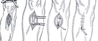
- When a port catheter is inserted, a cavity is created under the skin to accommodate it, and the incision is sutured with absorbable thread.
- Check the injection site (if it hurts, if there is bleeding or fluid discharge).
Care
Proper care of the central venous catheter is very important to prevent purulent infections:
- At least once every three days it is necessary to clean the catheter insertion hole and change the bandage.
- The junction of the dropper with the catheter should be wrapped in a sterile napkin.
- After injecting the solution, wrap the free end of the catheter with sterile material.
- Avoid touching the infusion system.
- Change infusion systems daily.
- Do not bend the catheter.
Immediately after the procedure, an x-ray is taken to ensure proper placement of the catheter. The puncture site should be checked for bleeding, and the port catheter should be washed. Wash your hands thoroughly before touching the catheter and before changing the dressing. The patient is monitored for infection, which is characterized by symptoms such as chills, swelling, induration, redness of the catheter insertion site, and fluid discharge.
At home, the patient should follow the doctor's recommendations and care for the catheter:
- Keep the puncture site dry, clean and bandaged.
- Do not touch the catheter with unwashed and undisinfected hands.
- Do not swim or shower with the instrument installed.
- Don't let anyone touch him.
- Do not engage in activities that could weaken the catheter.
- Check the puncture site daily for signs of infection.
- Rinse the catheter with saline solution.
Complications after CVC installation
Central vein catheterization can lead to complications, including:
- Puncture of the lungs with accumulation of air in the pleural cavity.
- Accumulation of blood in the pleural cavity.
- Puncture of the artery (vertebral, carotid, subclavian).
- Pulmonary embolism.
- Incorrect placement of the catheter.
- Puncture of lymphatic vessels.
- Catheter infection, sepsis.
- Heart rhythm disturbance when advancing the catheter.
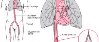
- Thrombosis.
- Nerve damage.
Reasons for performing the procedure
A central catheter is inserted when the patient needs:
- Administer medications or fluids long-term;
- During chemotherapy;
- For nutrition, if food intake through the digestive system is impossible;
- Periodic blood sampling;
- Blood transfusion;
- For intravenous administration of drugs when the veins in the arm are difficult to access;
- For dialysis.
The central catheter is usually inserted by an interventional radiologist or vascular surgeon. Once inserted, the catheter can be used for several weeks to months.
Peripheral catheter
A peripheral venous catheter is installed for the following indications:
- Inability to take liquids orally.
- Transfusion of blood and its components.
- Parenteral nutrition (administration of nutrients).
- The need for frequent administration of drugs into a vein.
- Anesthesia during surgery.
PVK cannot be used if it is necessary to administer solutions that irritate the inner surface of blood vessels, a high infusion rate is required, and also when transfusing large volumes of blood
How to choose veins
A peripheral venous catheter can only be inserted into peripheral vessels and cannot be installed into central ones. It is usually placed on the back of the hand and on the inside of the forearm. Rules for choosing a vessel:
- Well visible veins.
- Vessels that are not on the dominant side, for example, for right-handed people should be chosen on the left side).
- On the other side of the surgical site.
- If there is a straight section of the vessel corresponding to the length of the cannula.
- Vessels with large diameter.
PVC should not be placed in the following vessels:
- In the veins of the legs (high risk of thrombus formation due to low blood flow speed).
- On the bends of the arms, near the joints.
- In a vein located close to an artery.
- In the median ulna.
- In poorly visible saphenous veins.
- In weakened sclerotic.
- In deep-lying ones.
- On infected skin areas.
How to put
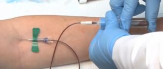
Placement of a peripheral venous catheter can be performed by a trained nurse. There are two ways to grab it in your hand: longitudinal grip and transverse grip. The first option is more often used, which allows you to more securely fix the needle in relation to the catheter tube and prevent it from going into the cannula. The second option is usually preferred by nurses who are accustomed to performing vein punctures with a needle.
Algorithm for placing a peripheral venous catheter:
- The puncture site is treated with alcohol or an alcohol-chlorhexidine mixture.
- A tourniquet is applied, after the vein is filled with blood, the skin is taut and the cannula is installed at a slight angle.
- Venipuncture is performed (if blood appears in the imaging chamber, it means the needle is in the vein).
- Once blood appears in the imaging chamber, the needle stops advancing and must now be removed.
- If, after removing the needle, the vein is lost, reinserting the needle into the catheter is unacceptable; you need to pull out the catheter completely, connect it to the needle and reinsert it.
- After the needle is removed and the catheter is in the vein, you need to put a plug on the free end of the catheter, fix it on the skin with a special bandage or adhesive tape and flush the catheter through the additional port if it is ported, and the attached system if it is not ported. Rinsing is necessary after each infusion of liquid.
Caring for a peripheral venous catheter follows approximately the same rules as for a central one. It is important to maintain asepsis, wear gloves, avoid touching the catheter, change plugs more often and wash the instrument after each infusion. It is necessary to monitor the bandage, change it every three days and do not use scissors when changing the adhesive plaster bandage. The puncture site should be carefully observed.
Although catheterization of peripheral veins is considered less dangerous than central veins, if the rules of installation and care are not followed, unpleasant consequences are possible
Complications
Nowadays, consequences after a catheter occur less and less, thanks to improved models of instruments and safe and low-traumatic methods of their installation.
Complications that may occur include the following:
- bruises, swelling, bleeding at the insertion site;
- infection in the area where the catheter is installed;
- inflammation of the vein walls (phlebitis);
- formation of a blood clot in a vessel.
Complications from intravenous catheters
The most common complication that occurs when a catheter is left in a vein for a long time is phlebitis or inflammation of the vein. Phlebitis may be accompanied by vein thrombosis. Most often, thrombophlebitis develops as a reaction to polyethylene catheters; in young children, this undesirable reaction can appear within a few hours. The most striking signs of phlebitis and thrombosis:
- the vein becomes hard and can be easily palpated;
- there is pain along the vein;
- the skin in the area where the catheter is located is hyperemic;
- There is a local increase in skin temperature.
Any signs of inflammation in the catheter area are an absolute indication for its removal.