Main signs of the disease
You can understand that pulmonary failure is developing if you know what kind of disease it is and how it manifests itself.
The main symptoms of the disease include:
- Shortness of breath (regardless of its intensity);
- Headaches that bother patients mainly in the morning;
- Increased frequency of contractions of the heart muscle;
- Insomnia;
- Reduced pressure levels;
- Vomiting, nausea;
- Blueness of the skin;
- Memory problems;
- Changes in breathing (the depth and frequency of inhalations/exhalations changes);
- Participation of auxiliary muscles in the breathing process.
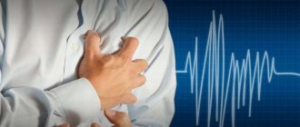
Patients complain of suffocation and severe shortness of breath.
Problems with consciousness and memory are caused by a lack of oxygen in the bloodstream and the accumulation of carbon dioxide in excessive quantities. In severe cases, this causes loss of consciousness or coma.
If such signs appear, you should understand how the disease is diagnosed and treated.
To determine the cause of the above symptoms, you need to:
If signs of pulmonary failure appear, the patient is sent to a medical facility for inpatient treatment.
First aid for acute cardiopulmonary failure
Forced pose with elbows on a chair
Call an ambulance immediately. If the patient cannot figure out how to breathe easier, we sit him on a chair (“on horseback” position) with his legs down. Your elbows should rest on the back of the chair.
Pulmonary edema is one of the most dangerous conditions in medicine; no folk methods, the power of self-hypnosis and reflexology can save you from it.
Regardless of your assumptions about the nature of the condition, the patient needs to put a nitroglycerin tablet under the tongue.
If the leading mechanism of failure is myocardial infarction, then timely prescribed nitroglycerin significantly reduces the area of tissue damage, i.e. reduces the risk of rupture (transmural infarction) of the heart muscle and improves the overall prognosis.
Ventilate the room
We open the windows in the room - the air should be saturated with oxygen. If there is oxygen in the room (in a container), let the patient breathe.
It is quite effective to apply a tight tourniquet to both legs - this will significantly relieve the load on the heart, because will reduce the volume of circulating fluid. This manipulation can save the patient’s life, however, with prolonged tourniquet, the patient dies from intoxication after decompression of the legs.
If you are afraid that the patient will not last until the ambulance arrives (foamy sputum from the mouth, the patient turns blue), you can burn the lower limbs.
Diagnostics
Diagnosis of this condition begins with examining the patient and asking about his well-being and complaints. For a more detailed assessment, an instrumental examination is carried out:
- X-ray of the lungs, which reveals both diseases of the respiratory system and changes in heart size;
- Breast CT is performed if necessary if the diagnosis remains doubtful after radiography;
- Ultrasound of the heart allows you to determine the degree of dysfunction of this organ;
- catheterization and invasive manometry accurately determines pressure in the pulmonary artery and heart cavities;
- The ECG reflects secondary changes in advanced cases.
Prevention
Prevention of PD - quitting smoking and alcohol abuse, a healthy lifestyle, adequate physical activity, and playing amateur sports. Frequent hypothermia and acute respiratory diseases increase the load on the respiratory system, which adversely affects the condition of the lungs in old age.
People whose professions involve working in hot shops and waste production with the formation of by-product dust must undergo a medical examination once a year by a therapist, pulmonologist, hematologist and phlebologist. Be tested for vital lung capacity, oxygen-carbon ratio of blood and its gas composition.
About the chronic form of insufficiency
Unlike the acute form of failure, the contractility of the heart in the chronic course decreases very slowly. Therefore, all of the above symptoms are not pronounced; they are often manifested by one sign, for example, shortness of breath during physical exertion.
An important sign of chronic pulmonary heart failure is an increase in pressure in the pulmonary circulation, and then in the large one. As a result, severe swelling occurs, primarily in the legs. The swelling is symmetrical, of a dough-like consistency, located on the ankles and back of the feet. In the initial stages of failure, they completely disappear at night, since it is easier for the heart to pump blood when the body is in a horizontal position. Then the swelling becomes permanent and the liver swells. Hypertension also occurs in the pulmonary circulation, since the blood entering there is retained there. This occurs due to the weak contractility of the main muscle - the myocardium of the left ventricle, as well as weakness of the left atrium. The result of this is a wet cough, which can lead to pulmonary edema or an attack of cardiac asthma.
Cardiac asthma appears as shortness of breath, abundant moist wheezing appears in the lungs, breathing becomes difficult when inhaling, and becomes bubbling. As pulmonary edema develops, pink foam appears and progressive suffocation occurs. One of the oldest ways to treat pulmonary edema and cardiac asthma is to sit, lower your legs into a basin of hot water, and apply tourniquets to your arms and legs for a short time. All these measures were aimed at reducing the flow of venous blood to the heart and reducing the manifestations of cardiac asthma.
In conclusion, it is worth mentioning the reasons for the development of chronic pulmonary heart failure. Their list is extensive: on the part of the heart - these are congenital and acquired defects, valvular defects, bacterial endocarditis. On the part of the lungs, these are chronic obstructive diseases, severe pneumosclerosis, effusion pleurisy and other conditions. If symptoms such as shortness of breath on exertion, pain in the heart, cough and swelling in the legs appear, you should immediately consult a therapist or cardiologist to find out the reasons. After all, untimely treatment leads to the appearance of chronic tissue hypoxia, which significantly worsens the prognosis.
Chronic cardiopulmonary failure
Denoted by the abbreviation CHF, this is a very common pathology associated with gradual decompensation (overload) of the left ventricle. It is this part of the heart that is responsible for the blood supply to the entire body, i.e. produces systolic ejection of blood.
As a rule, the cause of heart failure in this case is associated with a gradual narrowing of the lumen of the coronary arteries due to atherosclerosis. The heart is gradually “cut off” from nutrition, as a result of which the heart muscle first enlarges and then collapses, which leads to failure.
The second consequence of atherosclerosis is increased blood pressure, which aggravates the process of destruction of the vascular system.
Causes of CHF in the photo
Atherosclerosis
Overweight
Heart pathologies
Physical exercise
Another common cause of CHF is excessive physical activity and an increase in total body weight. Both bodybuilders and people simply prone to obesity increase the overall resistance in the vascular bed (more mass - more vessels). Simply put, there are more pipes, but the motor is the same. For some time, the heart works at increased speed - the muscle tries to produce normal pressure. Then the heart muscle is destroyed and failure occurs.
It is believed that stress and smoking are also factors that provoke CHF, since constant vasoconstriction enhances the effect of atherosclerotic changes.
Treatment of the disease
In acute forms of insufficiency, therapy is carried out in intensive care conditions, since the patient’s serious condition poses a threat to his life. Inhalation of an oxygen mixture is used through a mask or by installing a nasal catheter. This helps saturate the blood with oxygen molecules and mitigate the manifestations of hypoxia on body tissue. In severe cases, the patient is transferred to artificial ventilation.
Carrying out resuscitation measures in acute forms of cardiac failure
The following drugs are administered intravenously:
- thrombolysis drugs (streptocaniasis, actilisis) - for thromboembolism of the pulmonary artery trunk and its branches to dissolve the blood clot and restore blood flow;
- atropine relaxes the smooth muscle muscles of the bronchi, thereby improving respiratory function;
- papaverine reduces vascular tone, expands their lumen, normalizes pressure in the pulmonary circulation;
- anticoagulants (warfarin, heparin) prevent thrombosis of blood vessels and cavities of the heart, thin the blood;
- aminophylline normalizes the contractile function of the myocardium and reduces the manifestations of respiratory disorders.
In the case of a chronic form of incompetence, the underlying ailment is treated. Anti-inflammatory drugs, bronchodilators to dilate the bronchi, and hormonal drugs are prescribed. To treat pathologies of the heart and lungs, treatment is used that is used for heart failure:
- potassium-sparing diuretics (veroshpiron, triampur) remove stagnant fluid from the body;
- cardiac glycosides (digitalis) improve myocardial function;
- selective beta blockers (bisaprolol, atenolol) normalize high blood pressure;
- drugs that stimulate the vasomotor center (caffeine, camphor) are prescribed for respiratory depression;
- cardioprotectors (mildronate) protect myocardial and vascular cells from destruction as a result of hypoxia;
- potassium and magnesium preparations (panangin) improve metabolic reactions in the cells of damaged tissues.
In case of severe erythrocytosis, bloodletting is performed in an amount of 280-400 ml, followed by replacement of the blood volume with low-density solutions (saline solution, rheopolyglucin). They recommend giving up bad habits and prescribe a low-fat, salt-free diet. To maintain normal heart function, reduce the amount of fluid consumed, limit vigorous physical activity and stressful situations.
Heart failure with severe signs of pulmonary hypertension requires timely diagnosis and treatment. Constant monitoring and supportive courses of therapy help avoid severe complications and increase the life expectancy of patients.
Surgery
Sometimes surgical treatment methods are necessary to treat the initial disease, such as:
- Thrombembolectomy for pulmonary embolism
- Sympathectomy
- Removal of a lung tissue tumor
- Heart and lung transplantation
- Balloon atrial septostomy
Palliative treatment
In severe and advanced cases, existing treatment methods are not effective, and cardiopulmonary failure progresses.
In this case, it is necessary to take active steps to maintain a decent quality of life for a person in a special clinic (hospice) or at home.
Recommendations for lifestyle changes:
- Stop smoking
- Weight control and maintaining a healthy diet
- Regularly checking your feet and ankles for swelling
- Limiting salt intake
- Vaccination against influenza and pneumonia
- Limiting alcohol intake
- Reducing stress levels
- Physical activity
- Monitoring the implementation of your prescriptions (creating a schedule, carrying the required amount of medications with you)
- Discuss taking additional medications, dietary supplements, vitamins with your doctor
- Keeping a diary to monitor weight, diuresis, blood pressure, fluids drunk and medications taken, and other important notes.
Choice of treatment tactics
In 30% of cases, patients are admitted to the hospital with acute pulmonary failure.
It should be aimed at normalizing airway patency, eliminating hemodynamic disturbances and restoring perfusion and ventilation.
Information about emergency methods is given in the table.
| Action of medical personnel | Characteristic |
| Oral examination | Required to remove trapped foreign bodies, remove a sunken tongue, and perform aspiration of the respiratory tract |
| Oxygen therapy | Necessary to maintain proper blood gas levels |
| Vibromassage of the chest | Provides restoration of bronchial patency |
| Carrying out artificial ventilation of the lungs | Prescribed for diagnosis of stage 2 pulmonary insufficiency |
| Tracheal intubation | Necessary if there is a high risk of suffocation and there is no progress from first aid. |
| Drainage of the pleural cavity | Performed for hemo- and pneumothorax |
When pulmonary ventilation normalizes, treatment continues. If necessary, continue to supply humidified oxygen: this is done using a nasal catheter, through an oxygen mask or a tent.
If the problems were caused by bronchospasm, then glucocorticosteroids and bronchodilators are prescribed.
If the pathology is accompanied by painful sensations, then analgesics are given. To stimulate the work of the heart and blood vessels, cardiac glycosides are prescribed, and respiratory analeptics are prescribed. Infusion therapy can eliminate signs of intoxication and hypovolemia.
When restoring the functioning of the respiratory system, it is necessary to carry out simultaneous treatment of the underlying disease that provoked the development of pulmonary failure.
Treatment of acute left ventricular heart failure in a hospital
Such patients are taken either to the heart attack department or to the intensive care unit at the cardiology clinic or department. If the process is accompanied by arrhythmia, go to the cardiology hospital where there are arrhythmologists.
In the hospital, pulmonary edema will be relieved with hormonal drugs, and the patient will be given narcotic analgesics. If the problem was thrombosis of the coronary arteries, thrombolytics are prescribed: Actelise, Metalyse, Streptokinase, Urokinase, etc.
Action of thrombolytics
Thrombolytics are quite expensive, but they help save the patient in the first hours after thrombosis. In fact, when the blood clot breaks down, the situation resolves without serious consequences for the patient. Don’t skimp at this stage - if the clinic runs out of a tender thrombolytic, buy it.
In addition, doctors prescribe drugs that reduce the activity of the respiratory center, sedatives and stimulants of metabolism in the heart muscle (metabolic therapy).
If a patient's heart failure is accompanied by arrhythmia (atrial fibrillation, atrial fibrillation), antithrombotic therapy is prescribed.
Irregular heart rhythm caused by left ventricular heart failure is itself a factor provoking thrombosis. To prevent this common complication, the drugs Xarelto, Clexane and other fractionated heparins are prescribed.
After emergency care is provided, the patient is transferred to a regular ward and prescribed drugs that lower blood pressure (ACE inhibitors, diuretics) and drugs to normalize the heart rhythm.
Causes
Pulmonary hypertension leads to a violation of the enrichment of blood in the alveoli with oxygen. As a result, the myocardium of the right ventricle increases cardiac output in order to reduce tissue hypoxia (lack of oxygen). Over time, due to excessive stress, the muscles of the right side of the heart grow.
There are several groups of factors that cause the disease:
| Bronchopulmonary factors include: |
The disease can develop with tuberculosis and pulmonary sarcoidosis. |
| Vascular factors include: |
|
| The disease can cause deformities of the diaphragm and chest: |
|
Under the influence of vascular factors, the arteries narrow. This occurs due to blockage by a blood clot or thickening of the vascular walls due to the inflammatory process.
In medical practice, the disease most often develops against the background of:
- pneumosclerosis;
- pulmonary vasculitis;
- emphysema;
- thromboembolism;
- pulmonary edema;
- pulmonary artery stenosis.
| Symptoms of the disease may appear suddenly. In this case, they are distinguished by rapid development and a vivid clinical picture. In the acute form of the disease, emergency medical care and placement in the intensive care unit are required. Acute cardiopulmonary failure occurs:
Under the complex influence of unfavorable factors, hemodynamics are sharply disrupted. This manifests itself in the form of insufficient blood circulation of the “right heart”. The disorder is accompanied by the following symptoms:
The acute form of the disease may be accompanied by pulsation in the epigastric region of the dilated right ventricle. The x-ray shows an increase in the mediastinum to the right and upward; the electrocardiogram shows overload of the “right heart”. When listening to the heart, ri and muffled tones are clearly revealed. In case of acute blockage of the pulmonary artery by a thrombus, pulmonary edema and pain shock rapidly develop, which can lead to rapid death. | |
Symptoms depend on the stage of the disease. In the compensated form of the pathology, symptoms characteristic of high pressure in the pulmonary circulation are revealed. Chronic pulmonary heart failure can develop over several years. It appears as:
| |
| Decompensated form | It is accompanied by increasing symptoms and leads to irreversible consequences in all tissues and organs. Signs of a progressive disease include:
With increasing death of all tissues (terminal state), serious damage to the brain and kidneys develops. These processes are expressed in the form of lethargy, apathy, impaired mental functions, and cessation of urine output. In the blood, due to a lack of oxygen, the concentration of hemoglobin and red blood cells increases. |
Causes of cardiopulmonary failure
Causes of SLN:
- bronchial asthma
- obstructive bronchitis
- chronic obstructive pulmonary disease (COPD)
- pulmonary tuberculosis
- emphysema
- systemic connective tissue disorders (collagenosis)
- pulmonary embolism
- Deformations of the chest and spine (kyphoscoliosis)
- trauma, including pneumothorax
- arthritis
- polio
- botulism
- vasculitis
- pneumoconiosis (accumulation of solid particles of silicates and coal dust in the lung tissue)
- tumors
- major lung surgery
Risk factors for developing CHF:
- arrhythmias
- arterial hypertension
- overdose of certain drugs
- smoking
- injecting drugs
- valvular heart defects
- severe obesity
- immunodeficiency states
Diagnostic features
If this disease develops, the patient should be observed by two specialists: a cardiologist and a pulmonologist. Making a diagnosis begins with collecting a detailed history, when the patient talks about his complaints, bad habits, previous illnesses, working conditions and lifestyle, etc.
The next stage is listening to the heart, determining its boundaries using percussion, and measuring pressure. With right ventricular hypertrophy, muffled tones are detected, accompanied by increased pulmonary pressure, strong heartbeat and decreased blood pressure. If congestion in the lungs is observed, then signs of arterial hypertension may appear against their background.
After this, instrumental diagnostics are prescribed, the purpose of which is to accurately determine the nature of the pathology:
- X-ray of the sternum. Allows you to determine possible pathologies of the lung tissue when the mediastinum grows to the right.
- Echocardiography. One of the main diagnostic methods by which functional deviations in the operation of the valve apparatus are determined. Also, during the study, the specialist can identify changes in cardiac output and assess the correctness of myocardial contractions.
- CT. This procedure is used to in-depth study those areas of the lungs and heart that have undergone changes.
- Angiography. Necessary for visualizing the lumen of the vessel, its shape, identifying blood clots and various atherosclerotic changes.
- Electrocardiography. Allows you to determine the conductivity and excitability of the organ. In this way, areas of cardiac muscle hypertrophy, rhythm disturbances and ischemic foci are identified. If doubts arise, specialists additionally conduct research using the Holter apparatus.
- Catheterization with pressure gauge. Necessary for determining pressure in large vessels and cavities of the heart. The procedure is very important in the treatment of thrombosis, since in this way agents are introduced into the vessels to help break down blood clots.
- Spirometry, through which it is possible to determine the degree of respiratory failure.
Diagnosis is advisable in the early stages of the disease. With timely detection of pathology, it is possible to prevent the development of irreversible changes in the myocardium, kidneys, liver, lungs and brain. If a patient develops concomitant diseases that lead to cardiopulmonary disorders, then the examination should be carried out at the preclinical stage of pathology development.
Symptoms of acute heart failure
The patient begins to experience shortness of breath, gradually turning into painful suffocation. The patient takes a forced position
Posture for pulmonary failure
Due to the effusion of fluid in the lungs, sputum may be coughed up, which does not bring any relief to the patient. In severe cases, frothy pink sputum is discharged from the mouth and nose.
Swelling and pallor may appear quite quickly. The patient becomes very scared (and for good reason).
Acute cardiopulmonary failure may not have any special acoustic manifestations. Sometimes, using a phonendoscope, you can hear fine bubbly wheezing in the lower parts of the lungs (edema). With advanced edema, moist rales appear over the entire surface of the lungs.
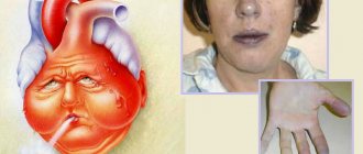
Symptoms of acute heart failure in the photo
Foamy pink sputum
Tachycardia
Feeling of fear
Heartache
Pressure in acute heart failure may be increased or decreased - this is not a diagnostic criterion. Due to severe stress in this condition, the heart rate increases. If the patient has had episodes of arrhythmia in the past, it may happen again.
Classification of the disease
Depending on the mechanism of development, there are 3 forms of pulmonary failure:
- Hypoxemic;
- Hypercapnic;
- Mixed.
In the hypoxemic form, there is a lack of oxygen in the tissues.
It is difficult to bring the condition back to normal even with the help of oxygen therapy. This type of pathology is characteristic of lesions of the respiratory system and those pathologies in which the lung tissue is replaced by connective tissue.
The alveoli, the peripheral nerve system responsible for the coordinated functioning of the respiratory organs, the muscles involved in breathing, the central nervous system, the chest, and the respiratory tract are affected.
In hypercapnic form, carbon dioxide accumulates in tissues
. This type of disease is also characterized by a lack of oxygen.
The disease develops against the background of weakness of the muscles that regulate the respiratory system, obesity, and chronic obstructive pulmonary disease.
The mixed form is characterized
a simultaneous combination of hypoxemia and primary hypercapnia.
Based on the rate of progression, the following forms are distinguished:
- Spicy
- Chronic
This is a rather dangerous pathology that threatens life. The condition can be normalized only with the help of intensive care in the intensive care unit.
The chronic form of the disease develops slowly
. The pathology can progress without threatening the patient’s life over many months or years.
Within the specified time, the body manages to turn on the adaptation mechanisms and ensures the optimal gas composition of the blood. This type is considered safe, because it can be detected in a timely manner and the patient’s condition can be normalized.
Experts distinguish 3 degrees of pulmonary failure, which occurs in a chronic form:
Depending on the severity of the pathology, 3 stages of the disease are distinguished:
- I (mild) stage:
the partial pressure of oxygen is in the range of 60-80 mmHg, the blood is saturated with oxygen at the level of 90-94%; - Stage II:
partial oxygen pressure drops to 40-59 mmHg, oxygen concentration in the blood varies between 75-89%; - Stage III:
the condition is critical, the oxygen pressure becomes less than 40 mmHg, the oxygen concentration in the blood drops to less than 75%.
When pulmonary failure develops, it is necessary to understand the reasons for its occurrence. Further treatment will depend on this.
With this pathology, the blood does not completely flow into the respiratory organs. Pulmonary regurgitation of the 1st degree is considered not dangerous; the stream of thrown blood is practically invisible.
In grade 4, only surgical intervention can preserve the patient’s health.
Varieties
Respiratory failure is classified according to several criteria.
According to the mechanism of development, the disease is:
- parenchymal;
- ventilation;
- mixed.
The first option is a violation of blood oxygen saturation to a state of hypoxemia. Occurs against the background of progressive pneumonia, alveolitis, pulmonary edema, hemosiderosis.
The ventilation form of the disease becomes a history of hyperkalemia, muscle weakness of the respiratory organs, mechanical injury to the thoracic region, and obesity. It is classified into the following subspecies:
- Centrogenic - breathing processes are suppressed after brain injury, ischemic attack, or drug poisoning.
- Thoradiaphragmatic - difficulty in working the chest and diaphragm is caused by kyphoscoliosis.
- Neuromuscular – malfunction of the spinal cord causes an imbalance in the supply of nerve impulses to the muscles of the respiratory organs, the development of polio.
- Bronchopulmonary – disruption of the respiratory channels, reduction of the respiratory surface and alveolar distensibility.
The mixed form of the disease is the result of combining the first two forms. According to the etiology of development, DN is divided into types:
- Obstructive – the external respiratory system is affected: the ability to fully inhale and exhale becomes more difficult. The breathing rate becomes limited.
- Restrictive - full inspiration is limited.
- Combined – combines the symptoms of the two previous types with a predominance of one of them. Occurs in cases of prolonged progression of cardiopulmonary diseases.
- Hemodynamic – due to the lack of blood flow or impaired oxygenation of part of the lung.
- Diffuse - occurs when there is a failure in the flow of gases through the pulmonary membrane, subject to its pathological thickening.
Based on the rate of worsening of symptoms, DN is divided into the following types:
- Acute respiratory failure (ARF) is dangerous for the patient and is characterized by rapid development from a couple of minutes to several days. Occurs against the background of a chronic type of illness.
- Chronic respiratory failure can last from several months to several years. Long-term pathogenic processes in the lungs cause failure of the respiratory and circulatory organs. The main factor in the formation of the acute form is hyperventilation, which ensures oxygen saturation of the blood. An increase in the level of hemoglobin in the blood allows the body to adapt to the condition.
Some time later, cardiopulmonary pathology develops - right ventricular heart disease is connected to the pulmonary disease. It is formed due to an increase in pressure in the pulmonary vessels, veins, arteries and the occurrence of cor pulmonale due to lung diseases.
Etiology
Extrapulmonary causes include:
- Disruption of the processes of neurogenic regulation of external respiration - consequences of severe intoxication, encephalitis, intracranial hypertension, compression and hypoxia of the brain as a result of stroke, tumor, edema, consequences of anesthesia, hypothyroidism, sleep apnea.
- Disruption of the functioning of peripheral nerves and muscles - botulism, tetanus, muscle wasting, polymyositis, status epilepticus, myalgia.
- Circulatory disorders in the pulmonary circulation and lungs - pulmonary embolism, heart failure, congenital defects, pulmonary hypertension, peripheral circulatory disorders.
- Respiratory failure is a complication of other diseases: scoliosis, flatulence, malignant neoplasms of the lungs and mediastinum, obesity, burns, poisoning, vasculitis, renal failure, cardiogenic shock, uremia, ketoacidotic coma, peritonitis, typhoid fever.
Alveolar hypoventilation and broncho-obstruction are the main pathological processes of respiratory failure.
In the initial stages of the disease, compensation reactions are activated, which eliminate hypoxia and the patient feels satisfactory. With pronounced disturbances and changes in the blood gas composition, these mechanisms fail, which leads to the development of characteristic clinical signs, and subsequently severe complications.
Treatment options
Currently, treatment of cardiopulmonary insufficiency is carried out:
- diuretics;
- cardiac glycosides
- beta blockers;
- surgical intervention;
- bloodletting;
- folk healing.
Diuretic drugs
Treatment with diuretics helps eliminate excess fluid that accumulates in the body as a result of decreased contractility of the heart. Hydrochlorothiazide is an effective and inexpensive diuretic. It stabilizes blood pressure and removes excess fluid.
Furosemide is an immediate and stronger drug. It is usually taken in the morning on an empty stomach with regular monitoring of electrolyte-salt balance. Since important microelements are removed from the body along with the liquid. The effect of the drug lasts 6 hours. It can be used even with weak kidney function. Furosemide helps to quickly relieve swelling and removes excess fluid well. Another effective diuretic drug that can help get rid of swelling and remove excess fluid is ethacrynic acid.
Beta blockers
Treatment of the disease with beta blockers improves the functioning of the left ventricle of the heart, normalizes blood circulation, and helps relieve swelling.
The most effective beta blockers are propranolol and timolol. They have adrenergic-selective properties and eliminate almost all the symptoms of this disease. Treatment with metoprolol is also considered effective. Because it has maximum cardioselectivity and eliminates all signs of the disease.
Surgical intervention
Cardinal treatment is applicable if the disease is severe. The most common treatments are atrial septostomy, thromboendarterectomy, or organ transplantation.
Atrial septomy is necessary to reduce pressure in the right atrium and pulmonary artery. Thrombendarterectomy is used to remove blood clots from the lungs. Transplantation is used if treatment by other means has not given the desired effect.
Bloodletting
This treatment involves removing a certain amount of blood from the bloodstream. Up to 400 ml of blood is released from the sufferer’s body. With this method of rescuing the disease, the patient’s blood pressure decreases, excess fluid is eliminated, and swelling disappears.
Glycosides
The most effective glycoside that is common in Russia is digoxin. Glycosides are positive inotropic agents that improve the quality of life of patients suffering from pulmonary-cardiac insufficiency.
Glycosides are prescribed in small doses. Using cardiac glycosides, patients seek hospitalization less often.
Folk remedies
Treatment with folk remedies should be carried out only after consultation and doctor’s prescriptions. Because this disease is very serious and dangerous.
The main remedy for this disease is simple wormwood. It normalizes blood circulation, eliminates pain, and removes excess fluid. You need to prepare a decoction of wormwood and take it before meals every day, three-quarters of a glass.
Another equally effective remedy is a decoction of nettles. Hand baths should be made with this decoction. Timed treatment lasts 10 minutes every day
Pumpkin juice is also an excellent remedy for this disease.
You must always remember that folk recipes alone cannot be used to treat heart and lung diseases; moreover, some medications cannot be used simultaneously with herbal ones due to a possible increase in side effects.
Treatment of chronic cardiopulmonary failure
Treatment should be lifelong under periodic medical supervision
Psychological phenomenon - no one wants to undergo long-term treatment (consciousness rejects an incurable disease). As a rule, patients take medications only 2-3 months after visiting a cardiologist. This is the main reason for the relatively rapid death from heart failure. Treatment of chronic heart failure is prescribed for life. Canceling a doctor’s prescription quickly leads to a transition to the next functional class of CHF.
Drugs that lower blood pressure are prescribed - sartans (Losartan), ACE inhibitors (enalapril, lisinopril), diuretics (hydrochlorothiazide, etc.). In addition to diuretics, patients are advised to strictly limit their fluid intake. To control heart rate, beta blockers (carvedilol, metaprolol, etc.) are recommended.
To prevent thrombosis, patients must be prescribed acetylsalicylic acid (aspirin). Sometimes it is combined with warfarin and other anticoagulants and antiplatelet agents.
Video: treatment of chronic heart failure
Differences between respiratory failure and heart failure with mild heart disease
| Symptoms | Respiratory failure | Pulmonary heart failure |
| 1. Shortness of breath | Shortness of breath is manifested by a feeling of suffocation or lack of air (impaired “breathing mechanics”), depending on the type of respiratory failure: with the obstructive type, deep breathing and almost normal respiratory rate, with the restrictive type, frequent shallow breathing. Shortness of breath changes throughout the day and worsens in the cold. Patients can tolerate long-term, uniform physical activity relatively easily, but do not tolerate “explosive” loads well. During sleep, patients take a horizontal position. | With the development of cor pulmonale, shortness of breath increases. There is a feeling of urgent need to inhale immediately at the end of the inhalation. Due to higher pressure in the vessels of the lungs, a “vascular framework” is created, leading to restriction. Therefore, breathing becomes more shallow and frequent. A paroxysmal cough appears, sometimes leading to loss of consciousness. During sleep, patients occupy the crtopnoe position. |
| 2. Cyanosis | Cyanosis is diffuse, warm, quickly decreases after inhaling oxygen, does not change color when the limbs are lowered into warm water. | Acrocyanosis increases (stronger staining of the tip of the tongue, nose, lips, fingers). Signs of cyanosis of cardiac origin appear. Cyanosis becomes cold and decreases only after prolonged inhalation of oxygen (since arteriolovenous anastomoses in the lungs, which open when blood circulation in the pulmonary circle is obstructed, play a role in its origin). |
| 3. Swelling of the neck veins | The neck veins swell only at the moment of exhalation, when intrathoracic pressure increases and less blood leaves the veins in the right atrium. | With the development of stagnation in the systemic circulation, pressure is increased throughout the venous system. Therefore, swelling of the neck veins persists in any position of the body and does not depend on the phase of breathing. |
| 4. Edema | Edema appears due to increased capillary permeability due to prolonged arterial hypoxemia of pulmonary origin. Edema may appear in the lower extremities as a result of the low position of the diaphragm (emphysema) and compression of the v. cava inferior as it passes through the formen quadrilaterum. More often edema of the 1st degree | Visible edema appears when systemic venous pressure increases by more than 2 cm of water and when fluid retention exceeds 5-6 liters. Before this, patients quickly increase their weight by 1-2-3 kg (hidden edema). According to the laws of gravity, swelling is first located on the feet, then gradually goes higher. |
| 5. Liver enlargement | No | This is one of the early signs of cor pulmonale decompensation. There is a feeling of heaviness in the right hypochondrium, the size of the liver increases. |
| Signs | Bronchial asthma | Chronic obstructive bronchitis |
| Allergy | Characteristic | Not typical |
| Cough | Predominantly paroxysmal | Constant, varying intensity |
| Dyspnea | Attacks of expiratory dyspnea | Constant without sharp fluctuations in severity |
| Daily changes in FEV1 | More than 15% of proper values | Less than 10% of proper values |
| Reversibility of bronchial obstruction | Characteristic | Not typical |
| Eosinophilia of sputum and blood | Characteristic | Out of character |
Pathogenesis
A decrease in the pumping function of the heart in CHF leads to secondary activation of the sympathoadrenal (SNS) and renin-angiotensin-aldosterone systems (RAAS) on the one hand and vasopressin (known as the antidiuretic hormone ADH) and atrial peptides on the other hand. The process mediates peripheral and renal vasoconstriction, causing a decrease in glomerular filtration rate, which, coupled with the already reduced blood filling pressure, leads to secondary activation of the RAAS. Activation of the RAAS results in an increase in aldosterone secretion, thereby ensuring proper perfusion pressure in the tissues due to increased reabsorption of sodium and water by the proximal nephron tubules.
Stimulation of the release of vasopressin from the posterior lobe of the pituitary gland occurs in response to the activation of baroreceptors that respond to a decrease in blood filling pressure. Increased ADH levels lead to myocardial fibrosis, hypertrophy and vasoconstriction, as well as increased water reabsorption in the nephron collecting ducts, despite the already existing overload of the heart with circulating blood volume in the form of stretched atrial tissue and low plasma osmolality.
Pathogenetically, water-electrolyte homeostasis is mainly under the control of renal regulation, which is the most vulnerable point of sodium-potassium homeostasis. The clinical effects of ADH release are thirst and increased water intake. Angiotensin II also contributes by stimulating the brain's "thirst center" and promoting the release of ADH. Under physiological conditions, hyperactivation of neurotransmitter systems is excluded by the multidirectionality of their action, and with CHF, conditions arise for the occurrence of edema, against the background of hypotension and hypoosmolality. It is true that it is worth noting that, according to experimental data, different mechanisms were noted underlying the activation of the RAAS and SNS on the one hand and the vasopressin and atrial peptide systems on the other hand
A transient decrease in renal function and direct renal damage inevitably contribute to an increase in potassium levels, which is leveled for the time being by taking loop and/or thiazide diuretics. However, it is worth noting that it would be preferable to choose the drug Torasemide, which, compared to Furosemide, promotes less activation of the RAAS and mediates less excretion of potassium from the body.
Acute heart failure
Acute heart failure (AHF), which is a consequence of impaired myocardial contractility and a decrease in systolic and minute blood volumes, is manifested by extremely severe clinical syndromes: cardiogenic shock, pulmonary edema, acute renal failure.
Acute heart failure is most often left ventricular and can manifest as cardiac asthma, cardiogenic pulmonary edema, or cardiogenic shock.
Pathogenesis of respiratory failure
Lung function can be roughly divided into 3 main processes: ventilation, pulmonary blood flow and gas diffusion. Deviations from the norm in any of them inevitably lead to respiratory failure. But the significance and consequences of violations in these processes are different.
Often, respiratory failure develops when ventilation is reduced, resulting in the formation of excess carbon dioxide (hypercapnia) and lack of oxygen (hypoxemia) in the blood. Carbon dioxide has a high diffusion (penetrating) ability, therefore, when pulmonary diffusion is impaired, hypercapnia rarely occurs; they are more often accompanied by hypoxemia. But diffusion disorders are rare.
An isolated violation of ventilation in the lungs is possible, but most often there are combined disorders based on disturbances in the uniformity of blood flow and ventilation. Thus, respiratory failure is the result of pathological changes in the ventilation/blood flow ratio.
A violation in the direction of increasing this ratio leads to an increase in physiologically dead space in the lungs (areas of lung tissue that do not perform their functions, for example, in severe pneumonia) and the accumulation of carbon dioxide (hypercapnia). A decrease in the ratio causes an increase in shunts or vascular anastomoses (additional blood flow paths) in the lungs, resulting in a decrease in oxygen content in the blood (hypoxemia). The resulting hypoxemia may not be accompanied by hypercapnia, but hypercapnia, as a rule, leads to hypoxemia.
1. Violation of alveolar ventilation.
a) restrictive
c) mixed
https://youtube.com/watch?v=_TVO5Rnvxio
Table 10
Symptoms
Failure of cardiopulmonary origin has pronounced symptoms that cannot be ignored.
- Symptoms of shortness of breath appear already at the initial stage of the disease. In most cases, shortness of breath occurs with any physical activity.
- Symptoms of cyanosis (cyanosis) also become noticeable immediately. This occurs due to a lack of oxygen in the arterial blood. Because of this, the skin of a sick person becomes ashen-gray.
- Symptoms of a compensatory reaction occur after the appearance of cyanosis. Blood deprived of the necessary oxygen begins increased production of hemoglobin and red blood cells. Therefore, when taking tests, the patient’s blood components are elevated.
- Symptoms of pain in the right hypochondrium also indicate insufficiency of a cardiopulmonary nature, as this is a sign of insufficiency of the right chambers of the heart.
- Symptoms of sudden hypotension may also be signs of this disease. Sometimes a person feels severe weakness and headache, darkening of the eyes.
Sometimes the symptoms described above may be signs of another disease.
Share the article on social media. networks:
Symptoms
Propaedeutics of pulmonology involves prescribing treatment depending on the manifestation of the syndrome. The more pronounced the symptoms, the more aggressive the treatment: strong drugs can inhibit the functioning of the peripheral nervous system. Symptoms of LN:
- shortness of breath;
- nausea in the morning and when tilting the head down;
- insomnia;
- cyanosis of eyelids, lips, cheeks;
- increase in heart rate to 120 - 150 beats per minute;
- memory impairment;
- chest pain;
- drowsiness at lunchtime;
- decreased concentration;
- short-term memory lapses;
- apathy and depression;
- shallow breathing.
In the hyperacute course of the disease, only specific symptoms associated with breathing appear. No hidden progression of FN has been observed; in children, symptoms may be mistaken for the initial stage of asthma development.