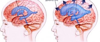
How does intracranial hypertension occur?
The skull is a hollow formation with free space inside. Most of it is filled with the brain and cerebrospinal fluid, called cerebrospinal fluid. The substance constantly circulates and is renewed approximately 7 times a day.
Intracranial pressure refers to the difference between pressure in the cranial cavity and atmospheric pressure. For newborns, the norm is considered to be 1.5-6 mmHg, while for children over 1 year old - 3-7 mm. Intracranial hypertension is a condition in which the indicator exceeds 14.7 mm (up to 6 years), 15 mm (7-10 years) and 15.6 mm (in adolescence).
Hypertension develops due to an imbalance of fluids circulating in the cranium. A constant level of pressure is maintained by the balance between cerebrospinal fluid and blood. If the concentration of one of the fluids increases, the child will develop symptoms of hypertension.
The appearance of ICH is a dangerous pathological condition. It is believed that the disease may be benign. However, cerebral hypertension in infants often provokes blood flow disturbances, atrophic and ischemic processes. The disease is included in the ICD 10 classification in the section of neurological pathologies.
Thus, ICP in children is a dangerous disease that can lead to irreversible consequences.
Consequences of ICH
The brain loses functionality, being in an unhealthy compressed state. This leads to atrophy of brain cells, which affects a decrease in intelligence and disruption of regulatory processes. If left untreated, compression of the brain provokes displacement or wedging of parts into the base of the skull
. This condition leads to death.
When compressed, the brain can shift to the occipital or cerebellar part, the process is accompanied by compression of the stem areas. In such a situation, the patient dies from respiratory arrest. When wedged into the temporal lobe, the pupil dilates, breathing becomes difficult, and the person falls into a coma.
If wedging occurs in the area of the tentorium, the patient becomes deaf, drowsy, and lethargic. Breathing slows down. An increase in intracranial pressure provokes a rapid decrease in vision, as the pathology leads to atrophy of the optic nerves.
Classification and symptoms
The clinical picture of high intracranial pressure is pronounced, but the infant is not able to tell about troubling ailments. The severity of symptoms depends on the severity.
Types of intracranial hypertension:
- Lightweight,
- Weakly expressed
- Expressed
- Heavy.
When benign intracranial hypertension occurs in children, complications such as hydrocephalus or deformity are not observed. Severe ICH is usually a consequence of neurological pathologies, tumors or injuries accompanied by hemorrhages.
Symptoms of ICP in infants:
- Poor healing of the fontanel. The cranial bones of a newborn are mobile and compress during childbirth to facilitate birth. In the first few weeks of life, the fontanel tightens. With ICP, the regeneration process becomes more difficult. There is a bulging of this area, but there is no characteristic pulsation.
- Sleep disorders. An infant does not sleep well, which is unnatural for newborns. Normally, a baby sleeps about 16 hours a day. With intracranial hypertension, the baby falls asleep for a short time and sleeps restlessly.
- Head enlargement. In severe forms of ICH, the structure of the child’s skull changes. It becomes disproportionate to the body and is constantly increasing. The disorder is associated with stagnation of fluid inside the skull due to difficulty in its outflow.
- Developmental delay. Normally, at the age of five months, the child holds his head up and focuses his gaze on objects of interest. One of the reasons why underdevelopment occurs is increased intracranial pressure. Hypertension affects certain brain structures, causing them to develop more slowly.
- In infancy, with ICH, children also experience a disturbance in the sucking reflex and a deterioration in appetite. There is susceptibility to changing weather conditions. When there is a sharp change in atmospheric pressure, the child cries for a long time. At the same time, the veins located on the scalp expand.
Signs of hypertension in children 1–12 years old:
- Headache. During exacerbations, ICH is moderate or intense in nature, without a specific localization. Often the syndrome spreads to the neck muscles, ears, and facial tissues. This indicates pressure on the nerve fibers.
- Nausea occurs due to the impact on the centers located in the medulla oblongata. Against the background of a severe form of hypertension, the child practically does not consume food, since its intake immediately causes nausea and vomiting. The symptom worsens with physical activity.
- Irritability. Against the background of ICH, children experience increased emotional sensitivity. In addition to the manifestation of nervous conditions, regular hysterics are observed, accompanied by mild convulsions.
- Fatigue. When performing physical activity, the child gets tired quickly. There is marked drowsiness that occurs even after a person has slept for a long period of time. Efficiency and performance at school decreases. The child becomes inattentive, quickly distracted, and gets tired when solving simple problems.
It should be noted that the signs of intracranial hypertension are in many ways similar to the symptoms of other diseases, and therefore special diagnostics are necessary.
Manifestations of hypertension in children
If infants experience an increase in head parameters, restlessness, systematic regurgitation, and sleep problems, this may indicate intracranial hypertension. With the intracranial form, the monthly increase in head circumference will be more than 1 cm. The pathological symptom is usually accompanied by dehiscence of the sutures of the skull, swelling of the fontanelle, and increased excitability.
If one-year-old babies often hold their heads, this may indicate intense pain, which can intensify with movement, sneezing, and the cough reflex. A characteristic sign of the disease is vomiting, not caused by overeating. The clinical picture is complemented by visual disturbances and decreased intelligence.
Provoking factors
The famous pediatrician Evgeny Komarovsky emphasized that ICP is not an independent disease, but is a symptomatic manifestation. Imbalance of organic fluids occurs due to many physiological abnormalities, and therefore there are different causes of intracranial hypertension in children.
The most common factors causing ICH are:
Congenital anomalies. They are caused by pathologies during intrauterine development. Often occur due to hypoxia associated with a lack of oxygen in the tissues. Congenital anomalies appear against the background of a complicated pregnancy, accompanied by prolonged toxicosis.
Difficult birth. With prolonged labor, the child experiences asphyxia. Strangulation can be fatal if not treated promptly. With prolonged deficiency, atrophy of brain tissue occurs, accompanied by high intracranial pressure.
Infections. Damage to the brain by bacteria or viruses is a common cause of hypertension. Due to infection, tissue swelling occurs, as a result of which the free space inside the skull decreases and fluid circulation is disrupted.
Nervous pathologies. ICH occurs due to increased neuronal conductivity. Against this background, autonomic dysfunction develops, accompanied by an outflow of cerebrospinal fluid.
Hormonal disorders. With increased secretion of certain substances, the production of cerebrospinal fluid increases. Against this background, an imbalance develops inside the skull and, as a consequence, hypertension.
Thus, the formation of ICP is associated with congenital pathologies and acquired diseases.
Methods of therapy
The human brain is unable to function properly under high blood pressure. This will lead to atrophic processes, decreased intellectual capabilities, and disruption of nervous regulation. Therefore, it is necessary to resort to treatment methods that will restore healthy blood pressure values.
Treatment of the syndrome includes the use of the following methods:
- non-drug therapy - lifestyle changes, menu adjustments, physiotherapeutic procedures, visits to a neuropsychologist;
- drug therapy - dehydration, taking sedatives, neuroprotective, metabolic drugs;
- surgical intervention used for severe cerebral hypertension that is not amenable to drug treatment.
Non-drug therapy can be used even after recovery. The patient must normalize nutrition and drinking regimen, perform feasible physical exercises, and use physiotherapeutic methods.
The basis of treatment for ICH is the need to reduce the synthesis of cerebrospinal fluid along with increasing its absorption. For this purpose, diuretics are prescribed that reduce the production of exudate (Diacarb). With prolonged use of diuretics and the absence of a therapeutic effect, the patient is prescribed glucocorticosteroids (Dexamethasone).
To eliminate hypertension syndrome, it is necessary to take medications that improve blood flow through the veins (Troxevasin). When the pain is intense, drugs from a number of non-steroidal anti-inflammatory drugs (Nimid) are used. In case of ICH that occurs against the background of infectious diseases, the victim is administered antibacterial drugs
.
With a pronounced increase in ICH, Mannitol is administered intravenously, which has dehydrating activity. For pathology that occurs as a result of neurosurgical intervention, drugs from a number of barbiturates (Thiopental) are used.
If cerebral hypertension progresses and painful symptoms are not eliminated by medications, then surgery is indicated for the patient. Lumbar puncture is often used, through which 30 ml of spinal fluid is removed. In many cases, such manipulation significantly alleviates the patient’s condition. Usually multiple procedures are required.
To level out pathological manifestations in severe cases, lumbar-peritoneal shunting is used, in which conditions are artificially created for the outflow of exudate. To do this, a special tube is inserted into the cerebrospinal fluid cavity, the other end of which is placed in the peritoneal area. This way, excess fluid is evacuated from the brain.
The most aggressive treatment method is cranial trephination, during which doctors deliberately injure the skull so that the brain matter does not rest against the bone tissue. This therapeutic method is used very rarely.
To treat visual disorders, decompression of the myelin sheaths of the optic nerve is used.
Diagnostics
Establishing a reliable diagnosis is an important procedure that affects the method of treatment. Examination of the child allows us to identify the cause of hypertension and determine the best method of eliminating it.
For diagnostic purposes the following are used:
- Inspection and survey. The pediatrician measures the circumference of the skull, compares the results with normal values, and assesses the condition of the fontanel. The pediatrician interviews parents about specific forms of behavior of the infant, his sleep, and nutrition.
- Ultrasound. Using ultrasound, the condition of the cerebral vessels is studied. With ICP, diagnostic results indicate a violation of the outflow of fluids in the veins.
- MRI. Tomography is aimed at studying the state of the brain. The procedure allows you to determine the location of the accumulation of cerebrospinal fluid, the area of compression of the cerebral vessels. Prescribed to confirm the diagnosis.
- X-rays are used for suspected tumors in the brain. If a tumor is present, X-ray examination allows you to determine its location and size.
- Fundus examination. With intracranial hypertension, the vascular pattern changes. The veins are greatly dilated, indicating a violation of the outflow of fluid. This symptom confirms the presence of hypertension.
To diagnose intracranial hypertension, several instrumental procedures are used, as well as visual examination and questioning for symptomatic manifestations.
Treatment
The goals of therapeutic measures are to treat the underlying disease, eliminate symptoms and improve the condition of the sick child. At the initial stage, certain groups of medications, physiotherapy and therapeutic massage most often help. In more severe cases, surgery may be required.
Drug therapy
Treatment in this case should be comprehensive. Most often, medications of various groups are prescribed. The most effective of them are presented in the table.
| Drug group | Peculiarities |
| Diuretics | They help remove excess fluid from the body’s cells, which helps normalize the volume of cerebrospinal fluid. |
| Nootropics | They improve memory and blood circulation, stimulate brain activity, and also replenish the lack of oxygen in the brain. |
| B vitamins | Improves nerve cell conductivity and helps replenish nutrient deficiencies. |
| Corticosteroids | Eliminate symptoms (in particular, headaches). |
| Barbiturates | Restores psycho-emotional state and normalizes sleep. |
| Anti-inflammatory drugs (non-steroidal) | They are used only for pathologies of an infectious nature. |
Important! The drugs, their combination and dosage are prescribed by the attending physician after a complete examination of the patient. The course of therapy depends on the severity of the disease and the individual characteristics of the body. Self-medication is strictly prohibited.
Surgical intervention
Surgical intervention in most cases is required if increased intracranial pressure is accompanied by hydrocephalus. To eliminate the pathology, shunting is performed (removal of excess fluid from the ventricles of the brain).
Using this procedure, it is possible to normalize blood pressure, restore cerebral circulation and prevent the risk of complications. The operation is quite complex, but in almost all cases it shows good results.
Most cases with surgical intervention end in positive dynamics
Causes
The main cause of intracranial hypertension in a child is an increase in the amount of free cerebrospinal fluid. This causes an increase in pressure on the cerebral vessels, which leads to disruption of the nutrition of its individual fragments. These processes develop for the following reasons:
- Anomalies of intrauterine development. The disease develops if the child experienced oxygen deficiency during the formation of organs and systems.
- Birth injury. The cause of the disease is disruption of labor or asphyxia of the baby.
- Early damage. The provoking factors of the disease are falls of a newborn baby or mechanical injuries to the head and cervical spine.
- Infectious lesions of the cerebral cortex. These include encephalitis and meningitis, which lead to brain damage and can cause the development of brain edema.
- Diseases of the nervous system. Pathologies that are accompanied by excessive neural conduction pose a threat.
- Blockage of the liquor ducts. Imbalance of hormones can lead to problems with the production of cerebrospinal fluid.
- Neoplasms in the brain. As they increase, the cerebrospinal fluid ducts are compressed, which leads to a decrease in the space between the parts of the brain.
Intracranial hypertension in adolescents often develops at the age of 15-17 years, when the balance of hormones in the body seriously changes. Also provoking factors are stressful situations, emotional stress, neurocirculatory dystonia and autoimmune pathologies.