Vertebrobasilar insufficiency is a chronic disorder of blood circulation in the vertebral and basilar arteries. The pathological process is accompanied by a lot of symptoms that are noticeable even at an early stage.
Progression is associated with the complication of the patient’s situation; the chances of recovery diminish as the disorder develops.
According to statistical estimates, approximately 5% of people suffer from this disease. The numbers are several times higher in patients with concomitant pathologies of the cardiovascular system and/or musculoskeletal system.
Treatment is conservative or surgical. Depends on the root cause of the disorder and the current condition of the patient.
The prognosis is directly determined by the moment the therapy begins, so delaying visiting a doctor is life-threatening.
Development mechanism
Vertebrobasilar syndrome is a polyetiological condition, that is, it develops for many reasons, often existing in parallel. The formation is based on the following factors:
Narrowing of the vertebral artery on one or both sides
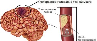
The so-called atherosclerosis. As a result of stenosis due to smoking, taking medications, and biochemical factors. Or as a result of the deposition of cholesterol, lipid structures and plaque blockage.
In any case, the lumen of the artery becomes smaller, the blood moves worse and poorly supplies the brain with oxygen and necessary substances. The result in the short term is vertebrobasilar insufficiency and ischemia in cerebral structures.
Congenital vascular anomalies
Slightly less common. However, they are no less difficult. Among such diagnoses, one can highlight hypoplasia of the vertebral artery (its underdevelopment) and, as a result, a smaller diameter of the vessel compared to normal.
Read more about hypoplasia of the right vertebral artery here, and of the left - here.
There are other fundamental factors (see reasons), regardless of them, the immediate moment is always associated with a narrowing of the lumen of the artery, and therefore a weakening of blood flow in the brain. As ischemia progresses, it becomes more severe.
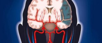
Vertebral vessels provide up to 40% of the total blood flow in nerve tissues and nourish a number of structures:
- Occipital and parietal lobe.
- Cerebellum.
- Hypothalamus.
- Brain stem.
- Pons.
- Visual thalamus.
That’s why the clinic is almost always obvious. Since the central nervous system does not receive enough nutrients and oxygen, changes begin that are irreversible in nature. For example, expansion of the ventricles.
In this way, the body tries to compensate for the poor blood flow.
As long as the body is able to cope, there is a clinic, but there are no critical violations. At a certain point, the intensity of nutrition becomes so low that everything gets out of control.
The terminal phase of the pathological process begins. Most often, it is accompanied first by transient (transient) ischemic attacks, and then by a full-fledged stroke.
Attention:
Mortality from necrosis of nerve tissue in the posterior regions of the brain is approximately four times higher, compared with damage to areas that are supplied by the carotid arteries. Therefore, the probability of death is enormous.
Causes
Osteochondrosis of the cervical spine is a chronic degenerative disease that occurs due to gradual damage to the intervertebral discs and then to the vertebrae. The cause of the destruction of cartilage tissue is a violation of diffuse metabolism, through which vitamins, microelements, and nutrients enter the vertebral structures. Under conditions of the resulting deficiency, destructive changes begin to occur in the cartilage discs. They become damaged, become thinner, and lose their shock-absorbing properties.
The X-ray image shows deformation of the vertebrae.
Then comes the turn of the vertebrae, which are injured under serious static and dynamic loads. With osteochondrosis of 2, 3, 4 degrees of severity, these bone structures shift, and displacement of the intervertebral disc provokes the formation of intervertebral hernias. The vertebrae become unstable, and processes are launched in the body to compensate for this disorder. The edges of the bone plates flatten and grow, forming bone growths - osteophytes. The transverse processes of the vertebrae are equipped with openings in which the vertebral artery is located. A large blood vessel supplies oxygen and nutrients to the following structures:
- spinal segments up to the 2nd thoracic vertebra;
- brain stem;
- inner ear and balance organs;
- cerebellum;
- posterior parts of the hypothalamus, thalamus;
- temporal, occipital lobes of the brain.
This is what osteophytes look like in the photo.
Formed osteophytes, as well as a narrowed intervertebral canal, prevent normal blood supply to these vital structures. The centers of the brain stem regulate breathing, swallowing, and the functioning of the cardiovascular system. No less important are the parts of the central nervous system that are supplied with blood by the vertebral artery. At the initial stage of osteochondrosis, with a lack of oxygen or nutrients, anastomoses, or natural connections of blood vessels, are involved. If one artery partially loses its ability to supply blood, the second begins to pump a larger volume of blood.
Classification
The disease is divided according to two criteria. Based on the severity of the clinical manifestations and organic disorders, they are called:
- Stage 1 VBI. It is accompanied by a minimal set of manifestations, which, however, are noticeable if you listen to the body. Focal neurological moments predominate. If the cerebellum is involved - dizziness, lack of coordination, occipital lobe - visual problems, etc.
Rarely in isolation. Usually from 1-2 structures at once. There is a good chance of a complete cure if started at this point.
- VBI stage 2. Irreversible changes accompany the patient, so one can no longer count on a complete recovery. But with complex therapy, it is possible to compensate for the disorder and reduce the risks of death and disability.
Symptoms become more severe. Almost 70% of patients experience transient ischemic attacks, which are also called microstrokes or dyscirculatory encephalopathy.
As it progresses, the chances of recovery, at least partial, become increasingly slim.
- VBN stage 3. Accompanied by critical circulatory disorders in the brain. This is a decompensated phase that does not imply cure. The end result is a stroke, usually extensive.
Since correction is no longer possible, a relapse will not be long in coming, which will become fatal. Help is mainly palliative, to improve well-being and relieve discomfort.
In parallel, the course of dyscirculatory encephalopathy in the terminal phase is observed.
In this case we are talking about staging, this is a generally accepted method of classification.
The syndrome can also be classified according to the neurological characteristics of the disorder.
Angiodystonic phase
Changes are just beginning. The clinic is minimal or completely absent (which is quite rare).
Signs are mainly represented by general points: headache, disturbance of orientation in space. Focal manifestations are possible, but are less common or much less noticeable.
Mixed phase
Signs of progression of the disorder, insufficient blood circulation and organic abnormalities, which can be detected by imaging methods, are included. For example, MRI.
The clinic includes general and focal symptoms.
Ischemic stage
Moments typical of insufficient blood circulation in cerebral structures predominate. Recovery is already challenging, but still possible.
There is a high risk of stroke. Without treatment, this condition develops, on average, over a period of 1-4 years.
The symptoms of pre-stroke are described in detail in this article.
Persistent phase
As the name suggests, this is a chronic stage of the pathological process with a constantly existing complete clinical picture.
It should be noted that this stage is not always equally difficult. Depends on the course characteristic of a particular patient. A complete cure is impossible; the condition must be corrected to help the patient. How much it will be possible to influence the situation is also controversial.
Both classifications are used equally by neurologists and specialized vascular surgeons. The main purpose is to determine the specific phase of the disorder, methods of therapy, and prospects for impact.
Clinical picture
The clinical picture (symptoms) that occurs with ischemia of the vertebrobasilar region can be very diverse. A common symptom is dizziness, which is often accompanied by nausea and vomiting. Vertigo is a phenomenon that is the initial symptom in approximately half of all cases when spinal circulation is disrupted. But more often it is accompanied by other manifestations associated with a disorder of venous blood flow, therefore, a deficiency of blood supply and a lack of tissue nutrition:
- visual impairment;
- diplopia;
- blurred vision;
- unilateral and bilateral homonymous hemianopsia;
- dysarthria;
- dysphagia;
- paresthesia - mild paralysis, numbness in the face;
- various combinations of weakness or lack of sensation in the legs.
Vertebro-basilar insufficiency, the symptoms of which often alternate, can only manifest as vertigo, which can occur as an isolated symptom. Long periods of relapses (more than 6 months) with dizziness without other accompaniments are not typical for VBI. Tinnitus and hearing loss are not common symptoms.
In approximately 40% of cases, attacks of vertebrobasilar insufficiency last more than 1 hour, although patients themselves often indicate a duration of several minutes. About 90% of TIAs last less than 2 hours. The difference between the 2 conditions is that VBI usually has a shorter duration than carotid TIA. The severity of symptoms is highly variable, ranging from mild to severe. The frequency of attacks varies from single attacks to multiple attacks throughout the day.
The condition of vertebrobasilar insufficiency is a combination of symptoms. According to one study group, 43% of patients had vertigo, 60% had ataxia, 39% had diplopia, 27% had dysarthria, and 37% had blurred vision. Manifestations may vary depending on the damage to the extra- or intracranial part. Damage to the extracranial vertebral part, mainly, there is dizziness, blurred vision, imbalance, involvement of the intracranial part is characterized only by vertigo. TIA due to basilar artery disease usually has 2 or more of the following symptoms:
- dizziness;
- slurred speech;
- double vision;
- dysphagia;
- unilateral or bilateral limb weakness.
With vertebrobasilar insufficiency, sudden short-term loss of muscle tone (drop attacks) may occur; the patient, while conscious, suddenly falls (mostly to his knees). With more severe damage, loss of consciousness occurs - syncope.
When examining patients, it is important to correlate subjective symptoms with objective neurological signs of the vertebrobasilar territory. Common and important objective findings include:
- nystagmus;
- oculomotor disorders;
- Horner's syndrome (ptosis and meiosis, sometimes anhidrosis on the affected side);
- numbness and impaired motor skills in the face and limbs;
- speech and coordination disorders.
Examination of posture and gait is important. In patients, only one symptom is indicated as a subjective sign, for example, vertigo; with an objective study, other manifestations are determined, in particular, ataxia of the limbs or trunk. For localized lesions, variable (crossed) syndromes are typical, when on the affected side - ipsilateral - there is a failure of the cranial nerve nucleus, Horner's syndrome or cerebellar syndrome, and on the contralateral side hemiparesis or hemihypesthesia is formed. Vertebro-basilar insufficiency is characterized by any combination of weakness, paresthesia, numbness of the upper and lower extremities, and face.
Symptoms
The clinical picture is extremely varied, which is associated with the many structures that are supplied by the vertebral vessels.
The syndrome of the vertebrobasilar arterial system develops gradually, the manifestations progress slowly, layering one on top of the other.
If we talk about an approximate list, we can make the following list:
- Dizziness. It appears almost as a highlight. Almost all patients, regardless of the stage of the disorder. But not always to the same extent. There are also options in terms of flow. For some, this is a constant sensation, for others - attacks of transient discomfort.
- Headache. Its intensity is also not the same. Depends on the characteristics of the human body and the stage of the disorder. The character is oppressive, pulsating. Localization is mainly the back of the head, but not always. There may be diffuse sensations, migration of discomfort to the forehead, temples (the so-called “legionnaire’s gesture”).
- Loss of coordination, unsteadiness of gait and uncertainty when moving. A person cannot normally navigate in space. This is one of the most common signs, present in almost 70%. The reason is damage to the extrapyramidal system, in particular the cerebellum. It does not exist all the time; periodically the discomfort intensifies.
- Decreased hearing acuity and tinnitus. Both symptoms go hand in hand.
- Nausea, possibly vomiting. At the peak of a headache attack or spatial disorientation.
These symptoms are characteristic of all stages of the pathological process.
If we talk about specific focal moments, they do not always occur and not in all patients:
- Violations of the visual analyzer. Flickering of floaters in the eyes, an increase in the number of floaters (look like translucent threads, worms, spots), photopsia (flashes of light in the form of dots, lines, simple visual hallucinations), the formation of scotomas (blind areas that look like black spots).
Increased eye fatigue, redness and dryness are also possible. These are parts of one whole. The result of irritation of the occipital lobe of the brain.
- Increase in the number of heart beats per minute. Tachycardia. An alarming manifestation. May be the result of insufficient trophism of the brain stem, also of the hypothalamus. The severity depends on the degree of ischemia. As a rule, this is a transient symptom that occurs without visible precipitating factors.
- Decrease in speed and productivity of thinking. Intellectual impairments develop later and are not critically severe. Sometimes it is impossible to detect the problem on your own. Memory and attention suffer slightly. However, in the final stages, the symptom is much more severe.
- Increased sweating. Or hyperhidrosis. Even outside of physical activity at rest.
- Weakness, drowsiness. There is not enough strength for work or leisure. The patient constantly wants to sleep. There is also heaviness in the limbs and insufficient muscle tone. This is the result of damage to the same extrapyramidal system represented by the cerebellum.
- Increased susceptibility to negative environmental factors. Insufficient oxygen levels in the room, high or low temperatures around, etc., also cause meteosensitivity.
- Behavioral disorders. Increased aggressiveness, touchiness, sudden outbursts of rage that end in severe fatigue. Psychasthenic phenomena in their purest form.
- Drop attacks are also often encountered as a calling card of an advanced pathological process. Temporary paralysis of the body with throwing back of the head and falling.
- In some situations, it is impossible to control natural bladder and bowel movements. Incontinence.
There are a lot of clinical signs; doctors combine them in various combinations to more accurately describe the form of the pathological process.
Risk factors for developing VBI
If a person has a congenital anomaly of vascular development, or osteochondrosis of the cervical spine, this does not mean at all that he will necessarily develop circulatory failure in this pool. But with certain risk factors, the likelihood of this increases.
- Dyslipidemia is increased blood cholesterol and a violation of the lipoprotein ratio.
- Untreated arterial hypertension.
- Increased blood viscosity.
- Diabetes.
- Alcohol abuse.
- Smoking.
- Heart rhythm disturbances.
- Heart valve defects with an increased risk of thrombosis.
The onset of an ischemic attack in the vertebrobasilar bed can be provoked by:
- Hypertensive crisis.
- A sharp decrease in blood pressure.
- Prolonged uncomfortable head position.
- A sharp turn or hyperextension of the neck.
- Stress.
- Non-professional massage or manual therapy.
- Head or neck injury.
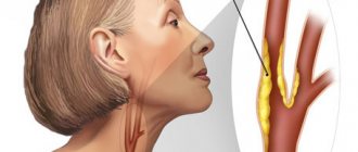
Double vision and blurred vision, tinnitus, dizziness are some of the symptoms of VBI
Causes of VBI
Vertebrobasilar insufficiency syndrome is a polyetiological disorder; a group of factors is involved in its development.
If we highlight the main ones:
- Atherosclerosis. As a symptomatic narrowing of the vertebral arteries or blockage of them with cholesterol plaques. Perhaps the main culprit, it accounts for up to 80% of recorded cases.
- Congenital disorders such as vascular hypoplasia.
Other factors are not so common, but they do occur:
- Previous neck injuries.
- Osteochondrosis. Accompanied by compression of the arteries. As it progresses, spinal disc herniations form, which leads to stabilization of the pathological process and the impossibility of its complete cure.
- Muscle problems. Chronic muscle spasm, previous inflammatory diseases (myositis) and others. Eliminating such violations is not easy; massage and physical therapy will be required.
- Hypertension or symptomatic increase in blood pressure.
- Angiopathy due to diabetes.
- Vasculitis is also an inflammation of the artery walls.
The reasons are taken into account when developing therapeutic tactics. Without knowing the culprit, the disease cannot be eliminated.
Causes
Experts identify the main causes of vertebrobasilar syndrome:
- Cervical osteochondrosis (degenerative changes in the vertebrae), which negatively affects the patency of blood vessels in the brain and leads to spasms and impaired blood flow in the main arteries. According to statistics, every third patient with osteochondrosis is at risk of developing vertebrobasilar syndrome.
- The syndrome can be congenital - pathologies during pregnancy and childbirth (trauma, premature birth, any violation of the integrity of the blood vessels of the baby's brain), congenital anomalies (dysplasia, congenital circulatory disorder - hypoplasia, Kimmerly's disease - a congenital defect in the area of the connection of the vertebrae and the skull).
- Acquired injuries of the cervical vertebrae (accidents, road traffic and sports injuries).
- Vascular diseases, inflammatory processes of the walls of blood vessels (autoimmune inflammation of the branches of the aorta, arteritis).
- Atherosclerosis (chronic blockage of blood vessels, deposition of cholesterol plaques), defects in the medial lining of blood vessels (aneurysms) and other vascular patency disorders (carotid artery stenosis, etc.).
- Diabetes mellitus type 2 (damage to small vessels of the brain).
- Hypertension, in which high blood pressure persists, resulting in disruption of the structure and basic functions of the arteries and heart.
- Phospholipid syndrome, which provokes the formation of blood clots in the veins and arteries, and other autoimmune pathologies (lupus erythematosus, Asherson syndrome, etc.).
- Damage (dissection) of arteries – dissection of the carotid and vertebral arteries.
- Thrombosis of blood vessels and veins (a pathology due to which a blood clot appears inside a vein or artery; the clot blocks and disrupts normal blood flow).
- Vertebral hernias, spondylosis and other pathological changes in the intervertebral discs compress the basilar or vertebral arteries, thereby impairing blood circulation.
Diagnostics
It is carried out under the supervision of a neurologist or vascular surgeon. Often both specialists work in tandem.
Standard set of measures:
- Oral interview to identify complaints and create a clinical picture.
- Anamnesis collection. When did the disturbances begin, is there any deterioration, lifestyle, bad habits, presence of chronic and current diseases. And other factors.
- Special functional tests. With hyperventilation (fast breathing), turning, throwing back the head, etc. They are found in almost all patients and are used as a reliable diagnostic method. A characteristic feature is that when performing tests, the symptoms of vertebrobasilar insufficiency increase significantly. This provides the basis for making a diagnosis.
- Dopplerography of neck and brain vessels, duplex scanning. To assess the speed and quality of blood flow.
- Angiography.
- Also an MRI if needed. Both of these methods are key and provide maximum information on the condition of the arteries.
- General blood test and biochemistry with an expanded picture of lipids. Allows you to identify excess cholesterol and other fats, detect atherosclerosis and its cause.
The diagnosis of VBI is made based on the results of instrumental and laboratory studies, taking into account symptoms.
In some cases, it takes more than one week to accurately understand the process, but these are relatively rare situations. Usually there are no problems with detecting pathology.
Diagnosis of VBN
- VBI can be suspected based on complaints and symptoms, especially if they develop acutely.
- During examination, a neurologist may reveal: Nystagmus - involuntary oscillatory movements of the eyeballs when looking to the side.
- Damage to the cranial nerves (facial asymmetry, strabismus).
- Check coordination samples and sensitivity.
- Compare limb strength.
- Identify pathological reflexes.
Treatment methods
Therapy is predominantly conservative. It is aimed at eliminating the root cause of VBI, relieving symptoms and preventing the progression of the disorder and stroke. For this purpose, medications are prescribed.
Which ones exactly:
- Nootropics. Accelerate metabolic processes in nerve tissues. Glycine. Phenibut. With caution, as they often provoke allergic reactions.
- Vasodilators. Betahistine, Cinnarizine, Pentoxifylline and others. Their function is clear from the name.
- Antiplatelet agents and anticoagulants. To normalize blood fluidity and prevent the formation of blood clots. Also partially adjust the quality of nutrition of cerebral tissues. This includes Aspirin in various modifications and other medications.
- Cerebrovascular. Improves microcirculation in the brain. These include Piracetam, Actovegin, Cavinton.
- It is possible to prescribe statins and related drugs to combat atherosclerosis.
- The use of antihypertensive drugs is practiced. To lower blood pressure. ACE inhibitors, beta blockers, calcium antagonists and others.
Massage and physiotherapy are a good help. Of necessity. The procedure is determined by the doctor.
Surgical treatment methods are prescribed in extreme cases. The essence of the intervention is to expand the arteries mechanically (stenting, ballooning), plastic surgery. Physical removal of hard cholesterol plaque.
In case of intervertebral hernia, the non-functional component is removed and replaced with a prosthesis. There are several options, but they are resorted to when conservative treatment is ineffective and if there is no other option.
To improve brain nutrition and prevent vascular stenosis and atherosclerosis, it is recommended to change your lifestyle:
- Give up cigarettes and alcohol.
- If possible, maintain an adequate physical activity regimen. Don't overwork yourself, but don't sit still either. One hour of walking a day is good for the average person.
- Have a full rest. Sleep 8-9 hours during the night.
- Consume less animal fat and salt (2 to 7 grams).
- Avoid stress if possible.
Diagnostics and examinations
In the diagnosis of VBI, imaging is the most important method, similar to other TIAs. This is an image of brain tissue using CT or MRI. For vertebrobasilar insufficiency, MRI is the preferred method, since small lesions in the brain, sometimes in the cerebellum, are not clearly identified on CT. However, CT may exclude other abnormalities (bleeding, tumor).
In addition to brain imaging, examination of the vascular system is important in making the diagnosis. The main non-invasive technique is ultrasound, especially visualization of the extracranial part. In the intracranial part of a. vertebralis cannot be examined in the traditional way (conventional duplex sonography); color-coded transcranial ultrasonography or transcranial Doppler sonography must be used.
In more than 50% of cases, atherosclerosis of the vertebral arteries affects their intervals, which often cannot be examined sonographically. Research may further complicate arterial hypoplasia. If stenotic changes or other abnormalities are suspected, angiography is recommended. Classic digital angiography today is receding a little into the background, because... It is possible to perform high-quality CT or MR angiography.
In addition to imaging studies, standard screening and identification of risk factors for the development of atherosclerosis are required. Cardiac testing is advisable because 25% of patients with TIA have symptomatic and 20% asymptomatic coronary artery disease. The mortality rate for patients with TIA due to myocardial infarction is 5% (overall mortality is 6%).
Possible consequences
The key complication is stroke. Death of nerve tissue. In addition, dyscirculatory encephalopathy and vascular dementia (dementia) often develop.
Ultimately, the patient becomes deeply disabled or dies.
Vertebro-basilar insufficiency is a complex pathological process, it is easily diagnosed, but extremely dangerous if not treated in a timely manner.
If symptoms develop that even remotely resemble manifestations of VBI, you need to urgently go to a neurologist. This will save your health and life.
Features in children and adolescents
Vertebrobasilar syndrome has recently become significantly younger due to congenital anomalies, trauma during childbirth (occurs in children 3–5 years old), a sedentary lifestyle, injuries, etc. (in children 7–14 years old).
Reference. The disease in childhood is easily treatable. Only in severe clinical cases is drug treatment and surgery performed. How to recognize signs of vertebrobasilar insufficiency in children?
- posture is disturbed;
- the child gets tired quickly, drowsiness, and performance decreases sharply;
- irritability appears, crying for no reason;
- dizziness, nausea, and frequent fainting are possible;
- reacts sharply to stuffiness and stale air;
- the child chooses the wrong position when sitting (sitting askew).
Important! If symptoms occur or after a recent injury to the spine or neck, you should immediately contact a neurologist for an initial evaluation. The sooner the cause is identified and a diagnosis is made, the more effective the treatment will be.
Treatment of vertebrobasilar insufficiency
After performing diagnostic measures and identifying vertebrobasilar insufficiency, the doctor will prescribe treatment. If arterial pathology is at an early stage of development, therapy can be carried out at home. In case of pronounced symptoms with signs of ischemic stroke, the patient is referred to the neurological department.
Medicines
Photo from the site irecommend.ru
There is no single treatment regimen for circulatory failure. For each patient, a different tactic is developed, which depends on the degree of dyscirculation and the severity of the course.
Most often, the following drugs are used to eliminate the symptoms of vertebrobasilar vascular insufficiency:
- to improve microcirculation and cerebral blood flow - Reopoliglyukin, Pentoxifylline, Reomacrodex;
- nootropics - Cerebrolysin, Pantogam, Vinpocetine, Phenibut, Seamax, Actovegin, Piracetam;
- osmodiuretics - Glycerol, Mannitol;
- for dizziness - Phezam, Bellataminal, Meclozine, Betahistine;
- to eliminate nausea and vomiting - Ondansetron, Domperidone, Metoclopramide.
In case of acute vertebrobasilar insufficiency and transient ischemic attack, long-term use of antiplatelet agents is necessary. Most often, Aspirin in minimal dosages, Bromcamphor, and Ticlopedine are used for this purpose.
Folk remedies
Medicines alone cannot cope with pathology. Therefore, doctors often advise supplementing drug treatment for vertebrobasilar insufficiency with folk remedies.
The healing composition of garlic gives a good blood-thinning effect. You need to take 4 heads of hot vegetable, peel, chop and place in a glass jar. Leave in a dark room until juice appears. Then filter, add a tablespoon of honey and the juice of one lemon. Take 1 tsp. after breakfast. The treatment regimen is a week break every 10 days.
Chestnut tincture helps with vertebrobasilar insufficiency. Fresh fruits (1/2 kg) are crushed and poured with boiled water (1 liter). Keep in a dark place for 7 days, filter and take 1 tsp. twice a day before meals.
For hypertensive patients with vertebrobasilar insufficiency, a herbal mixture is suitable:
- lemon balm - 20 g;
- corn silk - 40 g;
- valerian - 20 g.
Pour a liter of boiling water over dry plants, cover and leave until cool. Strain the finished infusion and mix with the juice of one lemon. Drink 100 ml three times a day, preferably before meals. After 7 days take a break.
The following folk recipe will help eliminate the vascular symptoms of vertebrobasilar insufficiency:
- hawthorn fruits - 20 g;
- water - 250 ml;
- honey - 1 tsp.
Dry berries are poured with boiling water and placed on low heat. After 10 minutes, remove, cool under the lid and filter. The warm infusion is combined with honey, divided into three servings and consumed half an hour before meals.
Gymnastics
Special gymnastics has a remarkable preventive and healing effect on vertebrobasilar vascular insufficiency. With its help, you can stretch the spinal column, relax cramped muscles and increase blood flow.
For patients with impaired brain activity, quiet exercises with a small amplitude are suitable. They need to be done daily, without allowing long breaks.
Below is a set of exercises indicated for patients with vertebrobasilar insufficiency syndrome:
- From a standing position. Tilt your head and try to touch your chin to your chest.
- Perform circular movements with your head, first to the right, then to the left.
- Bend your head to the side, trying to touch your right shoulder with your ear, then the left.
- Raise and lower your shoulders until a feeling of warmth appears in your muscles.
- Stand on one leg as long as you can. You cannot hold onto your hands or lean on the wall. If the movement is easy, you can close your eyes. Then change legs.
It is permissible to add forward bends and circular movements of the arms to the gymnastics complex for vertebrobasilar insufficiency. Swimming has an excellent healing effect. It’s great if you can go to the pool twice a week, but a one-time workout will be of great benefit.
Microstroke and TIA - you can’t wait!
Damage to any of the arteries supplying the brain leads to disruption of the function of parts of the brain in the basin of this artery. Lack of blood supply causes temporary dysfunction, and its cessation leads to the death of a part of the brain and a stroke. With the development of circulatory failure in the carotid arteries, paralysis of half the body and impaired speech functions occur. The basin of the vertebral arteries is responsible for the area of the brain that maintains balance and provides auditory perception. This area is supplied by the vertebrobasilar system and therefore one of the most important causes of such symptoms is a violation of blood flow in the vertebrobasilar system.
In 70% of cases, cerebral circulatory disorders are associated with atherosclerosis or tortuosity of the arteries in the neck - carotid and vertebral. Therefore, any microstroke or vertebrobasilar insufficiency should be a reason to study these vessels.
Vascular surgeons of the Innovative Vascular Center have significant experience in unique operations on the carotid and vertebral arteries. The advantage of our approach is the use of local anesthesia for operations on the carotid and vertebral arteries. Local anesthesia provides contact with the patient during arterial clamping and minimizes the risk of stroke during surgery.
Indications for operations on the carotid and vertebral arteries
Transient ischemic attacks
Usually they arise suddenly and reach a maximum degree within a few seconds or one or two minutes, they persist for 10-15 minutes, much less often - several hours (up to a day). Symptoms of brain damage are varied and are determined by the localization of cerebral ischemia in the carotid artery basin or the vertebrobasilar basin. Depending on the side and location of the brain lesion, weakness occurs in the arm or arm and leg on one side, often accompanied by speech disorders - “porridge in the mouth”, or “verbal hash” (aphasia), sometimes blindness develops in half of the visual field or complete blindness. These phenomena disappear after a few minutes or less often hours, but during the day they can be repeated more than once. An emergency doctor who arrives to a call can see a “healthy person”, although 10-15 minutes ago the patient could not say a word or move his hand. TIAs often develop with plaques in the carotid arteries, pathological tortuosity of the carotid or vertebral arteries. A transient cerebrovascular accident is not a completed stroke, which will happen sooner or later, and it is necessary to use this signal to eliminate its causes. If the cause of TIA is damage to the carotid or vertebral arteries, then surgery is necessary, since 25% of patients after TIA develop a major stroke within the first month. Typically, removal of an atherosclerotic plaque from the carotid artery or elimination of a pathological bend is performed.
Microstroke
With a microstroke, the focus of brain tissue death is usually very small, and the blood circulation of the surrounding tissues is restored. As a rule, after a mini-stroke, a person recovers quickly. But a microstroke is a very dangerous harbinger of an impending brain catastrophe. A major stroke after a microstroke develops in half of the patients during the first year. The cause of a microstroke in 70% of cases is an atherosclerotic plaque or tortuosity of the carotid or vertebral arteries. Detection of this pathology requires mandatory surgical correction.
Vertebro-basilar insufficiency
Vertebrobasilar insufficiency can cause dizziness that usually lasts for several minutes. Dizziness may be accompanied by other brainstem symptoms, such as headache, diplopia (double vision), vision loss, hallucinations, numbness around the mouth, and speech impairment. The patient may faint without loss of consciousness due to sudden weakness in the legs. These attacks often occur suddenly and are called a drop attack. Sometimes symptoms can be triggered by a change in body position, such as when the neck is stretched too far. Strokes in the vertebrobasilar system are characterized by a very severe course, the development of coma, swallowing and breathing disorders. The causes of vertebrobasilar insufficiency are often pathological bends of the vertebral arteries; strokes often develop due to their thrombosis.
Thrombosis (occlusion) of the internal carotid artery
Typically, in patients with large strokes, complete blockage (occlusion) of the internal carotid artery is found due to its thrombosis. Thrombosis develops in the presence of large atherosclerotic plaques. If compensation of cerebral blood flow during thrombosis through bypass pathways is not enough, then a major stroke develops. Sometimes thrombosis can go unnoticed. If carotid artery occlusion is detected after a stroke, then the goal is to prevent a recurrent stroke in this or the other hemisphere of the brain. The presence of a large plaque in the other carotid artery requires its removal, since the patient is unlikely to survive thrombosis on the other side. If, in the presence of occlusion of the carotid artery, the patient experiences repeated microstrokes on the same side, the question is raised about creating an additional blood flow path to the brain. This is an EICMA operation, which consists of creating a path from the external carotid artery directly to the cerebral vessels. Performed according to indications, this operation significantly improves the prognosis for life in patients who have suffered a stroke.
Don’t delay your examination and consultation with a vascular surgeon!
A microstroke is a precursor to a major stroke. Surgeries on the carotid and vertebral arteries are performed by experienced surgeons at our Center. The advantages of our approach are:
- Correct indications for surgery - we never operate on patients without symptoms of brain failure. Therefore, our operations truly achieve the goal of stroke prevention
- Using local anesthesia for operations on the carotid and vertebral arteries. Local anesthesia allows you to control brain functions during surgery and prevent possible complications
- A full range of surgical interventions - we operate at all levels of damage to the cerebral arteries - carotid, vertebral arteries, cerebral arteries.
- Low probability of postoperative complications. In our practice, complications of operations on the carotid and vertebral columns occurred in less than 1% of patients, while international statistics allow 3% of complications.
Vertebrobasilar syndrome can lead to stroke
Prevention
As you know, it is easier to prevent any illness than to treat it later. To prevent the occurrence of vertebrobasilar stroke, no specific preventive measures are needed. All you need to do is:
- completely stop smoking, since nicotine negatively affects the size of the lumen of blood vessels, and therefore blood flow;
- if the pressure is high, it is necessary to take medications to help normalize it;
- monitor cholesterol levels;
- swim, walk more in the fresh air;
- change your diet towards reducing animal fats, salt, and giving up alcohol;
- undergo a preventive examination at least once a year.
If you have the slightest suspicion of a vertebrobasilar stroke, immediately contact the nearest stroke clinic in your city.