Varicose veins of the lower extremities deservedly occupy one of the first places among all causes of disability. The leading symptoms of this pathology include ulcers on the legs due to varicose veins. The appearance of changes on the legs and feet is a consequence of disruption of the arteries and veins. As a result, the supply of blood to the surrounding tissues ceases, which leads to disruption of trophism. It is this process that is reflected in the very name of the disease - trophic ulcer of the lower extremities.
What is trophic damage to the extremities with varicose veins
Varicose veins are caused by many reasons; the disease progresses in stages:
- Stage 1 – primary subtle symptoms;
- Stage 2 formation of spider veins;
- Stage 3 – increased clinical signs of the disease;
- Stages 4 and 5 – trophic disorders of tissues and skin.
To a greater extent, the pathological process affects the veins of the lower extremities located below the knee (femoral vessels are less often involved).
Trophic disorder (defeat) is the final stage of venous stagnation.
According to experts, the formation of ulcers and wounds is one of the types of complications of the disease, which leads to fatal consequences in the absence of timely and adequate therapy.
Education mechanism
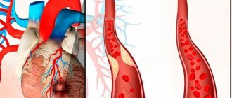
The wound surface of the skin is caused by a long-term inflammatory process of the affected blood vessel.
Impaired functioning of the valve apparatus of the vein leads to stagnation of blood. Over time, the wall becomes thinner, its elasticity is lost, and some of the plasma leaks into the surrounding tissue.
Lack of blood supply, prolonged deficiency of oxygen and nutrients leads to early death of certain areas of the muscle layer.
Externally, red, painful formations with gradual necrosis appear on the legs.
The pathological process can be stopped in the first stages of the disease. When trophic lesions occur, returning tissue to its normal state is almost impossible.
Folk remedies
In folk medicine, there are a large number of remedies based on medicinal plants that help in the treatment of varicose ulcers on the legs. But before using them, you should consult your doctor. This is necessary in order to exclude the possibility of complications. After all, not all folk remedies can be used on the open surface of a wound, since this results in rapid absorption of the drug components into the blood.
You need to be prepared for the fact that some medicinal products can cause allergic reactions. The main drugs in folk medicine that are indicated for use in case of varicose ulcers are:
- Aloe and Kalanchoe juice. Most often used in the form of compresses. Medicinal plants have antibacterial, anti-inflammatory and antiseptic properties.
- Honey and propolis. These products are famous for their unique healing properties. Honey contains a large amount of vitamins, microelements and other beneficial substances. Honey and propolis eliminate inflammation, relieve swelling, and nourish tissues. They can be used both for treating varicose ulcers and applying lotions, or added to ointments together with other components.
- Chicken and quail protein. It is an effective folk remedy for the treatment of ulcers. It completely covers the surface of the skin where the defect is located, thereby creating a protective film. Thanks to this, pathogenic bacteria do not enter the wound.
- Infusions and decoctions. Medicinal herbs such as chamomile, celandine, and calendula are used for preparation. These infusions have antiseptic, anti-inflammatory, analgesic, antibacterial, cleansing and decongestant properties. All components must be combined in equal parts. Method of preparation: one tablespoon of chopped herbs is brewed with 200 ml of boiling water and left for up to two hours. It is then strained and used as a lotion or to treat ulcers.
Causes
The occurrence of foci of violation of the integrity of the skin is due to the advanced stage of varicose veins of the lower extremities.
In addition, there are various risk factors that influence trophic changes:
- Associated vein pathologies: phlebitis, thrombosis.
- Hereditary predisposition.
- Lack of timely treatment or incorrectly selected therapy.
- Mechanical injuries of affected vessels.
- Impact of low temperatures on feet.
- Tendency to form gangrene of the legs due to endocrine disorders.
- Hypertension is a “provocateur” of the disease.
- Restriction in limb movement.
- Blood diseases.
- Alcohol abuse and other bad habits.
Despite the significant list of causes of TU, the basis is a violation of venous circulation.
The longer the patient does not receive treatment, the higher the likelihood of the disease progressing to advanced fifth stage, which is characterized by wound defects.
The essence of compression therapy
Compression treatment is carried out using an elastic bandage or therapeutic stocking, which can apply external pressure to the tissues of the lower leg, helping the functioning of the venous valves by improving regional blood flow. For bedridden patients, compression therapy is contraindicated.
The essence of the therapeutic effect is the elimination of the main cause of varicose veins. The main condition is continuity of treatment. If it is performed, then compression (special compression stockings or elastic bandages) has the following effect on the veins:
- normalizes the functioning of vein valves, thereby improving blood flow;
- removes excess blood volume that circulates in the vessels of the lower extremities;
- minimizes reverse blood flow in superficial and deep veins;
- reduces blood pressure in the veins of the legs.
At the same time, compression acts on the tissue, reducing pressure, inflammation, and stimulating regeneration.
Compression stockings and how to wear them
Therapeutic knitwear with a compression effect is divided into classes: from I to IV. Each class is effective in its own way, so the decision to wear stockings is made by the doctor, focusing on the level of venous insufficiency and the condition of the arteries of the legs. If the concomitant pathology is also arterial insufficiency, compression can provoke a deterioration in blood flow, nutrition and oxygen supply to the tissues of the legs.
Compression stockings are sold in specialized stores and are selected individually according to the doctor’s prescription, according to the sizes specified in the “prescription”. Putting on stockings, especially for the first time, is very difficult, so you need to try the process in the presence of the seller (maybe with his help), and, if necessary, purchase additional aids for pulling on the stocking. It is important to clarify the warranty period of the product.
When choosing compression stockings for the treatment of trophic disorders with varicose veins, you need to see the EU mark . This is a compression guarantee. In addition, you should check whether the upper edge of the stocking does not reach the groin (this is dangerous); if we are talking about knee socks, then the level of the socks should not be higher than two centimeters from the knee joint. Therapeutic stockings are fixed with a soft and wide edging, which does not allow them to go down. There may also be compression tights.
It is important to know that there are stockings on sale that are designed to prevent thrombosis; they are white and are not suitable for compression of trophic ulcers.
One of the reasons for patients’ reluctance to use compression to prevent relapses or treat ulcers with varicose veins is the difficulty of putting on stockings, tights and even knee socks. In fact, at the beginning of wearing the stockings are extremely tight, and the leg is swollen, so it is not possible to put the product on. But there are several aids to facilitate the putting on process: for example, a frame onto which the stocking is first pulled and then the leg is inserted into it. If the stocking is without a sock, use a bag with a sliding surface into which the foot is placed, and then pull the stocking by sliding. The bag is removed through the open toe of the stocking or knee socks. If the nose of the stocking is preserved, you need to turn the stocking inside out and gradually pull it up from the toes.
If all else fails, you can reduce swelling with compression bandages.
Compression bandages
Compression bandages are an alternative to stockings in the treatment of trophic ulcers with varicose veins: the products must be elastic with a width of more than 10 cm and a length of more than 7 meters. If the bandage irritates the skin, place a cotton bandage under it.
Put the bandage on the leg in the morning after waking up, while there is still no swelling on the leg. The dressing is done from the foot with as much pressure as possible, gradually rising up in a circle so that the next circle covers 2/3 of the previous one. The last circle is fixed with adhesive tape. The greatest compression should be in the lower third of the leg, and should decrease towards the heart.
Stages
There are 4 stages of the pathological process:
- During the first stage, pigmentation of a certain area of the skin occurs. On palpation, the area is painful with obvious hyperemia; as the pathological process develops, the area of skin acquires a lacquered sheen, and whitish, blurry spots form around it. At this stage, timely medical care helps prevent damage, since the nutrition of tissues and blood vessels is not completely impaired.
- At the second stage of the disease, primary disturbances in the nutrition of the deep layers of leg tissue occur. The patient has severe swelling of the affected limb, hyperemic areas merge into one enlarged spot. The integrity of the skin is compromised: peeling of the epidermal layer, increased redness and the formation of microcracks.
- The third stage is the stage of formation of the wound surface. Distinct wounds with clearly limited areas appear on the legs. The center of the wound is more pigmented, and the edges remain unchanged. Depending on the cause of the disease, the ulcerative defect differs in morphological structure. The speed of wound formation is influenced by provoking factors: weight, age and gender of the patient. Inflammation involves the subcutaneous fat layer, fiber and part of the muscles.
- The fourth stage occurs in two ways:
- If the patient has received assistance, the wound lesions are covered with connective tissue and scars appear. The process is influenced by many factors.
- In the absence of therapy, the wound deepens and new lesions form. A bacterial infection occurs and the patient risks losing a limb or even dying.
The stages of the disease make it possible to assess the extent of infection and damage to the legs in order to provide the correct treatment.
Possible complications
The addition of complications to varicose ulcerative defects sharply worsens the prognosis and can lead to amputation of the limb.
The course of a trophic ulcer can be complicated by:
- Sepsis.
- Varicose eczema.
- Gangrene.
- Varicose dermatitis.
- Erysipelas.
- Inflammatory pathology of the lower leg joint.
To prevent the development of complications, treatment should be carried out only in accordance with the recommendations of a highly specialized specialist!
Kinds
In addition to the stages of development of the disease, trophic wounds differ in morphological features and causes of pathology. Based on this, a classification of ulcers was created:
- Venous ulcers - trophic changes are caused by a malfunction of the valve system of the veins. Nutrient deficiency leads to cell death. According to statistics, venous wounds occupy the first place of trophic damage to the legs.
- Arterial type - the occurrence is associated with sclerotic diseases of the arteries. With this type, the wounds are localized mainly on the feet, toes or heels. Shapeless formations have different depths and diameters. Occur in most cases due to ischemic disorders.
- Neurotrophic type - caused by ischemia of nerve endings. The wound surface is distinguished by its depth and the presence of a “crater” of the ulcer. In advanced cases, fistulas form through which bone tissue can be seen.
The similarity of the types of wounds lies in the formation of fibrous purulent exudate with an unpleasant odor and particles of dead epithelium.
The more intense the pathological process, the higher the risk of infection of open wound surfaces with anaerobic microorganisms.
Surgery
The procedure is aimed at eliminating skin defects and the affected veins that provoked their appearance. In the first case, surgical manipulations are aimed at the most advanced cases that are not amenable to conservative treatment. In the second - to restore damaged blood vessels and prevent the appearance of ulcers.
The defect is eliminated by removing all necrotic tissue and sutured to the boundaries of healthy tissue, if the wound is not large.
We can talk about skin transplantation only for single, not deep defects.
If there are many ulcers, they have reached the level of the bone, the function of the limb is limited due to pain, intoxication and severe general condition, the question of amputation arises.
It will be more effective to eliminate damaged veins. There are several methods:
- Classic surgical with a scalpel (Phlebectomy). The method is very traumatic and technically complex. Patients after such manipulations need a long recovery period. It is used only in very complex cases when minimally invasive methods are ineffective and impractical.
- Stripping. Less traumatic and does not require special retraining or rehabilitation. The essence of the method is to invert and remove the vein using a probe that is inserted into its lumen.
- Microphlebectomy. Removal of damaged areas of the vessel through small holes.
- Laser coagulation. Using a laser knife, the doctor coagulates the vessel wall from the inside. Applicable only to small areas.
- Radiofrequency coagulation. It works similarly to the previous method, but can be used when larger branches and large areas are damaged.
Symptoms
The clinical picture of the disease depends on the stage and cause of the lesion. You can suspect something is wrong based on symptoms such as:
- swelling;
- soreness;
- burning and severe itching;
- feeling of frequent crawling “goosebumps”;
- convulsions;
- hyperpigmentation;
- chronic fatigue and heaviness in the legs.
There are specific signs indicating trophic disorders of the legs:
- increased swelling regardless of the time of day;
- formation of a pronounced capillary network;
- pain in the affected limb, which cannot be eliminated with standard analgesics;
- redness of the leg and the appearance of hyperemic areas;
- bulging vein;
- inability to walk normally, as when pressing on the foot, acute piercing pain occurs;
- joint pain;
- increase in body temperature.
In advanced cases, the skin ruptures, bleeding occurs, and infection cannot be ruled out.
The wounds become wet, the edges become dark burgundy, and the risk of necrosis or gangrene increases.
Intoxication and an increase in the volume of cellular breakdown products leads to loss of appetite and lethargy.
When and which doctor should you contact?
Phlebologists treat pathology, but often with trophic formations of the extremities they turn to dermatologists for help. Only a doctor is able to correctly diagnose the disease and establish the causes of trophic damage.
In order for the treatment to bring greater benefits, it is not advisable to postpone a visit to the doctor.
If the patient is prone to varicose veins or suffers from severe vasculitis or atopic dermatitis, visits to the doctor should be frequent.
Reviews
Reviews from patients report that varicose ulcers often manifest themselves in the absence of proper control over the course of the disease. You can get rid of it if you follow the rules described by the doctor, but then the patient must follow the instructions to ensure the prevention of relapse.
I have had varicose veins for a long time, but a year ago I noticed the first signs of trophic changes and immediately consulted a doctor. It turned out that it was an ulcer that was dangerous for my life - there was a risk of blood poisoning. I had surgery and the next day I was home. Then 2 weeks of dressings, now this problem is gone.
I work as a flight attendant, which means my legs are constantly under stress. Now I have advanced varicose veins, because I never have time for examination and rest. At the last appointment, the doctor said that there were changes for the worse. You will have to quit your job and focus on your health.
I have diabetes, varicose veins of the lower extremities and many vascular problems. That week, an examination revealed an ulcerative lesion. Fortunately, it can still be treated without surgery. I take medications, use ointments, and now I am choosing a traditional therapy for myself.
Diagnostics
To establish a diagnosis, the following diagnostic procedures are used:
- duplex scanning of veins;
- Ultrasound of the affected limb;
- CT or MRI;
- angiography and Doppler;
- visual inspection;
- laboratory blood test (general and biochemical analysis).
Often, a scraping is taken from the wound area for cytological examination (accurate determination of the stage of the pathological process) and identification of possible contamination with microbes (allows us to exclude bacterial infection).
Examples of patients with venous ulcers
The photographs show venous ulcers of patients we cured at the time of admission to the clinic and upon discharge.
Patient N., 24 years old Diagnosis: Varicose veins of the left lower limb
Patient A., 66 years old Diagnosis: Postthrombotic disease of the left lower limb
We examine all patients with venous ulcers for concomitant arterial pathology. And if it is present, we determine the ankle-brachial index. This is important for prescribing adequate compression therapy. Here is an example of such a patient.
- The head of the phlebology department of our clinic has 18 years of experience in treating lower extremity ulcers. During this time, he managed to cure more than 1000 patients with severe trophic leg ulcers of various origins.
Vascular surgeon, phlebologist, candidate of medical sciences, member of the European Society of Vascular Surgery.
He completed an internship in the best clinics in Western Europe: six months in Belgium - at the University Hospital of Brussels, a year in Milan, at a leading clinic for vascular surgery.
Author of more than 60 scientific papers and 5 Russian patents.
Treatment methods
There are two types of therapy: conservative (drug) and operative (surgical).
Conservative treatment includes the use of general and local pharmaceutical drugs. Patients are shown:
- ointments, creams and compresses that accelerate regenerative processes and prevent inflammation;
- ointments with an antiseptic effect and solutions for removing dead epithelial particles;
- antibacterial creams for external use;
- tablets are necessary to stimulate local immunity;
- antioxidants and multivitamin complexes;
- blood thinners and metabolites;
- venotonics to maintain the tone of the vascular wall.
Symptomatic treatment is carried out with anti-inflammatory, analgesic and antipyretic drugs.
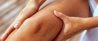
This same treatment group includes physiotherapy, massage, therapeutic exercises and hirudotherapy.
Surgery
It is possible to eliminate trophic changes using minimally invasive and abdominal operations.
There are 3 surgical methods: laser coagulation, sclerotherapy and removal of the affected vessel.
It is important to note that after removing the cause of the disease, restorative measures are necessary.
The ulcers are washed with special solutions, sterile bandages are applied, and sometimes they are tamponed.
Some doctors, after surgical treatment, stitch wounds, accelerating the regenerative process.
Traditional methods
Traditional treatment involves the use of compresses. A decoction of a mixture of medicinal herbs (chamomile, string) is applied to the wound surface, the main thing is that the components do not contain traces of alcohol.
For wet ulcerative processes, propolis and ointments based on birch tar are used.
Before applying an unconventional topical remedy, the wound surface should be washed and dried.
To reduce adverse effects, discuss therapeutic actions with your doctor.
The effectiveness of alternative treatment depends on the stage and cause of the disease.
Unconventional therapy is combined with surgical and medicinal methods for the treatment of lower extremity TU.
Physiotherapy
Treatment with physiotherapeutic procedures is considered an auxiliary method. These include the following types of therapy:
- Magnetotherapy. Improves microcirculation and metabolic processes, which allows tissues to recover faster.
- Laser irradiation. Carried out after mechanical cleaning, it reduces the risk of pathology progression.
- Infrared irradiation. Accelerates the healing process.
Physiotherapy is used in courses that include 10-15 procedures. The duration of exposure is determined by the patient individually and ranges from three to ten minutes.
Baths are considered an effective method; they can be air, sea, ozone and sodium chloride.
Prevention
To prevent trophic ulcers against the background of varicose veins, pathologies of blood vessels should be treated in a timely manner and the condition of the veins and arteries should be monitored.
It is important to reconsider your lifestyle, give up bad habits and give preference to proper nutrition.
If there are systemic diseases, take medications and monitor blood glucose levels or blood pressure readings.
At the first signs of varicose veins, do not self-medicate, but seek qualified help.
If varicose veins have progressed to the stage of formation of trophic ulcers, you should contact a vascular surgeon for medical help as soon as possible, and then strictly follow medical recommendations and prescriptions.
What should you be wary of before a peptic ulcer appears?
A trophic ulcer is a serious complication of varicose veins; its appearance is preceded by a number of signs characteristic of varicose veins:
- Feeling of heaviness and discomfort in the lower extremities, especially after physical activity.
- Skin itching.
- Pastyness of the lower extremities that does not go away after a night's rest, a mark from the sock elastic.
- Decreased sensitivity in the feet and paresthesia in the form of coldness and “pins and needles.”
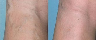
- Visualization of altered veins; with varicose veins, they become veiny, tortuous, and bluish.
- The initial manifestation of varicose veins is the appearance of a vascular capillary network on the skin.
- Cramps in the calf muscles, especially at night.
- Disappearance of skin patterns, cessation of hair growth due to the death of hair follicles.
- The appearance of pigmentation on the skin.