Inflammation of the heart muscle, occurring with severe rhythm disturbances, thromboembolic complications, severe circulatory decompensation and an irreversible increase in heart size, is named after the scientists who described this disease - Abramov-Fiedler myocarditis. Its exact cause is not known, but an autoimmune process is suspected. It most often affects young people.
The prognosis in most cases is unfavorable. A heart transplant may be required to save the patient's life.
Why does Abramov’s myocarditis develop?
Despite the fact that the disease was first described 130 years ago, the exact etiological factor has not been discovered to date. Therefore, one of the definitions of this pathology is idiopathic, that is, of unknown origin. Since the onset of myocarditis in many patients was preceded by an allergic reaction to medications, blood components, vaccines, an autoimmune process in the myocardium is considered one of the main causes.
A viral infection may be a trigger for the formation of antibodies against the heart's own cells. Every fifth patient has autoimmune lesions of blood vessels, thyroid gland, and intestines.
We recommend reading about chronic myocarditis. You will learn about the etiology and symptoms of the disease, its diagnosis, treatment principles, and doctors’ prognoses. And here is more information about the symptoms and treatment of viral myocarditis.
Etiology
Damage to the heart muscle during myocarditis can be caused by direct exposure to infection, parasitic infections, chemical and physical effects, and allergic damage to the myocardium. The leading role in the occurrence of myocarditis belongs to infectious diseases. The most common cause of myocarditis in recent times is viruses, which is associated with the spread of epidemics of viral respiratory tract infections in large populated areas, a decrease in natural immunity, and improved diagnostics to identify pathogens. Most often, myocarditis is caused by enteroviruses Coxsackie A and B, ECHO. In 20% of cases, the course of myocarditis caused by the Coxsackie B virus becomes chronic.
Myocarditis of bacterial origin takes 4th place, inferior not only to viral, but even parasitic lesions of the heart muscle in some regions. Streptococcal myocarditis accounts for no more than 5%. In this case, heart damage is caused either by the direct effect of bacteria on the myocardium, or by an infectious-allergic mechanism. Carditis can be caused by toxoplasma infection (especially congenital forms). Fungal myocarditis develops in patients with chronic diseases during massive antibacterial therapy. Allergic myocarditis (drug-induced, serum-related, post-vaccination) and myocarditis due to collagenosis are recorded. One should not lose sight of a possible hereditary predisposition to this disease.
Abramov-Fiedler classification of disease
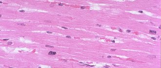
When examining the muscle tissue of the heart, the following changes are detected:
- dystrophy, flabbiness of the walls and overstretching of the chambers of the heart;
- thrombus formation in cavities;
- infiltration (impregnation) of myocardial leukocytes;
- extensive inflammatory process with replacement of myocytes by connective tissue;
- enlarged muscle fibers;
- the branches of the coronary arteries are inflamed throughout.
Dilatation of the wall of the left ventricle with focal fibrosis
Depending on which of these processes predominates, the following variants of the course of myocarditis are distinguished: destructive-dystrophic, with inflammatory infiltration, vascular and mixed. The first type of disease is accompanied by a malnutrition of cells (dystrophy), which leads to their destruction and the development of inactive connective tissue.
In the inflammatory-infiltrative form, the myocardium becomes edematous, accumulations of leukocytes (neutrophils and eosinophils) are noted, which are then replaced by giant cells with many nuclei.
With the vascular (vascular) variety, the coronary vessels of the heart are predominantly affected, and especially their small branches. The mixed form suggests the presence of signs of other variants of the disease.
The course of the disease can be:
- acute – from 14 days to 2 months;
- subacute – from 90 days to 1.5 years;
- chronic – over 18 months.
Conditional division according to the severity of clinical symptoms made it possible to identify the following forms of idiopathic myocarditis: dominance of asystole, arrhythmia, thromboembolism, pseudocoronary and combined.
Pathogenesis and pathological anatomy
The mechanisms of myocarditis development are due to the direct impact of the occurrence factor on the heart muscle, as well as their combination with immune disorders. With a decrease in the immunological reactivity of the body, the infection from the primary focus enters the bloodstream and spreads to various organs and tissues, where foci of secondary reproduction of the infectious agent are formed. The myocardium is most often affected. In this case, the pathogen has a direct damaging effect on the heart muscle, being a trigger for the development of the inflammatory process. In response to damage, special substances are released with the subsequent development of a hypersensitivity reaction. This increases the permeability of the vascular wall and damages the vascular walls.
Autoallergy in this process is one of the stages of the pathological process. In the chronic course of myocarditis, the pathogen does not play a significant role, and the disease is based on disorders of the immune system. All of these mechanisms lead to systemic myocardial damage, a malignant and relapsing course of the disease, resistant to traditional treatment.
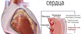
Changes in myocarditis are characterized by an increase in the mass and size of the heart, the thickness of the ventricles, the presence of focal or widespread replacement with connective tissue in the form of scars in the myocardium, and parietal thrombi.
Symptoms of idiopathic myocarditis
The acute form of myocarditis is characterized by a rapid increase in cardiac decompensation, mainly of the right ventricle, which quickly gives way to total decompensation. Main features:
- shortness of breath, worsening when lying down;
- fever;
- cyanotic skin tone;
- drop in blood pressure;
- frequent and irregular heartbeats;
- enlarged liver, spleen;
- swelling and fluid accumulation in the abdominal cavity;
- coughing attacks;
- pain in the heart like angina pectoris.
Sometimes the leading symptom may be thrombosis of blood vessels in the lungs, brain, kidneys, and spleen.
The fulminant form of the disease is always fatal; a latent variant of the course with a sudden stop of cardiac activity also occurs. With a long course, an increase in the area of cardiosclerosis, expansion of the cavities of the heart and an increase in circulatory failure are noted.
Clinical picture
Based on the occurrence of myocarditis, they are divided into viral (influenza (see), Coxsackie infection, ECHO, etc.), bacterial (scarlet fever (see), diphtheria (see), tuberculosis (see), typhoid fever (see)) , spirochetosis (syphilis (see), leptospirosis (see), relapsing fever (see)), rickettsial (typhus (see), Ku fever (see)), parasitic (toxoplasmosis (see), Chagas disease , trichinosis (see)), fungal (actinomycosis (see), candidiasis (see), coccidioidomycosis, aspergillosis (see)), infectious-allergic, idiopathic, medicinal, serum, nutritional, myocarditis in systemic connective tissue diseases ( systemic lupus erythematosus (see), scleroderma (see)), bronchial asthma (see), Lyell's syndrome (see), Goodpasture's syndrome (see), burns, transplantation, thyrotoxic, uremic, alcohol.
Based on pathogenetic characteristics, there are 3 forms of myocarditis: infectious and infectious-toxic, allergic (immune), toxic-allergic. According to the prevalence of the pathological process, myocarditis is divided into focal and widespread. According to the nature of the course, myocarditis can be acute and subacute, abortive, recurrent, chronic (latent).
Depending on the severity of clinical manifestations, oligosymptomatic myocarditis, pseudocoronary, decompensation (acute or chronic heart failure), arrhythmic, thromboembolic, pseudovalvular, mixed are distinguished.
Clinical and anatomical types of myocarditis can be systematized as follows: parenchymal, dystrophic, inflammatory-infiltrative, necrotic destructive, giant cell, mixed.
The first manifestations of myocarditis may be fatigue, increased sweating, increased body temperature, heart pain, palpitations and irregular heartbeats, shortness of breath with light exertion and even at rest. Persistent pain in the heart area is observed in almost all patients. The pain is constant, dull, and does not change in intensity with physical activity, negative emotions, or after taking coronary lytics. Often the above symptoms appear against the background of an acute respiratory viral infection. After some time, the patient periodically develops bouts of cyanosis, shortness of breath increases, wheezing is heard in the lungs, and a rapid heartbeat is noted. Urine output decreases, tissue swelling is detected, and the size of the liver increases (signs of right ventricular failure). The borders of the heart are moderately expanded. In patients with damage to the conduction system, heart sounds are more frequent than normal sonority. Disturbance of rhythmic activity during listening is defined as palpitations, tachyarrhythmia, bradycardia and bradyarrhythmia. The involvement of the cardiac conduction system in the process is indicated by persistent attacks of paroxysmal tachycardia (see). Cardiosclerosis of the atrioventricular junction leads to the constant functioning of the previously hidden bundle of Kent, which is manifested on the ECG by attacks of paroxysmal tachycardia. Tachyarrhythmias in non-rheumatic carditis can be persistent or transient, resulting from myocardial damage in combination with encephalogenic influences. With bradycardia, the heart rate ranges from 30 to 60 per minute. When the pulse is from 30 to 40, the patient may experience Morgagni-Adams-Stokes attacks. Heart failure is expressed in almost all patients with non-rheumatic myocarditis, but when the conduction system of the heart is damaged, its manifestations are minimal.
The ECG shows characteristic changes. On a chest x-ray, there is an increase in the pulmonary pattern with overflow of the venous bed (manifestation of left ventricular failure), a moderate increase in the size of the heart, the configuration of which, in an unfavorable course, becomes spherical within 1-1.5 months. Inflammatory changes are observed in the blood: an increase in the number of leukocytes, an increase in ESR.
Symptoms of myocarditis can be expressed to varying degrees, which makes it necessary to distinguish between mild, moderate and severe diseases.
Mild course (mild myocarditis): the general condition suffers slightly, but patients present a large number of complaints (heart pain, rapid heartbeat, elevated body temperature). The course is of moderate severity (moderate myocarditis): general health noticeably suffers, shortness of breath is pronounced, and severe weakness is observed. Symptoms of heart damage come to the fore and are accompanied by an increase in heart size, rhythm and conduction disturbances.
Severe course (pronounced myocarditis): the general condition of the patient is severe, complete heart failure is determined. Myocarditis can have an acute, subacute and chronic course. In acute cases, cardiac symptoms come to the fore in the clinical picture of the disease. The reverse development of clinical and instrumental data occurs within 6-18 months from the onset of the disease. In 50-60% of cases, complete recovery is recorded.
Subacute myocarditis is characterized by a gradual increase in heart failure over 4-6 months. In the acute phase of the disease, cardiac symptoms are pronounced, but gradually subside. Prolonged heart failure (despite treatment) and other signs are noted. In children, a heart hump develops. On the ECG, changes are characterized by a constant rhythm, deviation of the heart axis to the left, disruption of atrioventricular and intraventricular conduction, overload of the left ventricle and both atria (more than the left). Radiologically, a moderate increase in the pulmonary pattern along the venous bed and a change in the configuration of the cardiac shadow (trapezoidal or with an elongated left ventricle) are determined.
Chronic myocarditis is more often recorded in adults and older children. Often there is the presence of chronic inflammatory diseases of other organs (kidneys, lungs, nervous system). Chronic myocarditis can be primarily chronic or develop from acute and subacute myocarditis.
The disease is based on a significant change in the myocardium with the development of cardiosclerosis, enlargement of the myocardium and a decrease in the contractile function of the heart muscle. The course of chronic myocarditis is often characterized by a relative predominance of extracardiac manifestations: recurrent pneumonia (see), enlarged liver, attacks of loss of consciousness, fatigue, decreased performance, and in children there is a lag in physical development. Often, colds are provoking factors that intensify cardiac symptoms and lead to a deterioration in the patient’s condition.
Myocarditis idiopathic
Idiopathic Abramov-Fiedler myocarditis is currently considered as a multi-cause disease from the group of severe, irreversible forms of non-coronarogenic myocardial diseases. There are 6 clinical variants of the disease: decompensated, arrhythmic, infarction-like, thromboembolic, pseudovalvular, mixed. The age of patients is very different - from the first months of life to 70 years and older. The clinical picture is characterized by the sudden or gradual development of progressive heart failure of the total or right ventricular type. Patients complain of shortness of breath, sometimes paroxysmal in nature at night, cough (possibly hemoptysis), weakness, palpitations, chest pain of varying intensity. Body temperature is normal. The heart is enlarged. The tones are muffled, the third tone is often recorded when the dilated ventricle is filled, and an atrial gallop rhythm is noted as an early sign that appears before the development of circulatory failure. In some cases, a pericardial friction rub is heard, and in severe cases, embryocardia and alternating pulse are heard. Emboli may be the first manifestation of the disease. At the end of the disease, acute renal failure may develop.
Thyrotoxic myocarditis
Typical complaints of patients with thyrotoxic myocarditis are palpitations, shortness of breath, and constant rapid heartbeat (up to 90-140 beats per minute). Pain in the heart with this myocarditis is recorded much less frequently. An increase in the size of the heart is observed in severe thyrotoxicosis and is due to its expansion: the heart takes on a spherical shape, the arch of the pulmonary artery bulges, and the cardiac waist is smoothed out. When listening to thyrotoxic myocarditis, amplified or normal heart sounds are heard, the murmur is most pronounced along the left edge of the sternum. The ECG records nonspecific changes, the severity of which does not correspond to the severity of thyrotoxicosis. Characteristic is an increase in the functional activity of the myocardium.
The effects of thyroid hormones cause acceleration of blood flow, an increase in circulating blood volume and cardiac output due to increased heart rate or increased stroke volume, and a compensatory decrease in peripheral resistance as a result of vasodilation. Rhythm and conduction disturbances are represented by sinus tachycardia. The severity of tachycardia depends on the severity of thyrotoxicosis and inappropriately increases in response to physical activity. A characteristic symptom is the absence or weak response of the heart rate to the action of digitalis drugs (digitalis drugs). With thyrotoxic myocarditis, sick sinus syndrome may occur, which leads to paroxysmal tachycardia and paroxysmal atrial fibrillation. The second most common rhythm disorder is atrial fibrillation. Congestive circulatory failure in patients with thyrotoxic myocarditis is rare and occurs in the absence of adequate treatment.
Uremic myocarditis
Features of the clinical course and severity of uremic myocarditis (in chronic renal failure) are caused by persistent hypertension (see), enlargement of the left ventricle, heart failure, progressive anemia, disturbances in electrolyte balance and acid-base balance. Patients complain of severe weakness, pressing pain in the heart area, which intensifies in a horizontal position. Shortness of breath at the onset of the disease is recorded during physical activity, but quickly increases and occurs at rest, manifesting itself in attacks of suffocation. The condition of patients is usually severe, and there is lethargy. The skin is pale with a jaundiced or sallow tint; a characteristic symptom is a puffy face. An objective examination of the patient reveals severe tachycardia. Pulse of low filling and tension. The size of the heart is often significantly increased - the left border can reach the axillary line. The apical impulse is diffuse and intensified. Heart sounds are muffled, in some cases a pericardial friction noise is heard, which is determined by palpation and drowns out the heart sounds when listening. Symptoms of heart failure with this myocarditis may prevail over the manifestations of renal failure. Changes in the ECG are characterized by certain changes. In uremic myocarditis, they are characterized by pronounced sensitivity when using digitalis drugs.
Diphtheria myocarditis
The development of inflammatory damage to the heart muscle in diphtheria (see) is caused by the circulation of the toxin in the blood. Myocarditis can be early and late. Late myocarditis, which occurs in the 2-3rd week from the onset of the disease, has a more favorable course. The effect of diphtheria toxin on the myocardium leads to changes in the contractile myocardium and disorders of the conduction system of the heart. Conduction disturbances are the most common manifestation of diphtheria myocarditis and are accompanied by sinoauricular, intraatrial, atrioventricular and intraventricular blockades. With this disease, the symptoms of myocarditis are often combined with signs of severe vascular insufficiency, especially with early complications of diphtheria.
Myocarditis in typhoid fever
Inflammation of the heart muscle during typhoid fever is often combined with myocardial dystrophy and is observed as a result of inadequate therapy in the 2-4th week from the onset of the disease. Changes in the myocardium are characterized by weakness of contractile function and impaired tonic function of the heart muscle.
By the end of the 3rd week from the onset of the disease or 1-2 weeks after suffering from typhoid fever, the patient exhibits a pronounced rapid heartbeat (100-140 beats per minute), dullness of heart sounds, splitting of the first tone, expansion of the borders of the heart, and various rhythm disturbances. Extrasystole in typhoid myocarditis is rarely recorded, atrial fibrillation and complete atrioventricular block are almost not observed.
Myocarditis syphilitic
Syphilitic myocarditis is often combined with syphilitic aortitis. Chronic syphilitic myocarditis manifests itself in gummous and fibrous forms. With widespread syphilitic myocarditis with damage to the sinus node and the ventricular conduction system, combined rhythm and conduction disturbances are observed. With syphilitic lesions of the ascending aortic arch, anginal syndrome appears in the clinic. This form of myocarditis is characterized by a chronic course. Patients with syphilitic myocarditis in stages II and III of the disease often develop heart failure of the right ventricular type, which is a prognostically unfavorable sign.
Tuberculous myocarditis
The course of tuberculous myocarditis initially takes a protracted or chronic course. The effect of tuberculosis toxin on the myocardium at the onset of the disease is manifested by rapid heartbeat. Later, patients complain of palpitations, pain in the heart, a feeling of fullness and pressure in the chest, and loss of heartbeats. The borders of the heart are enlarged in diameter, with predominant damage to the right parts. This may be facilitated by hypertension in the pulmonary circulation. The apex beat is weakened and does not correspond to the filling of the pulse, the first tone is muffled, and the second tone is accentuated. With tuberculous myocarditis, a decrease in heart rate (usually in later stages of the disease) and a decrease in blood pressure may be observed, which is due to the course of the tuberculous process. Rhythm disturbances are observed in the form of extrasystolic arrhythmia and paroxysmal tachycardia. Conduction disturbances are recorded in all parts of the intramural nervous system of the heart. Characteristic symptoms are a decrease in myocardial contractility and manifestations of heart failure.
Myocarditis rickettsial
The course of rickettsial myocarditis can be acute and protracted and is accompanied by widespread damage to the heart muscle. Already at the beginning of the underlying disease, heart sounds become muffled, rhythm disturbances such as extrasystole and atrial fibrillation are recorded. In chronic rickettsial myocarditis, myocardial damage occurs as a type of cardiosclerosis (see) in combination with myocardial dystrophy (see). Thromboembolic complications and the formation of an aneurysm (see) of the heart in the apical region are possible. The progression of chronic rickettsial myocarditis often ends in death due to complete heart failure.
Myocarditis rheumatic
Rheumatic heart disease in most cases is characterized by involvement of all the membranes of the heart. With isolated myocardial damage, patients complain of pain in the heart. A characteristic sign is expansion of the predominantly left border of the heart. Listening also reveals characteristic changes. The ECG records a violation of the contractile function of the myocardium, repolarization processes, intraventricular conduction, and sinus arrhythmia. Similar heart damage is observed in subacute and primary protracted course of rheumatism.
Diagnosis of heart problems
Examination data and instrumental diagnostic methods reveal the following signs:
- swelling, enlarged liver, accumulation of fluid in the abdominal cavity;
- pulse weak, irregular, tachycardia;
- Auscultation – at the apex of the heart there is a systolic murmur, the rhythm resembles a gallop, the tones are dull; fine wheezing in the lungs;
- blood test - leukocytosis, high levels of ESR, C-reactive protein, immunoglobulins, fibrin, troponin, creatine phosphokinase activity above normal;
- the immunological profile does not reveal markers of the rheumatic process (differential diagnosis with systemic collagenoses), with repeated studies an increase in the titer of antibodies to myocardial cells is noted;
- X-ray - the heart is significantly larger than normal, there are signs of congestion in the lungs;
- echocardiography – fluid in the pericardium, dilation of the heart chambers, wall hypertrophy, parietal thrombi;
- ECG - atrial fibrillation, fibrillation, blockade of impulses, changes similar to infarction;
- coronary angiography – myocardial ischemia;
- cardiac biopsy – inflammatory infiltration of leukocytes.
Symptoms and signs
The clinical picture usually appears several weeks after the infection. It is important to exclude other heart diseases and non-cardiological pathologies occurring against the background of angina pectoris and hypertension. The patient may complain of:
- hyperthermia - a sharp increase in temperature;
- muscle pain, which is explained by concomitant inflammation of the skeletal muscles.
- interruptions in heart function;
- shortness of breath, which “lurks” a person at rest or with minimal physical activity;
- chest pain, for which taking Nitroglycerin does not bring relief;
- general weakness and sweating;
- cough, sometimes with hemoptysis - this indicates a complication of myocarditis, pulmonary embolism, pulmonary infarction and peri-infarction pneumonia.
Myocarditis can cause circulatory failure - acute (develops within two weeks) or chronic (phenomena increase gradually, over 3 months). If the myocardium of the left ventricle is damaged (left ventricular failure), the patient develops signs of stagnation in the pulmonary circulation:
- moist rales in the lungs on auscultation;
- shortness of breath at rest;
- attacks of suffocation.
When the function of the right ventricle deteriorates (right ventricular failure), swelling of the neck veins, swelling of the extremities, and enlargement of the liver appear. The clinical picture depends on the degree of damage, the activity of the inflammatory process, and the leading symptom.
Sometimes sudden death due to ventricular fibrillation may be the first and only manifestation.
Features in children
Pediatricians distinguish between congenital and acquired myocarditis. Clinical manifestations in children depend on age. They are often mistakenly interpreted as symptoms of other diseases - pneumonia, obstructive bronchitis, gastroenterocolitis.
In the neonatal period, myocarditis is characterized by a severe course. May be observed:
- breast refusal;
- shortness of breath when sucking;
- vomiting and regurgitation syndrome;
- pallor;
- swelling of the eyelids;
- apnea attacks;
- tachycardia;
- cough;
- noisy exhalation;
- retraction of intercostal spaces during inspiration;
- wheezing in the lungs on auscultation.
In preschoolers and primary schoolchildren, myocarditis can manifest itself as vomiting, abdominal pain, and hepatomegaly. In high school students – excessive weakness, increased breathing, and fainting.
Changes in the muscular structure of the heart during myocarditis
Treatment of Abramov-Fiedler myocarditis
Since the cause of this disease cannot be determined, therapy is only symptomatic. Strict bed rest and a light diet with limited animal fats, table salt and liquid are prescribed. The difficulty in treating myocarditis is that sensitivity to many drugs is reduced.
Main groups of medications:
- cardiac glycosides (Celanid, Strophanthin);
- diuretics (Britomar, Arifon retard, Trifas);
- blockers of angiotensin-converting enzyme (Enalapril, Capoten) and beta-adrenergic receptors (Bisoprolol, Egilok);
- long-acting nitrates (Kardiket, Izo-mac);
- aldosterone receptor antagonists (Aldactone, Veroshpiron);
- anticoagulants (Warfarin);
- non-steroidal anti-inflammatory drugs (Metindol, Voltaren).
When inflammation is highly active, hormones are used - Prednisolone, Metypred. In addition to basic medications, Riboxin, Panangin, and vitamin complexes are used. Transplantation is indicated for malignant disease, especially for giant cell myocarditis.
We recommend reading about what an ECG will show for myocarditis. You will learn about electrocardiograms for infectious, idiopathic, and rheumatic myocarditis. And here is more information about rheumatic myocarditis.
Myocarditis in children: how it manifests itself and how it is treated
More often, this disease is recorded in children 4-5 years old, and boys are the leaders in this regard. Myocarditis may also be diagnosed in adolescents.
The cause of such myocarditis can be any infection, but the virus dominates the list of pathogens. And most often, childhood myocarditis is caused by adenoviruses, influenza viruses, and Coxsackie enteroviruses. If we talk about bacterial pathogens, then most often the disease provokes rheumatism, as well as such “half-forgotten” diseases as diphtheria and scarlet fever.
Inflammation of the thick layer of the heart is also noted in patients during allergic reactions, under the influence of various toxic agents. There is also a congenital variant, which develops in a baby in utero if his mother becomes infected during gestation. Autoimmune myocarditis is a disease in which the body produces antibodies to its own cardiomyocytes, and they deform the myocardium.
Myocarditis in children
Prognosis for the patient
The rapid progression of cardiac decompensation, severe forms of arrhythmia and thromboembolic blockage of blood vessels in the lungs, kidneys and brain often leads to death. In acute cases, patients die within a few
days, the subacute form lasts up to six months.
A long-term and latent disease is accompanied by irreversible processes of hemodynamic disturbances and expansion of the heart with a decrease in contractility.
And Abramov-Fiedler diopathic myocarditis is a disease with an unfavorable prognosis and an incompletely studied cause . The main symptoms reflect the progression of heart failure, disturbances in the formation and conduction of electrical impulses in the heart, decreased coronary blood flow and thromboembolic complications. Treatment is symptomatic, and heart transplantation may be required.
Diet for illness
The essence of dietary nutrition comes down to a competent choice of products, which allows the body to obtain a supply of energy and plastic material.
Diet for myocarditis - recommendations:
- Raw vegetables are healthy - salads of tomatoes, cabbage and cucumbers, seasoned with olive oil;
- Prepare food without salt, but already prepared food can be slightly salted;
- You can eat soups, ideally vegetarian;
- 150 g of bread is the maximum norm per day;
- 30 g of vegetable oil is also the limit, but do not fry or stew with it, but make salads;
- No more than 20 g of butter per day, and only in the morning;
- The total daily fat intake is 80 g, taking into account the fact that 50% of this mass is already included in prepared foods.
Raw vegetables
The patient will not go hungry, since the diet involves split meals, five to six times a day.
Alcohol, fast food, confectionery products, canned food and marinades, spicy, salty and spicy foods are excluded.
Myocarditis caused by Toxoplasma
This rare form of protozoal myocardial damage most often develops in young people with weakened immune systems. Congenital toxoplasmosis is more common, but myocarditis is not a distinguishing feature. Myocardial damage can lead to cardiac dilatation, pericarditis, and the appearance of pericardial effusion. Heart failure, arrhythmias, and conduction disturbances may occur. This disease is difficult to diagnose, so its true incidence in clinical practice is apparently higher than is generally believed. Treatment is with chloridine and sulfonamide, but their effectiveness is highly variable.