Causes and mechanisms of disease development
Doctors divide encephalitis in children into two large groups: primary and secondary. The characteristics of these groups are presented in the table:
| Characteristics of encephalitis in children | ||
| Primary | Secondary | |
| Development mechanism | The pathogen, entering the human body, primarily affects brain cells. It is an independent disease. | It occurs as a complication of an existing disease. |
| Reasons for development |
|
|
Brain development from conception to adolescence
When my first child was born, I, as befits a zealous but young mother, collected a bunch of books about caring for babies and various progressive methods of education - so that my child would grow up to be a genius, and a happy one at that, I desperately needed authoritative advice. Unfortunately, it quickly became clear that most books were not particularly interested in explaining the biological basis of brain development. Let's try to figure out what science knows about the brain today and how modern pedagogy uses this knowledge.
Brain and its development
What is interesting in the development of the brain and what we, in fact, will observe at each stage of such development is the enormous interaction of genetically predetermined factors and environmental factors, which in the case of human development become factors of the social environment.
Embryonic development
In the human embryo, the brain begins to form from embryonic ectoderm tissue. Already on the 16th day of intrauterine development, the so-called neuronal plate can be distinguished, which over the next few days forms a groove, the upper edges of which fuse and form a tube. This process is the result of complex coordinated work of a number of genes and depends on the presence of certain signaling substances, in particular folic acid. A deficiency of this vitamin during pregnancy results in failure of the neural tube to close, leading to severe abnormalities in the development of the child's brain. Once the neural tube has closed, three major brain regions form at its anterior end: anterior, middle, and posterior. During the seventh week of development, these regions divide again, a process called encephalization. This process is the formal beginning of the development of the brain itself. The growth rate of the fetal brain is amazing: 250,000 new neurons are formed every minute! Millions of connections are formed between them! Each cell occupies its specific place, each connection is neatly organized. There is no room for arbitrariness and chance.
The fetus develops different sense organs. Peter Hepper writes about this in detail in the article Unravelling our beginnings: The first reaction to touch appears is tactile sensitivity. At the eighth week, the fetus reacts to touching the lips and cheeks. At week 14, the fetus responds to touching other parts of the body. The next thing to develop is taste - already at 12 weeks the fetus feels the taste of amniotic fluid and can react to the mother's diet. The fetus reacts to sound starting from 22-24 weeks of life. At first it picks up low-range sounds, but gradually the range expands, and already before birth the fetus recognizes different voices and even distinguishes between individual sounds. The uterine environment, where the fetus develops, is quite noisy: the heart beats here, the flow of fluids and peristalsis is noisy, various sounds come from the external environment, although muffled by the mother’s tissues, however - interestingly - the range of the human voice of 125-250 Hz is just weakly muffled . Consequently, outside conversations form the majority of the fetal sound environment.
The reaction to pain attracts special attention from researchers. Determining whether the fetus feels pain is difficult - pain is largely a subjective phenomenon. However, the unconscious response to painful stimuli begins around 24-26 weeks of development, when the neuronal response pathway is first formed. From the moment the first sense organs develop, information begins to flow from them to the brain, which in itself acts as a factor in the development of the same brain and leads to learning. The question arises, how important is the information obtained in this way and can we influence the fetus in a certain way, encouraging the brain to develop and promote learning? The fruit can learn to recognize taste and smell. For example, if a mother consumes garlic during pregnancy, the newborn infant will show less aversion to the smell of garlic than an infant whose mother did not consume garlic. Newborn babies will also favor music they heard in the womb over music they hear for the first time. All this has already been established by science. But it is still unclear whether the phenomenon of prenatal learning has at least any lasting effect. It is known that the “musical taste” for a certain piece, in the absence of reinforcement, disappears within three weeks. However, the fetus's ability to "learn" has led some people to believe that fetal brain development may be stimulated by a prenatal stimulation program. However, there are no solid scientific studies about this.
Newborn brain
At the time of birth, the baby's brain has virtually all the necessary neurons. But the brain continues to grow rapidly and over the next two years reaches 80% of the adult brain size. What happens during these two or three years? The main increase in brain weight occurs due to glial cells, which are 50 times more numerous than neurons. Glial cells do not transmit nerve impulses, as neurons do; they ensure the vital activity of neurons: some of them supply nutrients, others digest and destroy dead neurons or physically hold neurons in a certain position and form the myelin sheath. From the moment of birth, the baby’s brain receives a huge number of signals from all senses. The baby's brain is more open to the modeling hand of experience than at any other time in a person's life. In response to the demands of the environment, the brain is a sculptor of itself.
Vision and the brain
Understanding the peculiarities of the formation of the visual cortex began with the famous experiments of David Hubel and Torsten Wiesel in the 60s of the last century. They demonstrated that if kittens temporarily close one eye during a certain critical period for brain development, the brain does not form a certain connection. Even when vision is later restored, characteristic binocular vision will still never be formed. This discovery began a new era in understanding the role of critical periods of development and the importance of having the appropriate stimulus at this moment. In 1981, researchers received the Nobel Prize for this discovery, and now we can play with our brains and vision on David Hubel's page. What was done with kittens is obviously not humane to reproduce in humans. But these experiments allow us to extrapolate knowledge to a certain extent and thus understand the features of the development of the human brain. There are also examples of congenital cataracts in children, indicating that humans also have critical periods in brain development that require certain external visual stimuli for proper brain development.
What is known about the vision of a newborn?
A newborn child sees 40 times less separately than an adult. By observing and contemplating, the child’s brain learns to analyze the image and within two months he is able to distinguish primary colors, and the image becomes clearer. At three months, qualitative changes occur, the visual cortex is formed in the brain, and the image becomes close to how an adult will subsequently see. After six months, the child is already able to distinguish individual details and sees only 9 times worse than an adult. The visual cortex is fully formed by the 4th year of life. The first three years It is logical to assume that such a critical period concerns not only the development of the visual cortex.
No one can deny the obvious fact that in the first three years of life the most important stages in the formation of the brain occur. A serious confirmation can be the phenomenon of hospitalism, which Spitz described in 1945. We are talking about symptoms that develop in children in the first year of life, raised in medical institutions, ideal from the point of view of medical and hygienic care, but in the absence of parents. Starting from the third month of life, a deterioration in their physical and mental condition was observed. Children suffered from depression, were passive, inhibited in their movements, with poor facial expressions and poor visual coordination; even generally non-fatal diseases often had lethal consequences. Starting from the second year of life, signs of physical and mental retardation appeared: children could not sit, walk, or talk. The consequences of long-term hospitalization are long-lasting and often irreversible. Today they also describe the phenomenon of family hospitalism, which develops in children against the background of the emotional coldness of the mother. However, what exactly happens at this time in the child’s brain is not known exactly.
The fact that these first three years of life are clearly critical for the development of a child's brain has prompted subsequent research by scientists and a vigorous campaign by educators and politicians in support of stimulating a child's brain during the first three years of life. It all started from the statement that, obviously, the brain is formed from zero to three years, after which it is too late to do anything. In America, the “I Am Your Child” and “Better Brains for Babies” campaigns were launched with government financial support. The result is a mountain of books, educational programs for parents and articles in the press. The main message of these programs can be formulated as follows: since we already know from the work of neurophysiologists that neuronal connections are formed under the influence of external stimuli and completely in the first three years, then this environment needs to be strengthened as actively as possible, and the mental stimulation of the newborn’s brain must be activated accordingly. This approach is called science-based enriched environments. Parents rushed to buy CDs with Mozart for babies, flash cards with bright images and other toys that they should develop. But it turned out that teachers were somewhat ahead of scientists. In the midst of the campaign, one journalist called neuroscientist John Brewer, author of The Myth of the First Three Years: A New Understanding of Early Brain Development and Lifelong Learning, and asked: “Based on neurophysiology, what advice would you give to parents regarding their choice?” kindergarten for their children? Brewer replied: “Based on neurophysiology, nothing.” The truth is that science doesn't know what enriched environments should actually look like for optimal brain development during the first three years. John Brewer never tires of repeating: there are still no reliable studies that would clearly indicate what strength, intensity and quality the stimuli should be, nor are there relevant studies that would confirm the long-term effect of such stimuli over time.
The phenomenon of enriched environment was studied in rats. The rats were divided into two groups, one was simply put in a cage, and in the other, relatives and toys were placed with the rats. In the enriched environment, the rats actually formed many more synapses in their brains. But, as researcher Dr. logically notes. William Greenough, What is an enriched laboratory environment for rats may simply be a normal environment for a child. Babies are not left alone, they have the opportunity to explore a lot right at home - simply crawling around the apartment, exploring books pulled out of a bookshelf, or overturned baskets of clothes. However, the experiment with rats has already found its own special way in the press and has seriously worried parents who were inspired by the development of their babies. For parents who are concerned that they did not have enough time to develop their child in the first three years, scientists have a reassuring argument: brain development continues after three years. Neural connections are formed in the brain throughout life. Although this process is not entirely linear, it is also genetically programmed, and also depends on the acquired experience and environment. It is more intense in some periods of life than in others, and the next period of major brain restructuring is adolescence.
The teenage brain is a construction site
Scientists have long studied the human brain, mainly observing a variety of developmental abnormalities, or brain injuries, that lead to changes in function that manifest themselves in characteristic clinical pictures. But real progress began with the use of magnetic resonance imaging technology. This technology allows you to visualize active areas of the brain, which are called functional. We are talking not just about identifying an area, but about identifying exactly those areas that are activated in response to a stimulus. At the American National Institute of Mental Health, under the leadership of Dr. Jay Giedd began a large-scale project to study the brain of adolescents. The brains of 145 normal children were scanned at intervals of two years and examined which areas of the brain process information and whether the topography of functional areas changes compared to those in adults and as they grow older. What did scientists discover?
Prefrontal cortex
The first discovery involved a major restructuring of the prefrontal cortex. Giedd and his colleagues found that in an area called the frontal cortex (prefrontal cortex), the brain appears to grow again just before puberty. The prefrontal cortex is the area that is located just behind the frontal bones of the skull. The restructuring of this area is of particular interest because it is the brain's CEO, responsible for planning, working memory, organization, and a person's mood. As the prefrontal cortex matures, adolescents become more intelligent and develop more impulse control. The prefrontal cortex is the region of “sober assessment of decisions.” Until the prefrontal cortex has matured, the processing of emotional information remains immature and is carried out by other parts of the brain that are less specialized for such work. This is why teenagers are prone to unjustified risks and, in general, have difficulty distinguishing between the different emotional states of other people. I don’t know about you, but for me, as the mother of a teenager, this discovery explains a lot of things.
Use It or Lose It
If at the age of up to three years the development of neuronal pathways can be compared to the growth of tree branches, then in adolescence two opposite processes occur - additional growth of new pathways and simultaneous pruning of old ones. Although it may seem that having many synapses is a good thing, the brain thinks otherwise, and in the process of learning it reduces the remote synapses, while the white matter (myelin) goes to stabilize and strengthen those connections that are actively used. The selection is based on the principle of use it or lose it: “Shall we use it? Let's leave it! Don't use it? Let's get rid of it!" Accordingly, playing music, sports and any study in general encourages the formation and preservation of some connections, while lying on the couch, watching MTV and playing computer games - others. The same applies to learning foreign languages. If a child learned a second language before puberty, but does not use it during the major “adolescent” restructuring, then the neural connections that serve it are destroyed. Accordingly, the language that was studied after brain restructuring will occupy a special place in the language center and will use completely different connections than the native language.
Corpus callosum and cerebellum
Another discovery sheds light on other adolescent characteristics. We are talking about active restructuring in the corpus callosum, which is responsible for communication between the cerebral hemispheres and, as a result, is associated with the learning of languages and associative thinking. A comparison of the development of this region in twins demonstrated that it is only to a small extent determined genetically and is predominantly formed under the influence of the external environment. In addition to the corpus callosum, the cerebellum also undergoes serious restructuring, and such restructuring lasts until adulthood. Until now, it was thought that the cerebellum's function was limited to motor coordination, but magnetic resonance imaging findings have shown that it is also involved in processing mental tasks. The cerebellum does not play a critical role in the implementation of these tasks; rather, it serves as a coprocessor. Everything we call high-level thinking—math, music, philosophy, decision-making, social skills—passes through the cerebellum.
Conclusions: Despite the seriousness and amount of research conducted, scientists continue to claim that they still know little about the relationship between brain structure and function, as well as the development of behavior. It is also little known which factors are most significant for optimal development and what reserves for development we potentially have. However, we can say with confidence that a normal person, from the moment of birth to death, needs attention, communication, a normal living environment and sincere interest in himself.
Source: psy-practice.com
All articles
Clinical picture of primary encephalitis
Affected brain tissue
Encephalitis in children almost always begins acutely, with a pronounced intoxication syndrome. Depending on the cause of occurrence, the symptoms of the disease may differ in severity and duration of manifestation.
Main symptoms of encephalitis:
- High intoxication syndrome. Body temperature can rise to 41 degrees.
- Severe headaches. Observed in all types of diseases. They are difficult to relieve with painkillers. At the height of such pain, vomiting may occur, which brings absolutely no relief to the sick child. Typically, such vomiting is not accompanied by nausea or other gastrointestinal symptoms.
- Impaired consciousness. The depth of the impairment of consciousness depends on the volume of brain tissue affected. Symptoms and manifestations of impaired consciousness can be different, and include the following clinical forms:
- sopor;
- psychomotor agitation;
- visual, gustatory, sound hallucinations;
- comatose state of varying depth.
- Generalized convulsive syndrome. Such seizures are often mistaken for epileptic seizures.
- Focal disturbances. They depend on the localization of the inflammatory process in the tissues of the brain. They can manifest themselves as paresis, paralysis, visual impairment, hearing impairment, etc.
- Meningeal syndrome is characteristic of encephalitis, in which inflammation is localized near the meninges or affects them. Observed:
- rigidity of the muscles of the back of the neck and back of the head;
- position of the patient in the form of a “pointing dog”: on his side, with his head thrown back and his knees pressed to his stomach;
- positive symptoms of Kernig, Brudzinsky.
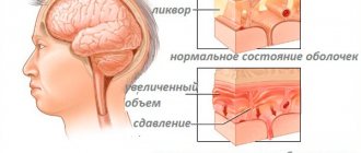
Symptoms of ischemia in newborns
Signs of cerebral ischemia in infants include:
- increase in head volume;
- enlarged fontanel;
- lethargy;
- muscle hypotonicity;
- weakening of conditioned reflexes;
Cerebral ischemia in adults and newborns is a serious condition that should not be taken lightly
- convulsive contractions of the limbs;
- tremor of the arms, legs, chin;
- coma;
- restless sleep;
- causeless crying.
In the first moments of a baby’s life, a neonatologist assesses his condition using the Apgar scale and identifies possible physiological complications. If the doctor identifies the prerequisites for coronary disease, the child is sent for examination using a neurosonogram.
Differences between different types of encephalitis
Encephalitis in children usually manifests itself with similar symptoms. But you also need to take into account the different features of encephalitis. Depending on the cause of occurrence, the clinical picture may differ. The main differences between the symptoms of the main types of encephalitis are presented in the table:
| Symptoms of the main types of encephalitis in children | ||
| Type of encephalitis | Characteristics and features of the flow | Other individual characteristics of the disease |
| Kleshchevoy | The incubation period lasts on average 2 weeks. Encephalitis is often combined with meningitis and peripheral nerve conduction disorders. Sensitivity disturbances on the skin are possible. | In more than 50% of cases, the disease ends in recovery at the stage of fever, without morphological damage to brain tissue. |
| Japanese | It begins acutely with an increase in temperature. Very often accompanied by visual impairment in the form of diplopia. Often the disease is accompanied by paralysis, paresis and convulsive syndrome. | Carriers are mosquitoes. In areas of infection, the population is vaccinated. |
| Herpetic | If left untreated, it leads to complete necrosis of the brain matter. Mortality rate – 50-80%. Most often, patients die from cerebral edema. | The causative agents are herpes viruses of the first or second type. Most often it occurs in newborns whose mothers had an exacerbation of herpetic infection during pregnancy. In adults it develops with immunodeficiency. |
| Measles | It may appear 3-5 days after the appearance of skin rashes. Paralysis, paresis, and dysfunction of the pelvic organs are often observed. | The severity of the disease directly depends on the course of measles. Mortality rate – 25%. Prevention for people who have been in contact with such patients consists of administering special gamma globulin. |
| Flu-like | There is pronounced swelling of the brain and hemorrhages in it. The symptoms are pronounced. | Prevention of this disease is possible. It involves getting vaccinated during the flu season. |
| For chicken pox | Characterized by convulsive syndrome and disturbances of consciousness. | The prognosis is favorable, mortality is low. |
List of consequences caused by the disease
The consequences of cerebral ischemia in newborns can be completely different, based on the severity of the disease.
A mild degree of coronary artery disease occurs without causing negative consequences; the development of children occurs similarly to healthy children
Even if treatment of the disease was started in a timely manner, patients may subsequently experience disturbances in sleep and attention, headaches, epileptic seizures may appear, and mental development may occur with some deviations
The consequences after the 3rd degree of the disease are directly dependent on in which part of the brain the damaged areas are localized and the area of dead tissue. These may be problems with the functioning of the musculoskeletal system, sometimes the patient remains completely paralyzed.
Possible complications in ischemic disease can be predicted based on how severe the oxygen deprivation was, what area of the brain was affected, and how timely professional medical care was provided.
First degree
The first degree of the disease, as a rule, ends favorably for babies. Their development follows the same pattern as that of their peers. Only in rare cases is the appearance of excessive activity and malnutrition observed.
May lead to the following complications:
- From 10 to 20% of patients subsequently experience a slight increase in blood pressure and frequent regurgitation;
- From 30 to 50% of patients have some disturbances in their mental development.
Third degree
Complications arising after the third degree of ischemia:
- Up to 50% of cases of the disease end in death within the first days or a little later, when the cause of death is a severe form of pneumonia or another infectious disease;
- Up to 80% of children suffer complications that are irreversible. The child may develop dementia or become autistic;
- In 10% of children, mental development occurs with minor deviations from the norm;
- In 10% of cases, the disease occurs without any negative consequences for the child.
All types of cerebral ischemia in infants should be treated in a hospital setting under the constant supervision of doctors. Based on the results of the examination, the doctor chooses one of the most suitable methods of treating the disease.
Basic principles of treatment of encephalitis in children
Infusion therapy
Treatment of such patients should begin before hospitalization. Emergency doctors begin infusion therapy, reduce body temperature, and, if necessary, administer corticosteroids and antibacterial drugs to the child. Such patients are hospitalized in intensive care and intensive care units at infectious diseases hospitals. There they immediately undergo a spinal cord puncture to obtain cerebrospinal fluid. Then treatment begins immediately. It consists of the following components:
- Etiological treatment. If there are suspicions or the diagnosis is known, drugs are prescribed that specifically affect the pathogen. For example, for herpetic encephalitis, the child is given Acyclovir, and for bacterial encephalitis, antibiotics are started.
- Decrease in body temperature. For children, the drugs of choice are two drugs: Paracetamol and Ibuprofen.
- Corticosteroids. Prescribed for infectious-toxic shock.
- Drugs for the protection and restoration of the central nervous system. These include B vitamins and Pirocetam.
- Infusion therapy. Doctors maintain the water-electrolyte composition of the patient's blood, while preventing brain edema.
- With the development of paralysis, Proserin is prescribed.
- For convulsions, use Sibazon or Seduxen.
What is perinatal CNS damage?
Perinatal lesions of the central nervous system, abbreviated as PPCNS, are a number of pathologies that are related to disruptions in the functioning of the brain and developmental abnormalities in its structure.
Similar deviations from the norm are observed in children during the perinatal period, the time frame of which ranges from the 24th week of pregnancy to the first 7 days of life after birth, inclusive. Currently, PPCNSL in newborns is a fairly common phenomenon. This diagnosis is established in 5-55% of children. The wide range of indicators is due to the fact that often lesions of the central nervous system of this kind resolve easily and quickly. Cases of severe forms of perinatal damage occur in 1-10% of children who were born on time. Premature babies are more susceptible to the disease.
Disease prevention
Mosquito encephalitis
Encephalitis in children can be caused by diseases for which vaccinations are available. For example, children are routinely vaccinated against measles, rubella and chickenpox. Typically, encephalitis in children caused by such diseases develops if the child is not immunized.
You should not refuse to vaccinate your children, because they can save their lives!
Encephalitis occurs many times more often in children than in adults. The reason for this is the failure of the children's immune system and the blood-brain barrier. Treatment of such children is carried out in intensive care units under the constant supervision of an anesthesiologist and infectious disease specialist.
Source
Brain diseases in infants
Content
Modern medicine identifies various brain diseases in infants. Next, we will look at their most common symptoms.
- Tumors. They arise due to uncontrolled cell division. Tumors often form during the period of active development of the body; in adults they are much less common. They can be either benign or malignant. In the first case, they develop very slowly, without affecting healthy tissue. In the second, their development can be called rapid, during which intact tissues are affected. In the early stages, the disease does not manifest itself in any way, which allows the tumor to develop to large sizes. Symptoms of the disease: frequent headache, vomiting, squint, stupor. Treatment is carried out only by surgery. Some tumors can be completely removed, others partially.
- Ischemia. This disease has complex consequences, and at the third stage it is not treatable at all. It can occur due to a significant reduction or complete cessation of oxygen supply to the brain. Factors that contribute to the development of cerebral ischemia in infants include problems with the cardiovascular system in the mother during gestation, late labor, early birth, too early or late age of the mother (less than 18, more than 35 years). Ischemia is characterized by constant baby convulsions, restless sleep, decreased muscle tone, and excitability. The goal of treatment is to restore normal blood circulation through massage and medication.
- Hydrocephalus. In advanced cases, it leads to the death of the baby. An obstruction occurs inside the brain, after which excess fluid begins to accumulate. It can put pressure on the brain, damaging tissue. The disease occurs not only in newborns, but also in older children. Treatment is performed mainly surgically. However, since bypass surgery entails many complications, this method has now begun to be replaced by more effective and safe endoscopic operations.
Main symptoms, signs and diagnosis of the disease
Hypoxic-ischemic brain damage in newborns is divided into degrees of severity. The longer a baby’s brain cells experience hypoxia, the brighter the clinical picture.
General signs:
- Febrile muscle cramps (twitching);
- Asymmetry of the skull, the volume of the head is sharply increased - vascular disorders cause a failure in the processes of cerebrospinal fluid dynamics, the newborn is diagnosed with congenital hydrocele with increased intracranial pressure;
- Increased excitability or, conversely, depression of the central nervous system - in both cases, muscle atony is noted. The newborn shows signs of anxiety - cries, sleeps poorly, and the chin trembles when screaming (tremor). When the central nervous system is depressed, muscle weakness is noticeable visually - the newborn has disproportionate facial features. The sucking reflex is weakly expressed or completely absent, and swallowing is difficult;
- Development of strabismus;
- Comatose state (no consciousness, all reflexes are suppressed).
Signs of cerebral ischemia appear immediately after the birth of the child. The neonatologist assesses the condition of the newborn using the Apgar score. Low scores (less than 7 points upon re-evaluation) are an indication for diagnosis.
Laboratory and hardware diagnostic methods help to find the reason why an insufficient amount of blood enters the central nervous system.
Comprehensive examination:
- Assessment of neurological status;
- Neurosonography with duplex scanning – the brain structures of a newborn are examined through the fontanelle, the vascular bed is assessed in 3D mode;
- CT or MRI - one of the methods is used to confirm the results of ultrasound, or when visualization was difficult;
- EEG – assesses overall brain activity. With irreversible changes in brain structures, the impulse is weak or completely absent (with localized damage);
- Laboratory blood diagnostics - the rate of blood clotting and gas exchange is assessed.
Based on the diagnostic results, the degree of ischemia in the newborn is determined and effective therapy is selected.
Cerebrovascular diseases in children
- Vegetative-vascular dystonia. It is considered to be a disease of adults, but it can manifest itself at an early age. The walls of blood vessels do not suffer as much from the disease as the child himself. After all, the symptoms can be very unpleasant: severe headaches, vomiting and nausea, increased heart rate, sweating.
- Migraine. Another disease that is so widespread in the “adult” world, but can cause an extreme headache for a child. This type of migraine is also called dysphrenic, which is accompanied by increased aggressiveness in the child’s behavior.
- Among cerebrovascular diseases in children, aneurysm should be highlighted. One section of the blood vessel may become enlarged and filled with blood. It will grow, putting pressure on nerves and tissue. In addition, there is a high risk of hemorrhage. The disease may be congenital. Treatment depends on the size of the area, its type and location. Not all cases require surgical intervention.
John Medina on how to develop your baby's brain
The popular book by the American author can be purchased or downloaded on the Internet. In his work, the researcher states that “all children are different and all parents are different, so there is no advice for all occasions.” Taking as a basis the individuality of both babies and their parents, Medina encourages spending as much time together as possible and turning this time into a fascinating process of learning about the baby, his needs, his already developing character.
In his book, the scientist pays great attention to the methods and techniques of developing the brain and thinking in young children, and warns against typical mistakes of inexperienced parents.
Infectious diseases affecting the brain and the body as a whole
- Encephalitis. It can be caused not only by infection, but also by parasites and fungi. Viruses are able to penetrate the nervous system, gradually destroying its cells.
- Meningitis. With this disease, the membranes of the brain become inflamed either on their own or as a result of an illness as a complication.
Other infectious diseases that affect the brain and the body as a whole include polio and arachnoiditis.
Pediatrics. National leadership / ed. A. A. Baranova. — 2014
The structure of concomitant pathology of the central nervous system in various forms of hydrocephalus in children / Abramova O. A., Basina M. A., Balashova E. A. // Practical Medicine. - 2010. - 7(46)
Source
Meningoencephalitis accounts for approximately 20% of all neuroinfections. For newborns, this infection is characterized by a high frequency of neurological pathologies and deaths. This is often due to the failure of the immune system and the inability to independently fight pathogenic microorganisms.
Brain treatment in Saratov, Russia
Sarklinik provides treatment for a number of diseases, diseases of the central and peripheral nervous system in Saratov, in Russia for children, adolescents, adults, boys, girls, guys, girls, men, women, brain treatment in Saratov . Hardware and non-hardware treatment methods make it possible to restore the functioning of the human nervous system.
Sign up for a consultation. There are contraindications. Specialist consultation is required.
Text: ® SARCLINIC | Sarclinic.com \ Sarlcinic.ru Photo: © pixologic / Photobank Photogenica / photogenica.ru The people depicted in the photo are models, do not suffer from the diseases described and/or all coincidences are excluded.
Related posts:
Claude H Claude syndrome, alternating peduncular syndrome
Accentuation of character in adolescents, types, test, methodology, correction in Saratov
Decerebrate rigidity: causes, symptoms, treatment
Opisthotonus: causes and treatment
Depersonalization: causes, symptoms, treatment of depersonalization in Saratov, Russia
Comments ()
Causes
- Bacterial meningoencephalitis. The causative agents of the pathology can be streptococci, staphylococci, gram-positive and gram-negative microorganisms, meningococci, pneumococci, treponema pallidum.
- Viral meningoencephalitis. The causes may be tick-borne encephalitis virus, cytomegalovirus, herpes virus, adenovirus, influenza viruses, measles, rubella, Epstein-Barr, Coxsackie, chickenpox, HIV infection, mumps virus, enteroviruses.
- Fungal meningoencephalitis.
- A disease caused by protozoan microorganisms . For example, toxoplasma, malarial plasmodium.
- Autoimmune meningoencephalitis. When certain malfunctions occur, the body begins to produce antibodies against its own brain cells and tissues.
- Tuberculous . Develops against the background of pulmonary tuberculosis. The causative agent is mycobacterium (Koch bacillus).
- Associations of several pathogens.
With delayed diagnosis and untimely treatment of this disease, there is a risk of developing serious consequences and multiple complications.
Kinds
The International Classification of Diseases does not contain OPGM. Therefore, the diagnosis is formulated as “unspecified brain damage”, “unspecified encephalopathy”. Nevertheless, there is a classification of pathologies united by the term “organic damage”:
- hydrocephalus;
- congenital developmental anomalies;
- cerebral palsy;
- disorders due to intrauterine infections (syphilis, cytomegaly);
- diseases affecting the central nervous system.
OPHM are also divided into diffuse - for example, due to encephalopathy, and localized - due to a tumor or brain injury.
Possible consequences
- Swelling of the brain due to the accumulation of exudate, which can cause displacement of brain structures. Herniation of the brainstem into the foramen magnum may occur. Swelling of the brain stem can cause death in the newborn.
- Purulent meningoencephalitis.
- Increased intracranial pressure . Signs: tension, bulging and pulsation in the area of the large fontanel, throwing back of the head, a piercing and monotonous cry.
- Sepsis, infectious-toxic shock. Due to the proliferation of bacteria and the entry of their toxins into the bloodstream.
- Kidney failure.
- Swallowing disorder.
- Respiratory dysfunction : breathing is shallow and frequent.
- From the heart: arrhythmic, rare sounds.
- Epilepsy.
- Paresis of limbs and paralysis.
- Facial asymmetry.
- Postnecrotic cysts of the brain . Often occurs after a history of herpes viral meningoencephalitis.
- Petrifications and cysts in the thalamus.
- Hemorrhages into the brain parenchyma, disseminated intravascular coagulation syndrome.
- Extensive intraventricular hemorrhages, atrophy of the brain substance.
- Optic nerve atrophy, conjunctivitis, anisocoria, inflammation of the choroid.
- Development of asthenic and neurotic syndromes.
- Loss of sensation, hearing and vision.
- Brain abscess.
- Deafness.
- At an older age, there may be speech disorders, delayed mental and mental development, and dementia.
- Coma with fulminant course of the disease.
- Widespread skin necrosis.
- Ischemic stroke, as a result of prolonged increase in intracranial pressure.
Diagnostics
The diagnosis of OPGM is carried out by a neuropathologist. After examining the patient, he prescribes the following brain tests:
- CT scan – the functional state of the organ is studied, structural changes are revealed;
- neurosonography – the cerebrospinal fluid spaces are examined and neoplasms are identified;
- REG – vessels are examined;
- Doppler ultrasound is an ultrasound of the brain cavities;
- MRI – structural changes in brain tissue, blood vessels, nerve fibers, and the degree of damage are revealed.
Recommendations
- If even minor symptoms of the disease appear, immediately seek help from a pediatrician or infectious disease specialist for timely diagnosis and adequate treatment.
- After suffering from meningoencephalitis, children should be regularly observed by their attending physician, registered at the dispensary and follow all recommendations.
- In some cases, after suffering a viral or bacterial disease, sanatorium treatment is necessary.
- It is necessary to monitor the newborn’s sleep and rest patterns and regularity of nutrition.
- You need to walk in the fresh air every day.
- After the birth of a child, immunoprophylactic measures should be taken.
- Be sure to treat foci of infection.
- If you come into contact with a sick person, take a course of antibacterial therapy.
Treatment and prevention of ischemia in infants
Despite the fact that at the moment, grade 1 and 2 cerebral ischemia in children responds well to treatment, no one can guarantee that the disease will go into remission and have minimal impact on the condition of the newborn.
In practice, treatment tactics depend on the degree of pathology and are based on restoring the supply of oxygen to the damaged area of the brain and stimulating its performance:
With mild cerebral ischemia, newborns are given a massage of the head and collar area, without prescribing medications.
- Stage 2 ischemia is treated with medication, and drugs are selected individually based on the underlying disease. Usually these are nootropics, blood circulation stimulants, anticonvulsants, restorative drugs to improve the overall health of the child. For thrombophilia, anticoagulants and vasodilators are prescribed.
- The most severe degree of ischemia requires serious medical procedures, since the life of the newborn often depends on it. These include artificial ventilation, surgical interventions for blockage and serious impairment of vascular patency. If ischemia was caused by other diseases, such as hydrocephalus, or there is swelling of the brain, then diuretics and other methods of draining fluid from the structures of the central nervous system, including brain shunting, are prescribed.
To minimize the risk of developing cerebral ischemia in a newborn, future parents should take care of their health in advance and take all measures to prevent the development of pathology
This is why it is so important for a woman to change her lifestyle for the better long before pregnancy, to exercise regularly and spend more time outdoors.
Also, to prevent and prevent the occurrence of serious complications in a newborn, a pregnant woman should follow all the appointments of a gynecologist and should not shy away from undergoing scheduled examinations on time.
conclusions
Meningoencephalitis is a fairly serious and dangerous disease that poses a threat to the child’s body. The mortality rate of newborns without treatment reaches 100% . But with timely diagnostic measures and properly selected therapy, there is a chance to avoid negative consequences and restore the child’s health.
Source
Meningoencephalitis in children is a dangerous and severe disease in which inflammation of the tissues of the brain and meninges develops.
There are many reasons for the development of this inflammatory disease. When its first symptoms appear, you must immediately consult a doctor, and this is 100% a reason to call an ambulance and emergency hospitalization. Only in this way can the development of severe complications be prevented, and treatment will be successful. In no case should you try to get such a child to a clinic or hospital on your own, since illiterate transportation can significantly worsen the patient’s condition.
What are some ways to activate your brain?
I want to repeat for the hundredth time that there is a close connection between brain activity and finger dexterity. Based on this, in childhood, especially early childhood, you need to start stimulating processes in the hemispheres by training your fingers. A lot has been written about this, but almost all of my materials are, to one degree or another, devoted to issues of motor skills, sensory skills, etc. But hand exercises are only one component of a set of activities for children.
The main, in my opinion, areas of work related to enhancing intelligence:
- Finger gymnastics;
- Logorhythmics;
- Kinesiological training;
- Self-massage;
- Neuroscience;
- Articulation and breathing exercises;
- Creative activities;
- Didactic games and tasks;
- Mnemonics;
- TRIZ;
- Educational activities in mathematics, speech development, etc.
As for finger gymnastics, creative activities, didactic games, mnemonics, logorhythmics (gymnastics with recitation), TRIZ and educational activities, I write about this all the time. If interested, look for articles.
To help you, I give excellent literature on strengthening the work of the child’s convolutions and memory:
- “Developing attention, memory and speech” - convenient cards on a paper clip will appeal to both teachers and parents;
- “We develop attention and memory. For children 5-6 years old" - an interesting selection of exercises that will help keep your child occupied and educated;
- - a great idea for family evenings. There is a deliberate mistake in the verses, you need to find it by ear;
- “We develop memory and thinking. For children 4-5 years old" is a unique benefit that will make your child an excellent student in school in the future;
- “Training memory and attention” from the “365 days before school” series – it’s time for active preparation for future first-graders;
- “Speed Memory” is the author’s manual on mnemonics, addressed to those of any age who want to improve their memorization abilities.
Causes and classification
Meningoencephalitis can be caused by various infectious agents: bacteria or viruses, fungi or protozoa. In fact, this inflammatory process of brain tissue becomes a consequence of many infectious diseases or local inflammatory reactions.
More often, inflammation is caused by viruses (for example, herpetic meningoencephalitis). In rare cases, the disease is caused by toxic effects or an infectious-allergic process.
It is in children that meningoencephalitis is extremely severe and is accompanied by a high incidence of consequences, disability and death.
Children of all ages, even newborns, can get meningoencephalitis. The following pathogens or diseases can provoke its occurrence:
- viruses: tick-borne encephalitis, herpes, cytomegalovirus, influenza, West Nile fever;
- bacteria: streptococci, meningococcal infection, Escherichia coli or Hemophilus influenzae, other gram-positive and gram-negative microorganisms;
- protozoa: toxoplasma, amoeba, etc.;
- autoimmune reactions;
- fungal infections (rare);
- purulent dental and ENT ailments;
- measles;
- rubella;
- chickenpox;
- tuberculosis.
The following conditions and pathologies increase the risk of developing this disease:
- immunodeficiency;
- chronic diseases;
- poor nutrition;
- bad habits;
- overwork, etc.
Experts divide meningoencephalitis into two types:
- primary – provoked by inflammation directly in the brain tissue;
- secondary - becomes a complication of a previous disease (for example, sinusitis).
The course of meningoencephalitis can be acute, fulminant or chronic. Depending on the nature of the damage to brain tissue, the disease occurs in the form of a serous, purulent or hemorrhagic variant.
Treatment of meningoencephalitis should begin immediately after the first signs appear.
The severity of the consequences of meningoencephalitis depends on many factors, including the shape, type and nature of brain lesions.
GM development during the first years of life
The newborn's brain is anatomically and functionally immature. During the breast period, it grows rapidly, the number of glial cells increases, and hydration decreases.
At the end of the 1st year, the weight of the GM doubles. The development of a child’s brain occurs intensively over the years; in the process of ontogenesis, innate unconditioned reflexes are replaced by conditioned ones.
At 3 years of age, the brain weighs approximately 3 times more than at birth. The ability of abstraction, learning, and memory is used; the child realizes his personality and becomes a social creature.
The first years of life are a critical stage for a child's brain development and provide the neurological foundation for intellectual development in adolescence and adulthood.
In the early years, children are very open to science through play. For young children, play is a way of preparing for a successful future life. Therefore, during this period, parental love and plenty of time spent together are important.
At 6 years of age, the weight of the brain is almost equal to the weight of the adult human brain (1250 g). The hemispheres are expressively grooved. Branching of neurons, myelination (creation of a covering that protects large nerve fibers from damage) are completed, memory and the ability to recreate memories improve. In the activity of the GM cortex, the ability of internal blocking is used; the child distinguishes between what he says, thinks, and reads.
During the first 8 years of life (especially the first 3 years), there are several critical periods for the acquisition of certain types of intelligence. If these “periods of opportunity” are closed, learning becomes more difficult, sometimes even impossible.
In adolescents, the GM grows mainly in the anterior lobes, its weight is about 1400 g.
Symptoms
Usually the disease develops acutely.
The fulminant course of meningoencephalitis is especially dangerous for a child’s life.
Children experience the following symptoms:
- headache;
- fever and chills;
- general anxiety;
- swelling of the fontanel in young children;
- nausea;
- vomit;
- tension in the muscles of the neck and back: difficulty tilting and turning the head, neck pain;
- photophobia;
- impaired coordination of movements;
- difficulty speaking;
- movement disorders;
- disorders of swallowing, speech, etc.
To identify symptoms of the disease, the doctor performs a series of tests when examining the child. If meningoencephalitis is present, the Kernig and Brudzinsky symptom complexes are positive.
The most important symptom of meningococcal infection is a characteristic rash in the form of blue-purple spots that do not disappear with pressure, quickly spread throughout the body and tend to merge. These spots are nothing more than hemorrhages. Moreover, such a rash may be the first and only sign of infection - the remaining symptoms simply do not have time to develop, and the child dies from severe infectious-toxic shock.
That is why, if even a single element of this nature is detected, even though the child is feeling well, it is necessary to immediately call an ambulance. Meningococcal infection is a reason to place a child in intensive care.
The same applies to various types of bleeding, including nosebleeds, that develop in a sick child. Any sick child with bleeding should be immediately evaluated to rule out meningococcal infection.
After the baby is confirmed to have this disease, quarantine is declared in all institutions he visits; children who have been in contact with him require vigilant monitoring, and ideally, emergency vaccination prevention. The disease is highly contagious and extremely life-threatening.
How to develop a child's brain correctly?
Most parents are very concerned about the correct and comprehensive development of their baby. Every family dreams of healthy and smart children. For the correct formation of brain functions in the offspring, it is important to follow simple rules.
Correct and timely development is primarily influenced by the surrounding reality. Parents and loved ones should take part in the upbringing and education of their child and broadening his horizons from birth.
From birth to 1.5 years, children learn about the world through sounds, tastes, and tactile sensations. During this period, it is important to improve fine motor skills, which will help the baby speak more quickly and learn to perform basic actions, for example, holding a spoon in his hands.
The child is developing speech, so it is important to constantly talk to him from birth, gradually processing sounds in his subconscious, he will learn to connect them into images, and then he will begin to speak himself.
From 1.5 to 4 years old, the baby learns to receive, process and show emotions. During this period, close emotional contact with parents is very important. Research has proven that the lack of close communication with parents during this period forms an incorrect emotional picture of the world in children. Children who did not receive positive emotions and love in childhood become aggressive or passive adults who do not know how to love, empathize, and they develop the wrong emotions.
In the period from 4 to 7 years, the formation of imaginative thinking occurs. It is during this period that it is important to intensively work with a preschooler: read to him, teach him to draw, sing, dance, read, play intellectual games. All this will allow him to learn to form complete images, remember and reproduce them.
From 7 to 9 years old, the child develops logic. He begins to make his own conclusions based on information received verbally and non-verbally. During this period, parents should not rely on school, but rather take part in the formation of logical thinking in their child. To do this, you can simply ask your son or daughter problems from life, and ask what he would do in a given situation. Find out from him why his choice is this way, help and correct if necessary. It is important that the child himself learns to understand the logic of processes occurring in life and learns to analyze them.
With the formation of the frontal lobe, which usually ends at age 8, the child becomes part of society. His world expands along with his social circle and he defines for himself the framework of behavior in society. At this time, it is important that the family shows by example how to behave in society. If a mother is rude, screams and swears, most likely her child will behave the same way, because he will consider this behavior to be the norm.
From 9 to 12 years of age, a student learns to process information in a complex manner. That is, it is at this time that he learns to obtain complex knowledge and uses various methods for this. Parents should control that the student does not get stuck on one source of information and teach him in every possible way. If he loves animals, then he should read about them, watch videos, communicate with them (petting, feeding, etc.). That is, obtain information about a subject of interest from different sources. This will give him the opportunity in the future to react correctly to various situations and evaluate them comprehensively.
From the age of 12, a hormonal surge begins, often called the transition period. Most children at this time do not get along well with their parents, and conflicts and misunderstandings arise in the family. The main thing during this period is not to lose touch with the teenager, and if there is no way to cope with the situation on your own, it is better to turn to psychologists.
How to develop the left hemisphere of the brain?
Development of the left hemisphere of the brain
The left side is responsible for logic. Language skills, solving logic circuits, mathematical operations, calculating integers and fractions, keeping track of time - all this is the work of the left half of the brain.
Therefore, to improve children's performance in school or to improve overall development, we must develop the left side of our “computer”.
So, how to develop the left hemisphere of the brain?
The following must be done:
- Work on developing logic - study mathematical algorithms, practice logical thinking
- Solve complex geometric problems. Studying a science like mathematics is impossible without a well-developed left half of our brain
- Solve puzzles and crosswords - the left hemisphere works better than the right when analyzing actions and composing words
- A left-handed person needs to do everything with his right hand: write, draw, eat. It is necessary to use the right side of the body
- Take vitamins to improve brain function
Important: Try not to focus on the development of one half of the brain. It is the work of both halves of our brain “computer” that is valuable. Develop the left and right hemispheres of your brain at the same time.
How to develop the right hemisphere of the brain?
Development of the right hemisphere of the brain
Above, we figured out what the left half of the brain is responsible for; now we need to work on developing intuitive abilities and creative thinking.
The right side of the brain processes data that comes from the senses. It allows you to synthesize existing data in your head, so a person can solve puzzles and act according to his own plans, and not according to compiled templates.
By developing the right half, we cultivate a holistic view of the world and reality. How to develop the right hemisphere of the brain? Do the following:
- Listen to music - it should be classical music, not rock or pop
- Dream, meditate alone. It is better to do meditation on the seashore or in the forest, where you connect with nature
- Draw, write poetry, compose music. Any creativity helps the development of the right half of the brain
Advice: Get creative, believe in your capabilities, and the result will not take long to arrive.
Complications
Bacterial and viral meningoencephalitis are often complicated, especially against the background of reduced immunity. The consequences of the disease include the following conditions and diseases:
- epilepsy;
- paralysis and paresis of the limbs and body;
- hydrocephalus (excessive fluid in the brain);
- delayed mental and mental development;
- death.
The development of complications can cause disability in young patients and requires parents to comply with all doctor’s instructions.
The first symptoms of meningitis or meningoencephalitis are a reason to call an ambulance.
To eliminate the consequences, rehabilitation courses in specialized centers are recommended. In addition, classes with the child are also carried out at home. To do this, the doctor teaches parents special techniques and exercises.
Symptoms
Deviations in children may indicate ischemia. Signs of central nervous system pathology differ; obvious developmental delays cause close attention from doctors and parents. In newborns, behavior may change dramatically, appetite may worsen, and tearfulness may increase . Constant regurgitation and changes in weather conditions should worry parents.
Let's list the main features:
- The size of the newborn's head increases, and the fontanel expands due to the large volume of fluid in the head. ICP increases. The baby is unconscious and his brain function is impaired.
- The central nervous system is depressed, muscle tissue weakens, sucking and swallowing reflexes worsen. Rarely does strabismus appear with asymmetrical proportions.
- Increased excitability of a child is characterized by increased or decreased muscle tone, tremors of the arms and legs, and tearfulness.
- The child often shudders, jerks his head, arms and legs.
Causes and treatment of cerebral ischemia
- acute heart failure;
- heart rhythm disturbances (tachycardia or bradycardia);
- increased blood viscosity (occurs as a result of anemia or erythrocytosis, as well as as a result of carbon monoxide poisoning).
Have you been struggling with HYPERTENSION for many years without success?
Head of the Institute: “You will be amazed at how easy it is to cure hypertension by taking it every day...
As for newborns, the main cause of ischemia in them is considered to be insufficient oxygen supply to the brain (hypoxia), which occurs during childbirth or pregnancy.
Cerebral ischemia in newborns
Depending on the results of medical examinations, ultrasound of the brain and symptoms, there are 3 degrees of cerebral ischemia:
- Mild or grade 1 cerebral ischemia. It is characterized by excessive excitement or, conversely, depression of the child during the first days of life.
- Moderate degree (cerebral ischemia grade 2). Manifests itself in the form of seizures and a longer period of disturbances.
- Severe 3rd degree of ischemia. As a rule, such children require a stay in the intensive care unit. The prognosis of the disease depends on its severity, concomitant diseases and all rehabilitation measures prescribed by a pediatric neurologist.
Basically, the first mild degree of ischemia is diagnosed in newborns, in which children recover almost within a few days.
Treatment of cerebral ischemia in newborns consists of restoring normal cerebral circulation, as well as creating all conditions for the full functioning of undamaged areas of the vessels of the head. The initial stage of treatment is quite simple. Only massage is used, and it can even do without the use of medications. Methods and methods of subsequent treatment are carried out only on the recommendations of a neurologist.
Symptoms of cerebral ischemia
One of the earliest symptoms of cerebral vascular ischemia is fatigue, especially during active mental work. Gradually, the patient's condition worsens, memory weakens and forgetfulness appears.
Clinical symptoms of cerebral ischemia:
- various types of headaches;
- sudden changes in blood pressure;
- dizziness;
- nausea and vomiting;
- frequent fainting;
- sensitivity disorder;
- speech and vision impairment;
- general malaise, weakness.
Treatment of cerebral ischemia
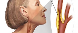
To date, there are still no sufficiently effective drugs and methods for treating chronic ischemia. The only way out for correcting atherosclerotic vascular damage is surgical intervention, which has been increasingly used in medical practice since the middle of the last century.
Its essence is to remove a blood clot or atherosclerotic plaque from the surface of a cerebral artery, which ensures the restoration of normal blood supply to the brain with oxygen and nutrients. Such operations are performed under local anesthesia.
Prevention of cerebral ischemia
Cerebral vascular ischemia is a disease that can be prevented if you take care of your health in childhood. Therefore, all measures that need to be taken should be carried out from a very early age.
Experts recommend protecting yourself as much as possible from the main provoking risk factors. This includes excess weight, physical inactivity, smoking, alcohol abuse, and stressful situations.
Treatment
Modern pediatrics is making significant progress in the treatment of cerebral ischemia in newborns.
Treatment of chronic cerebral ischemia in newborns includes restoration of blood circulation in the brain and timely creation of all the conditions necessary for the full functioning of areas of the brain undamaged by the disease, which includes the active intake of antioxidant complexes.
To treat the initial stage of this disease, a fairly simple course of treatment is used; most often, doctors do not prescribe any medications to children, making do with ordinary massages. For moderate and severe stages of the disease, therapy is selected by the doctor individually for each child.
Prognosis and consequences of the disease
The prognosis depends on how severe the infant’s cerebral ischemia is, as well as on the presence of concomitant pathologies and the effectiveness of rehabilitation procedures prescribed by the attending physician.
The consequences of the disease can be quite severe, so treatment should always begin as quickly as possible.
Possible consequences of chronic cerebral ischemia in newborns:
- headache;
- constant irritability;
- mental retardation;
- attention deficit, learning difficulties;
- epilepsy;
- sleep disturbance;
- silence.
Only an experienced doctor can correctly assess the entire set of important diagnostic signs. He will immediately take all necessary measures to minimize losses or completely eliminate brain hypoxia in a newborn.
Attention!
Types of treatment
Treatment of mild forms of ischemia begins with restorative procedures, such as massage, exercise therapy, and walks. Additionally, drug treatment from the following groups of drugs may be recommended:
- drugs that improve blood circulation in the brain;
- drugs that relieve swelling in the brain caused by ischemia;
- drugs that eliminate and mitigate the effects of hypoxia (antihypoxants).
Treatment of more severe stages of ischemia requires the selection of an individual treatment package for a specific patient, taking into account all the features of the ongoing disease. The basis of therapy is the creation of favorable conditions for the restoration of damaged brain structures and for the normal functioning of healthy areas.