Ischemic cerebral stroke occurs predominantly in older people, but the disease is increasingly affecting 30-40 year olds. Pathology is assigned several codes in ICD-10. It is based on a severe disruption of the blood supply. A stroke occurs unexpectedly and it is impossible to predict its occurrence. Lack of treatment in the first three hours leads to irreparable consequences and often death.
Classification of ischemic strokes
Since ischemic stroke is often the outcome of one of the previous cardiovascular diseases, several types are distinguished in neurology:
- Cardioembolic is a type of ischemic stroke that occurs due to arrhythmia, previous myocardial infarction, or existing valvular heart disease.
- Lacunar is a type of ischemic stroke caused by minor occlusion of the arteries.
- Atherothrombotic - its cause is atherosclerosis of large arteries, which causes arterial embolism.
- An ischemic stroke of unknown origin is an ischemic stroke, the root cause of which has not been established; it is possible that there are several reasons that could lead to the development of this disease, so it is difficult to immediately make an accurate diagnosis.
- Ischemic stroke, which is related to rare causes: dissection of arterial walls; blood hypercoagulation or non-atherosclerotic diseases.
Ischemic stroke is also classified depending on the time of occurrence. For example, there are 5 periods of ischemic stroke:
- Acute: occurs in the first three days. It should be said that in the first three hours after a stroke, the patient can benefit from thrombolytic drugs that are administered systemically. If the symptoms begin to regress, then on the first day of the disease a diagnosis of transient ischemic attack is made.
- Acute: lasts up to 4 weeks.
- Early recovery period: can last up to 0.5 years.
- Late recovery period: lasts up to two years.
- Time of residual effects: manifests itself after 2 years from the moment of illness.
It should also be noted that in practice, minor stroke is also common, which is characterized by regressive symptoms observed within 3 weeks from the onset of the disease.
Classification
The variety of symptoms, causes, and manifestation features allows us to create a classification of ischemic stroke.
By the nature of the lesion
Taking into account the nature and characteristics of the course of the disease, there are several types of stroke:
- Transient ischemic attack. The damage is concentrated in a small area. Signs of pathology last no more than a day.
- Minor stroke. Functional recovery is noted within 3 weeks.
- Progressive. Symptoms increase gradually over several hours or days. The normalization of the condition is not complete; residual effects persist.
- Total. Characterized by brain damage and developing functional deficits.
Because of
Types of diseases are described based on the reasons that led to them:
- Atherothrombotic stroke occurs during sleep. It is characterized by slow development - cholesterol formations gradually reduce the diameter of the cerebral vessels.
- The lacunar form does not appear immediately. On the first day, signs of this disease cannot be detected. With this disease, spasm occurs in the cerebral arteries passing through several meninges. It is associated with arterial hypertension, atherosclerosis and embolism. With lacunar syndrome, small capillaries that bring blood to tissues located deep in the brain are affected.
- On the contrary, a cardioembolic stroke occurs suddenly. A broken clot blocks blood flow in medium-sized arteries.
- Hemodynamic is caused by a sharp decrease in pressure or a decrease in the filling volume of the heart. The impact is not associated with physical activity.
- Hemorheological develops due to a blood clotting disorder.
For reference. A disease of unspecified origin occurs for unclear reasons. This type of ischemic stroke is also spoken of when several factors lead to the disease. In particular, an increase in pressure and an increase in lipid plaques.
On the side
An ischemic stroke on the right side is called right-sided. The processes of processing sensory information, perception, and fantasy are disrupted. Creative abilities are narrowed. The patient loses the ability to sympathize, memory deteriorates. If the patient is left-handed, his speech suffers. It turns out that the left side is paralyzed, the arm and leg cannot move, sensitivity disappears.
With an ischemic stroke on the left side, paralysis covers the right side of the body. The patient is prone to depression, short temper, and impulsiveness. Systemic and motor memory and speech suffer. A person loses the ability to analyze and learn new things.
By localization
The violation zone allows us to distinguish several types of stroke. Vertebro-basilar syndrome is characterized by atrophy in the area of the brainstem, cerebellum, and occiput.
When a disorder develops in the basilar part of the brain stem, the functioning of the heart and respiratory organs worsens. Death occurs most often due to the lesion located in this department.
For reference. A common form is the death of nerve tissue in the cerebellum area. This occurs due to blocking of blood flow in the superior basilar artery and its basin.
Patients suffer from coordination, tremor appears when trying to make a purposeful movement, vomiting, and coma develops. The most common outcome is death, and drug therapy rarely helps.
A circulatory disorder in the vertebrobasilar region is characterized by confusion, nausea, and inability to swallow.
With a stroke of the middle cerebral artery, sensitivity is impaired and paralysis develops. Patients are distinguished by their fixed gaze. Often they do not understand their illness.
In some cases, an ischemic crisis causes a disruption of the blood supply to the spinal cord. In this case, patients have paralyzed arms and legs, and have problems with bowel control.
For reference. Separately, they talk about an extensive cerebral infarction. This pathology develops in the event of a disruption of the blood supply in large arteries and their basins.
The researchers identified the volume of the lesion:
- extensive atherothrombic – 115 cm3;
- cardioembolic – 62 cm3;
- hemodynamic – 32 cm3;
- lacunar – 2 cm3.
A rheological lesion is characterized by an area of 1.5 cm3.
Etiology and pathogenesis of ischemic stroke
Since in medical practice ischemic stroke is not considered as an independent disease, there is no need to talk about any etiological cause that causes it.
However, doctors mention risk factors that affect the incidence of this disease, including:
- Modifiable factors: arterial hypertension, diabetes mellitus, asymptomatic lesions of the carotid arteries, myocardial infarction.
- Non-modifiable factors: heredity.
In addition to these two large groups of risk factors, there are also factors caused by lifestyle: stressful situations, prolonged psycho-emotional stress, poor standard of living, low physical activity, excess weight, bad habits.
A special sequence of biochemical changes occurring in the patient’s body, and in particular in the brain, is usually caused by focal cerebral ischemia (acute form), which entails various kinds of disorders at the tissue level, which causes the death of brain cells or cerebral infarction .
If we talk about the nature of the changes that occur during an ischemic stroke, it depends on the level and duration of reduced activity in the cerebral blood flow, as well as on how sensitive the brain substance is to ischemic processes.
It is interesting that the degree of reversibility of changes occurring in the tissue will be determined at each pathological stage by the level of decrease in cerebral blood flow, as well as by such an indicator as its duration, together with those factors that affect the sensitivity of the brain to damage of a hypoxic nature.
There are several medical terms that are controversial in their definition. In order to debunk all doubts, let's name them.
In neurology, the core of the infarction is the zone of irreversible damage, while the penumbra is usually called the zone of ischemic damage, which is characterized by a reversible nature. In the study of a particular case of ischemic stroke, doctors pay considerable attention to the duration of the existence of the penumbra, since over time those changes that were previously reversible become irreversible.
Another important term is the “oligemic zone,” which is an area with a preserved balance between the needs of the tissue and the processes that provide it with these needs, even despite reduced cerebral blood flow. Such zones usually exist for a very long time, without moving into the core of the infarction. This term should not be confused with the term "penumbra".
Pathogenesis and causes of development
The pathogenesis centers on the formation of an area of dead brain tissue around which swelling occurs.
Attention. It takes approximately 8 minutes for irreversible consequences to occur in the area where the blood flow rate has decreased significantly (less than 10 ml/100 g per minute). This area is called the core or core of ischemia.
For several hours after the impact, the core is surrounded by still living tissue. However, she had already begun to experience nutritional deficiencies. The rate of cerebral blood flow here is 20 ml/100 g per 1 minute.
This zone is usually called the penumbra or ischemic penumbra. In the cells of this area, a metabolic disorder occurs, as a result of which it loses functionality due to a lack of nutrients.
Clinical picture of ischemic stroke
Clinical manifestations of ischemic stroke are quite varied and depend on the location of the lesion and the area of brain damage. Most often, the lesion is located in the carotid region (accounting for more than 80% of cases of ischemic stroke), less often in the vertebrobasilar region.
The main feature of a heart attack in the area of blood supply to the middle type cerebral artery is the presence of a collateral blood supply system. If we talk about such a phenomenon as occlusion of the proximal part, then in the cavity of the middle cerebral artery it contributes to the subcortical type of infarction, while the cortical part will not be affected during the blood supply. If collateral data are missing, a large infarction may develop in the portion supplied by the middle cerebral artery.
If a heart attack occurs in a part characterized by blood supply to the superficial branches of the middle type cerebral artery, then the development of deviation of the eyeballs in the direction of the affected part of the hemisphere cannot be excluded. If the dominant hemisphere is affected, then the development of the ipsilateral form of ideomotor apraxia begins. In addition, total aphasia can be observed. If the subdominant hemisphere was affected, then we are talking about anosognosia. In addition, the development of dysarthria or aprosody cannot be ruled out. A contralateral type of space neglect may also occur.
Symptoms
Stroke is one of the main causes of disability and mortality in the population. According to neurologists, if a cerebral infarction is diagnosed in a timely manner and medical care is provided within the first three hours, then, as a rule, complications are excluded and the patient fully recovers.
Clinical symptoms differ depending on the lesion. According to statistics, left-sided stroke is more common. Acute circulatory disorders of the left hemisphere of the brain are characterized by the following symptoms.
- Muscle paralysis or loss of sensation on the right side of the body. Muscle strength in the right limbs is completely lost or reduced compared to the left side. Facial asymmetry - the right corner of the mouth does not rise.
- Problems understanding the meaning of words heard and speech disorders. The patient expresses himself in fragmentary phrases or completely loses the ability to speak clearly and mumbles.
- Loss of memory of recent events while maintaining the ability to remember the past.
- Difficulty walking, loss of coordination.
A stroke on the right side of the brain is accompanied by the following symptoms.
- Paralysis of the left side of the body. The left arm does not rise at all or falls limply when raised. There may be a drooping of the corner of the mouth if you ask the person to smile.
- Vision problems. Loss of the ability to see with one or both eyes at once, darkening of the eyes, double vision.
- Movements are unstable and fast.
Problems with speech during a right-sided stroke occur only in left-handed people, since their speech center is located in the right hemisphere.
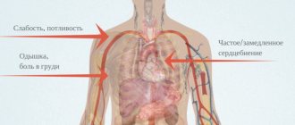
Common symptoms encountered in pathologies associated with brain damage are intense headaches, dizziness, and attacks of nausea. The signs of a hemorrhagic stroke are more pronounced. With a cerebral hemorrhage, sudden vomiting without nausea, convulsive seizures, changes in breathing and heart rate, decreased vision and hearing, and loss of consciousness may occur.
Even if the symptoms seem mild and short-lived, you should seek medical help immediately. Every minute counts. The longer a person goes without help, the more serious the brain damage and further complications. The patient may simply fall into a coma and die.
Infarction of the subdominant hemisphere
If an infarction of the subdominant hemisphere occurs, then the disease will be characterized by emotional disturbances and spatial non-response.
How much paresis will spread during a heart attack into the cavity of the blood supply (striatocapsular arteries) will depend on the location and area of the lesion itself (usually the upper part of the body: face, limbs or cotralateral part of the body). Speaking about an extensive infarction of the striatocapsular type, the main symptom is occlusion of the cerebral (middle) artery, which is expressed in aphasia or homonymous lateral hemianopia.
Symptoms of lacunar infarction
If we talk about lacunar infarction, then clinically it is manifested by the development of lacunar syndromes, which is reflected in hemihypesthesia, hemiparesis, both individually and in combination.
Most often, a heart attack that occurs in the blood supply of the anterior cerebral artery manifests itself symptomatically in the form of movement disorders. With ongoing occlusion of the cortical branches, the development of motor deficits begins in the area of the feet or in the lower extremities. In addition, there is an implicit paresis of the upper extremities, which also manifests itself in severe damage to the tongue and face as a whole.
How to restore speech?
Speech disorders cannot go away on their own, and mandatory sessions with a speech therapist are required. The process of speech restoration is long and complex.
If the patient’s condition is assessed as satisfactory, then speech therapy classes begin 7-20 days after the stroke. The sooner it is possible to begin this rehabilitation, the better the result will be.
The duration of classes at first does not exceed 20 minutes. As the person’s well-being improves, they are extended to 40 minutes. The entire period of speech restoration can take up to 4 years.
Infarctions in the vertebrobasilar blood supply
The cause of infarctions that occur in the vertebrobasilar blood supply is the occlusion of the perforating branch of the artery, which is called the basilar. The main symptoms of such infarctions are lesions of the CN from the ipsilateral part. The process of occlusion of the vertebral type of artery, as well as branches, which are often called penetrating, that is, directed from the distal part, often contributes to the occurrence of lateral medullary syndrome, which in neurology is called Wallenberg syndrome.
Diagnosis of ischemic stroke
Immediately before starting to collect anamnesis, doctors determine changes in cerebral circulation that have occurred since the onset of the disease. Experts determine the speed and level of progression of the onset of symptoms.
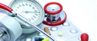
Ischemic stroke is characterized by a sudden onset of neurological symptoms. Important risk factors that you should pay attention to when diagnosing ischemic stroke are: atrial fibrillation, diabetes mellitus, atherosclerosis, arterial hypertension.
Diagnosis of ischemic stroke involves a physical examination, which is carried out according to accepted rules. When assessing the patient’s neurological status, attention should be paid to the presence of so-called cerebral symptoms:
- headache;
- generalized seizures;
- disturbance of consciousness.
In addition, patients may present with meningeal and neurological symptoms. If we talk about the types of laboratory tests, they are represented by urine and blood analysis (biochemical), coagulogram.
The main methods of instrumental diagnosis of ischemic stroke are CT and MRI, which also help to make a differential diagnosis, distinguishing ischemic stroke from various forms of intracranial damage or monitoring the dynamics of tissue changes during the treatment of this disease.
The most important ischemic symptom indicating damage in the area of the middle cerebral artery, which can be detected with CT diagnostics, is the weak expression of the lenticular nucleus and cortex, which subsequently leads to the development of cytotoxic edema.
In addition, during the course of an ischemic stroke, the doctor may detect changes associated with hyperdensity of the areas of the posterior or middle artery located on the affected side (for example, this could be thrombosis or embolism of the vessel).
During the first week of the disease, the area of gray matter with ischemic damage is characterized by an increase in the level of density, up to an isodense or slightly hyperdense state. The latter indicates the beginning of the development of the process of neovasogenesis or restoration of blood flow. Doctors also call this process the “fogging” effect, since at this time it is not always possible to determine the boundaries of ischemic damage, especially if the cerebral infarction has reached the subacute period.
MRI can accurately identify diffusion-weighted images of the affected area. Due to the resulting cytotoxic edema, water molecules usually move into the intracellular space (from the extracellular space), which leads to a slowdown in the rate of diffusion. All these changes that occur in the human body can be detected on the diffusion-weighted image obtained during MRI. It is this type of diagnosis that helps to identify the irreversibility of damage occurring in the structures of the brain.
Diagnostics
If there is a suspicion of a blood supply disorder, a special technique is used to determine ischemic stroke. If a person with an acute cerebrovascular accident is conscious, his ability to speak and hold his hands up is checked. A characteristic sign of ischemic stroke is drooping of one of the corners of the mouth.
For reference. To confirm the diagnosis of ischemic stroke, CT or MRI is performed. These studies make it possible to detect the boundaries of the lesion and the approximate time of its appearance. The resulting tomogram allows one to distinguish between ischemia and hemorrhagic stroke.
Angiography is indicated to determine the exact site of the lesion. The patient is given an electrocardiogram and their blood pressure is measured.
To diagnose ischemic stroke, blood tests are performed - general, cholesterol, sugar, coagulability.
Differential diagnosis
First of all, ischemic stroke should be distinguished from the hemorrhagic type of stroke. Neuroimaging techniques for studying the disease play a significant role in differential diagnosis. In some cases, it may be necessary to distinguish between ischemic stroke and acute hypertensive encephalopathy. Ischemic stroke should also be distinguished from toxic or metabolic types of encephalopathy, from brain tumors, abscesses, encephalitis and other brain lesions of an infectious nature.
Treatment of ischemic stroke
A person with even the slightest suspicion of an ischemic stroke should be hospitalized in a special department as quickly as possible. If the first symptoms of an ischemic stroke were detected no later than 6 hours from the onset of their manifestation, the patient is sent to the intensive care unit. Typically, the patient is transported in a reclining position, with the head raised to 30 degrees.
Hospitalization is not indicated for those patients who are in a state of terminal coma, suffer from oncology, or have a history of dementia with pronounced disability.
Treatment for ischemic stroke is usually:
- non-pharmacological (involves carrying out measures aimed at caring for the patient, as well as regulating the swallowing reflex, treating infectious diseases, which often manifest themselves in the form of pneumonia or genitourinary tract infections);
- medicinal (this type of therapy will be most effective only at the initial stage of the disease, in the first 6 hours from the onset of symptoms; medicinal treatment involves a multidisciplinary approach, which can be carried out within the walls of a special vascular department, usually located in the intensive care unit, where at any time you can perform a CT, MRI or ECG, as well as blood and urine tests.
Stages of development
In the process of developing an ischemic stroke, there are several stages. The first of them, acute, lasts up to 21 days from the onset of the attack. It can be divided into the most acute stage, lasting five days, when symptoms increase. This is the stage where most people die.
The second lasts up to six months. Reversible changes occur in cells during this period. Compensation for disturbances begins, blood circulation around the affected nuclei is restored. The stage is called the early recovery stage.
The third conditionally begins 6 months after the impact and lasts up to a year. During this period, cysts and glial formations form in the affected areas.
For reference. A year after the attack, the fourth stage begins. It is called the stage of residual manifestations. It lasts for the rest of a person’s life.
Maintaining body functions is the main stage of treatment
The main goal of any type of treatment for ischemic stroke is the correction of particularly important body functions and the mandatory maintenance of homeostasis. To achieve this goal, monitoring of all important physiological components is carried out, as well as measures aimed at maintaining hemodynamics, respiratory processes, and water and electrolyte balance. When treating a patient, one should also normalize his intracranial pressure in order to get rid of cerebral edema in the future. In addition, regular prevention of this disease and therapy aimed at combating the possible consequences of ischemic stroke should be carried out.
The main infusion solution during the treatment of this disease is sodium chloride solution. As for the use of glucose-containing solutions, in recent years doctors have abandoned them due to the possible development of hyperglycemia.
If an ischemic stroke develops against the background of diabetes mellitus, then the patient is given subcutaneous injections of insulin (short-acting). The exception is when specialists establish glycemic control while the patient is clearly conscious and does not suffer from impaired swallowing function.
In the first 2 days from the onset of the disease, doctors regularly measure the level of hemoglobin oxygen saturation in the blood. If the figure reaches 90-92%, then doctors administer oxygen therapy (usually starting with 2 liters per minute).
If the patient’s level of consciousness decreases to 8 points or less (calculated on the Glasgow scale), then this indicates an indicator for tracheal incubation. The decision whether to use mechanical ventilation in the future or not will be made based on the existing resuscitation provisions. If the patient's level of wakefulness is markedly reduced, there are clinical and other neuroimaging characteristics indicating cerebral edema (or increased intracranial pressure), then it will be necessary to maintain the head in an elevated state so that the neck does not flex. Coughing or signs of an incipient epileptic seizure should be minimized, as well as muscle excitation should be curbed. It should be remembered that infusions of hypoosmolal solutions are contraindicated.
Complications
In ICD-10, the consequences of ischemic stroke are assigned code 169. If the diagnosis is incorrect and thrombolysis is performed during an attack, the course of the latter may worsen.
For reference. With right-sided and left-sided lesions, disturbances occur in the motor area. The limbs are paralyzed, muscle weakness develops, which does not allow the patient to care for himself or move around. Discoordination of movements, especially voluntary ones, is noted.
Sensitivity suffers, and it takes longer to restore it than to normalize motor activity.
Most patients experience consequences such as psycho-emotional disorders. Ischemic stroke is characterized by a tendency to depression, emotional lability, in many cases, aggressiveness, and the appearance of fears.
Impairments in the intellectual sphere with left-sided lesions lead to memory impairment, inability to analyze the situation, childishness, and disorientation in space and time. With right-sidedness, fantasy suffers.
Speech disorders are manifested by incoherent speech, repetition of the same phrases or words. In left-handed people they are observed with pathology of the right hemisphere.
In approximately 10% of cases, victims develop epilepsy. There is often a risk of a second strike.
Nutrition of patients with ischemic stroke
The main task of therapy for ischemic stroke is considered to be proper nutrition of the patient and control of water-electrolyte balance. This requirement must be implemented regardless of where the patient is being treated: be it in the intensive care unit or in the neurological department.
The main indicator for enteral tube feeding will be the progress of swallowing function. The amount of dose of administered nutrients is calculated based on data on the metabolic needs of the patient’s body, as well as its physiological losses. If food is administered orally (or using a tube), the patient should take a semi-sitting position.
To prevent deep vein thrombosis (in the case of ischemic stroke), the use of compression bandages or stockings is prescribed. This method also helps prevent the development of pulmonary embolism. In addition, direct anticoagulants can be used.
Consequences for humans
The consequences of an ischemic stroke directly depend on the size of the affected area of the brain and the timeliness of assistance. When help is provided in a timely manner and adequate treatment is prescribed, complete or at least partial restoration of functions is possible. Sometimes, despite the prescribed treatment, symptoms increase, this can lead to serious consequences.
Let us highlight the following types of complications:
- infectious complications (arise due to prolonged lying in a supine position in combination with the addition of an infection, which leads to infections of the urinary system, pneumonia, bedsores, etc.);
- deep vein thrombosis of the lower leg area;
- pulmonary thromboembolism;
- cerebral edema;
- cognitive impairment;
- disorders of defecation and urination;
- epilepsy (develops in about 20% of cases);
- motor disorders (unilateral, bilateral), severe weakness and paralysis;
- mental disorders (changeable moods, irritability, depression, etc.);
- pain syndrome.
Consequences of different brain damage: right and left side
Left and right hemispheres of the human brain
Consequences of ischemic stroke on the right side of the brain. In addition to the usual disturbances for a stroke in the form of facial asymmetry, hypertonicity on the left side (upturned corner of the lips), a smoothed nasolabial fold on the right, paralysis and paresis of the left half of the body, there are other features.
- Loss of memory for recent events, with retained memories of the distant past (such as from childhood).
- Concentration is impaired.
- Speech impairment.
Consequences of ischemic stroke on the left side:
- Severe mental abnormalities - the patient does not orient himself in time and space, does not recognize close people, a peculiarity is positioning himself as a healthy person.
The most common causes of death from ischemic stroke in the first week are:
- cerebral edema and dislocation of the focus of necrosis of the brain stem with damage to the respiratory and cardiovascular centers, development of coma;
- hemorrhagic transformation of cerebral infarction with the formation of secondary hemorrhage;
- secondary ischemia of the brain stem with the formation of infarcts.
Neuroprotection
Speaking about the treatment of ischemic stroke, one should also mention a type of therapy such as neuroprotection. Its main focus is the use of special drugs that have neurotrophic and neuromodulatory functions. The most famous such drug today is Cerebrolysin (it belongs to the neurotrophic series of drugs). As is known, the central nervous system and spinal cord lack such an important property as storage. In other words, if you stop blood flow in these areas for 5 minutes, the neurons will begin to die. Therefore, the need to introduce neuroprotectors is mandatory. It should be carried out in the first minutes after the onset of an ischemic stroke.
Treatment of ischemic stroke will be successful with an integrated approach: basic therapy, rehabilitation, neuroprotection and reperfusion.
Treatment
After a stroke occurs, the nerve cells in the brain die within the first three to four hours. Only immediate hospitalization gives a chance for full recovery of the body.
First aid
First of all, you should make sure that the patient can breathe. To do this, you need to remove vomit from the mouth if the person vomited during an attack. To ensure sufficient air flow, it is necessary to free the person’s neck from constricting clothing and jewelry. It is advisable for the patient to take a horizontal position. But you need to make sure that your head lies flat: when you bend your neck too much, the blood flow through the vertebral arteries worsens.
If possible, you should measure your blood pressure and record the time of onset of the attack so that you can provide information to doctors when they arrive. There is no need to reduce high blood pressure on your own, as this is a normal protective reaction of the body during a stroke.
Diagnostics
The treatment approach depends on the causes of the acute cerebrovascular accident. The patient is immediately examined. MRI and CT scans of the brain are done to determine the factor provoking the disease and the extent of vascular damage. An ECG is performed to analyze the functioning of the cardiovascular system. Next, blood tests are carried out to find out the level of cholesterol, blood sugar, etc. The time required for recovery depends on each specific case. Inpatient treatment lasts from two weeks to several months, the rehabilitation period can last for more than a year.
Treatment of ischemic stroke
In the event of a stroke caused by a blocked artery, it is necessary to restore blood supply to the affected areas of the brain as soon as possible. To dissolve blood clots, intravenous administration of anticoagulants (plasminogen, heparin, nadroparin, warfarin) is recommended. Also, taking regular aspirin helps thin the blood and dissolve blood clots.
However, if more than half of the middle cerebral artery region is affected, blood pressure remains high, and there is a history of liver disease, kidney disease, or stomach ulcer, then thrombolytics can provoke internal hemorrhages, including hemorrhagic stroke. In this case, mechanical removal of the thrombus through surgery is necessary.
Carotid endarterectomy is a method that involves removing cholesterol plaque from the carotid artery.
Angioplasty and stenting. A thin catheter is inserted into the artery affected by atherosclerosis. There is a balloon at the end. When approaching the affected area of the vessel, it inflates, as a result of which the blocked lumen of the vessel expands. A catheter can also help break up a blood clot.
Treatment of hemorrhagic stroke
The actions of doctors are aimed at stopping the bleeding. Patients taking medications to prevent blood clots may be given medications to counteract the effects of anticoagulants or receive a blood transfusion. If the area of hemorrhage is large, surgery may be required to remove the blood to reduce intracranial pressure levels. To reduce the risk of relapse, surgical reconstruction of damaged vessels may be prescribed.
If the stroke was caused by a ruptured aneurysm, vascular malformation, or other vascular pathology, one of the following methods of surgical vascular repair may be prescribed:
- Clipping of a cerebral aneurysm. The purpose of the operation is to stop the flow of blood in the pathological area by applying a tiny clamp to the neck of the aneurysm. This way, the risk of complications such as aneurysm rupture and re-bleeding is eliminated.
- Endovascular embolization. Does not require craniotomy. The technique is effective if the aneurysm is difficult to access. A catheter is inserted into the vessel and guided to the brain under X-ray control. A catheter is used to inject material into the aneurysm, which causes thrombus formation and blocks blood flow in the aneurysm.
- Surgical removal of vascular malformation. The method is used for small sizes and superficial location of pathological connections of blood vessels.
- Intracranial shunting is the creation of a path for blood flow to bypass the pathological area with the installation of a shunt. When working in an area with intense blood flow, part of the patient's leg artery is used as a shunt. If a small vessel is required, choose one that nourishes the scalp.
- Stereotactic radiosurgery. High-frequency radiation is used to remove pathological formations. This is a new method with minimal damage to surrounding tissue.
Surgery
If we talk about the surgical type of treatment for ischemic stroke, then first of all, it includes surgical decompression, which implies a decrease in intracranial pressure and at the same time an increase in perfusion pressure against the background of preserved cerebral blood flow. According to statistics, the mortality rate from ischemic stroke has now been reduced to 30%.
The rehabilitation period after an ischemic stroke will be aimed at restoring previously lost motor and speech functions of the patient. This is the main task of neurologists who perform electromyostimulation, exercise therapy, regular massage of paretic limbs or mechanotherapy. To correct the patient’s speech, he is also referred to a speech therapist.
Hemorrhagic stroke
The main factor provoking this condition is hypertension. Increased pressure reduces the elasticity of the anterior walls, making them fragile and vulnerable. Another important risk factor is incorrect treatment with anticoagulants. A stroke can also occur as a result of a ruptured aneurysm. A much rarer cause of intracranial hemorrhage is arteriovenous malformation. This is a congenital abnormal interweaving of arteries and veins in the form of a tangle without the participation of capillaries. The walls of these arteries lack a full muscle layer and can rupture at any time.
Hemorrhagic stroke
There are two types of hemorrhagic stroke.
- Intracerebral hemorrhage (bleeding in the brain). It ranks second in prevalence among all types of stroke (after ischemic). This condition is caused by a rupture of an intracerebral vessel. This leads to blood filling the tissues surrounding the burst vessel and damaging them.
- Subarachnoid hemorrhage. Occurs when a vessel located close to the surface of the brain ruptures. Blood penetrating into the subarachnoid space (between the brain and skull) causes an inflammatory process and an increase in intracranial pressure. Secondary vascular spasm often develops, which leads to a lack of blood supply to brain tissue.
Prognosis for ischemic stroke
The prognosis in the case of ischemic stroke will be determined by the location and area of brain damage. In addition, important indicators here will be the patient’s age and the severity of concurrent diseases. The most difficult period for ischemic stroke is the first 5 days. It is at this time that there is an increase in cerebral edema in the area of its damage, after which a time of stabilization begins, when the impaired functions of the central nervous system gradually return to normal. To date, the percentage of deaths in patients with ischemic stroke is no more than 20%.
Forecast
How long a person will live after an ischemic attack depends on the location, size of the damage, time of initiation of treatment and concomitant diseases.
For reference. According to statistics, up to 25% of patients who have suffered a major ischemic stroke die within the first month. These are mainly those people whose attack occurred due to atherosclerosis or blood clot rupture.
The main cause of death in cerebral infarction is cerebral edema and its displacement. Sometimes exacerbation of cardiovascular diseases leads to death.
In the next 14-21 days of acute ischemic stroke, death occurs due to heart failure, the formation of a blood clot in the pulmonary artery, and pneumonia.
Survival rates for ischemic stroke are up to 70% at the end of the first 12 months. After 5 years, about 50% are still alive, after ten – a quarter. With a repeated attack, the prognosis worsens.
It is noted that the recovery of patients is best in the first three months, and the strength and movements of the leg muscles are normalized faster and better than in the arms.
For reference. The prognosis for complete paralysis is unfavorable. If recovery does not occur within a year, after this period normalization of the condition is unlikely. The exception is speech.
Doctors use several scales that allow them to assess the prospects for recovery.
NIHSS scale
The grading system is based on the manifestation of symptoms of the disease at the acute stage. The degree of consciousness, speech preservation, ability to control movements and their coordination, muscle strength, facial expressions, attention, eye movements, field of vision are analyzed. Moreover, if a person scores few points, the prognosis is satisfactory; if a person scores high, the prognosis is unfavorable.
For reference. If the patient scores up to 8-10 points on this scale, the probability of recovery is 70%, the observed disorders are characterized as mild. From 9 to 12 – medium; up to 15 – heavy; up to 33 – extremely severe. Indicator 34 characterizes coma. The probability of recovery for a patient who scores more than 20 points is no more than 16%.
Rankin scale
The Rankin scale is also often used. Based on various pathological symptoms, it describes the levels of the patient’s condition and the prognosis for the preservation of functions. A patient diagnosed with grade zero does not have any limitations or residual symptoms.
In the first degree, minor disturbances associated with speech, movements, and sensitivity are noted. The person remains able to work.
With the second, the patient loses some functions and cannot take care of himself fully.
With the third, a person walks independently with the help of a frame or a cane, but cannot fully take care of himself - cook, clean, go to the store. In some cases of left-sided lesions, a decrease in intellectual functions is observed.
The fourth characterizes patients who need help every day with dressing, moving, and eating.
With the fifth, the patient needs help and care constantly, he cannot be left alone.
Prevention of ischemic stroke
The most important thing in the prevention of ischemic stroke is considered to be measures to prevent thrombosis of blood vessels, which is formed when cholesterol plaques appear in the blood. For this purpose, doctors prescribe a set of procedures to maintain a healthy lifestyle, eliminating smoking, alcohol and fatty foods. As is known, those at risk of developing ischemic stroke are primarily those patients who suffer from chronic diseases of the cardiovascular system, diabetes mellitus or arterial hypertension.
Secondary prevention of ischemic stroke includes the implementation of a comprehensive program in the form of: antihypertensive therapy with the use of inhibitors, diuretics; lipid-lowering therapy with statins; surgical intervention (carotid endatherectomy is performed); antithrombotic therapy (antiplatelet agents and indirect anticoagulants).
Causes
A stroke occurs when the arteries supplying blood to the brain rupture or ischemia. As a result, cells lack oxygen and nutrients and begin to die.
If there is rupture of the arteries and bleeding in the brain, a hemorrhagic stroke is diagnosed. If a disruption in blood circulation occurs due to narrowing and blocking of the lumen of the arteries of the brain, an ischemic stroke is diagnosed.
Risk factors for development:
- elderly age;
- smoking;
- alcohol abuse;
- taking cocaine and methamphetamine;
- poor diet high in saturated fat and salt;
- physical inactivity;
- obesity;
- suffered a heart attack;
- arterial hypertension;
- diabetes;
- increased blood viscosity;
- vascular atherosclerosis;
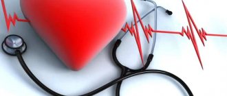
- cardiac ischemia;
- pregnancy;
- sickle cell anemia.