Osteoporosis is a progressive disease of bone tissue, which is characterized by decreased density, destruction and brittleness of bones. The disease is most often observed in older people and postmenopausal women.
To diagnose pathology you need to undergo a number of examinations:
- radiography;
- densitometry;
- ultrasound examination;
- magnetic resonance therapy.
But the most informative indicator is osteocalcin. It allows you to diagnose osteoporosis even in the early stages.
Functions of osteocalcin
Osteocalcin performs a number of specific functions.
These include:
- Formation of bone structures due to osteogenic regulation of metabolism;
- Bone mineralization;
- Maintaining an optimal level of calcium, which takes part in osteosynthesis;
- Stimulation of insulin secretion through a direct effect on Langerhans cells (pancreatic cells);
- Increased levels of adiponectin (a hormone) that is released from fat cells.
Bone tissue remodeling. Osteoclasts remove old bone tissue, osteoblasts form new one, releasing the protein osteocalcin, which then enters the bloodstream
Other indicators are also important for an accurate diagnosis
So, a blood test for osteocalcin is prescribed:
- For osteoporosis (the cause is conditions that create conditions for its development or the appearance of signs indicating that the pathological process has already begun);
- In order to determine the effectiveness of antiresorptive treatment in patients suffering from osteoporosis;
- In cases of hypercalcemia syndrome (increased ionized calcium in the blood).
Despite the fact that GLA-protein is a very sensitive marker of the metabolic activity of osteoblasts, an isolated determination of its level in plasma (serum) does not always provide a complete amount of information, therefore the study of osteocalcin is often complemented by other tests:
- Alkaline phosphatase (ALP), since it takes part in phosphorus-calcium metabolism;
- Ionized calcium (if a calcium metabolism disorder is suspected);
- Calcitonin (thyroid hormone, which reduces the level of Ca 2+ in the blood), which is considered as a tumor marker;
- PTH (parathyroid hormone is a hormone of the parathyroid glands that increases the concentration of Ca 2+ in the blood plasma by reducing the reabsorption of phosphorus (P) in the renal tubules;
- DPID (deoxypyridinoline) is an indicator of bone metabolism, determined in urine.
The concentration of osteocalcin is determined most often using a chemiluminescent immunoassay, the principle of which is a one-step “sandwich”.
The methods for preparing and collecting blood samples also do not have anything unusual, and therefore do not differ in this regard from other biochemical tests. It's simple: on an empty stomach, excluding fatty foods and alcohol the day before, as well as vigorous physical activity an hour before the test.
Blood protein level
The rate of osteocalcin depends on:
- age category;
- gender;
- menstrual cycle - in women.
The normal range for one bone marker varies depending on age and gender. Also, its indicators may change in women, taking into account the phase of the menstrual cycle. A couple of days before the onset of menstruation, osteocalcin is moderately increased.
During pregnancy, this protein is reduced. In adolescents, this indicator also exceeds the norm due to physiological characteristics.
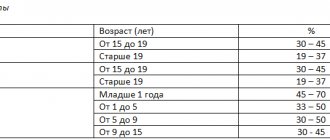
| Age category | Normal levels of osteocalcin in the blood, ng/ml | |
| Female | Male | |
| 6 months-6 years | 44-130 | 29-121 |
| 7-9 years | 73-206 | 66-182 |
| 10-12 years | 77-262 | 85-232 |
| 13-15 years old | 33-222 | 70-336 |
| 16-17 years old | 24-99 | 43-237 |
| 18-30 years old | 01.11.1943 | 24-70 |
| 30-50 years | 01.11.1943 | 14-42 |
| Over 50 years old | 15-46 | 14-46 |
There are several reasons that may affect the analysis performance:
- In a patient with renal failure.
- In women during menstruation.
The level of osteocalcin presence in human blood varies depending on age and gender. In girls before adolescence it is higher than in boys. In adulthood, the situation changes - the level of OC in the blood of men is higher than that of women.
| Age range | Women | Men |
| 6 months - 7 years | 45–130 | 30–120 |
| 8–10 | 75–200 | 65–185 |
| 11–13 | 75–260 | 85–230 |
| 14–16 | 35–220 | 70–335 |
| 17–18 | 25–100 | 45–235 |
| 19–29 | 10–45 | 25–70 |
| 30–49 | 10–45 | 15–40 |
| over 50 years old | 15–45 | 15–45 |
Careful consideration should be given to the study of bone markers when examining children. In them, especially in adolescence, due to intensive growth, the content of GLA proteins in the blood is greatly increased. During the examination, for an objective assessment in women, not only age, but also the current phase of the menstrual cycle is taken into account, otherwise the result of the analysis will mislead the doctor and frighten the patient. At the end of the luteal phase (2–3 days before the onset of menstruation), osteocalcin increases. During pregnancy, the level of this protein, on the contrary, decreases.
Osteocalcin can be brought back to normal by following the recommendations of doctors:
- Give up bad habits. Smoking reduces OC levels by interfering with genes responsible for the production of bone protein. Alcohol has the same effect, which leads to a long recovery of bones after fractures and cracks, and worsens the condition of teeth.
- Lead an active lifestyle. Studies conducted on a group of adolescents have proven a direct relationship between OC levels and physical activity.
- Watch your diet. A low-calorie diet increases bone protein content in older people. Obesity, on the contrary, helps to reduce it.
- Consult your doctor about the dosage of glucocorticoids. Reducing the dose of medications taken for rheumatoid arthritis proportionally increases the level of GLA protein.
- Normalize iron levels in the blood. A deficiency of this microelement reduces bone mineral density and reduces their strength.
- Treat vitamin K deficiency. The antihemorrhagic vitamin also causes osteocalcin deficiency. This increases the risk of fractures, especially in older people.
- Balance your vitamins. A sufficient amount of vitamin D, zinc, manganese, and ellagic acid guarantee a normal level of bone markers in the blood in the absence of pathologies.
Type 1 diabetes can also cause skeletal changes, especially if glucose is poorly controlled. It is possible to avoid bone problems by treating with insulin. This increases OA content, which improves bone structure and flexibility.
In women, the norm is 5.6-6.3 ng/ml, and in men – 6.3-7.3 ng/ml. Physiologically, about 15% of total osteocalcin is found in the serum, which has not been taken up by the bones.
A result above normal may indicate:
- primary bone tumors;
- metastasis of tumors to bones;
- Paget's disease;
- hyperparathyroidism;
- osteoporosis;
- rickets;
- osteomalacia.
A result below normal may indicate:
- hypoparathyroidism;
- hypofunction of the thyroid gland;
- liver failure.
Osteocalcin, as a marker of bone formation status, is used to recognize bone diseases.
Osteocalcin level testing should be performed in patients with malignant tumors when metastases appear. It is also very useful for older people, in whom bone diseases are much more common.
Studies of protein content reflect the rate of formation of new tissue, since when it is destroyed (resorption), it is not released. If it is normal, then the processes of osteosynthesis and osteoresorption are balanced, and new tissue is formed in place of dead tissue. At a low level, as well as at an excessively high level, destruction predominates.
Bone Formation Process
Reference (average normal values) values depend on the gender and age of the subject. When deciphering the results, it is necessary to take into account the influence of internal factors; the doctor may recommend additional diagnostics to clarify the diagnosis. High and low levels indicate a violation of bone metabolism.
Indicators are normal
For the chemiluminescent method (the most common), the values (ng/ml) indicated in the table are taken as the norm.
| Age | Men | Women |
| 2-16 years | 2,8-41 | 2,5-43 |
| 17-50 | 3,1-17,3 | 0,4-8,2 |
| After 50 | 1,5-11 | 0,3-7,8 |
When assessing the data obtained, the doctor takes into account the following factors:
- phase of the menstrual cycle - 2-3 days before the start of the next menstruation, the protein level is maximum;
- pregnancy – there is a physiological decline;
- the level of urine filtration in the kidneys - when it decreases, the excretion of osteocalcin is slowed down;
- the use of medications: vitamin D, biotin, anticonvulsants increase, and anticoagulants and Tamoxifen decrease.
Level reduction
Protein concentration decreases in the following diseases:
- rickets in early childhood;
- increased cortisol: Itsenko-Cushing's disease, long-term treatment with corticosteroids. The degree of decrease in this protein reflects the severity of hypercortisolism;
- cirrhosis of the liver;
- lack of growth hormone;
- insufficiency of hormonal activity of the parathyroid glands.
If a high or low level of osteocalcin is detected, an examination by a therapist and an endocrinologist is required; they can refer you for a consultation to a gynecologist, orthopedist, oncologist, and also recommend additional diagnostics: bone tissue densitometry, computed tomography or magnetic resonance imaging.
What indicators are considered normal?
The rate of osteocalcin depends on:
- age category;
- gender;
- menstrual cycle - in women.
Note. A few days before the onset of menstruation, a sharp increase in OC levels occurs. This circumstance is absolutely normal and should not frighten a woman. It is due to the fact that during menstruation the body becomes vulnerable to any irritants, since it loses many trace elements along with menstrual blood. For this reason, it requires additional protection, which is the hormone osteocalcin.
Normal test data in women
The norm of osteocalcin in women by age group is given below (ng/ml blood):
- Girls 18 years old and women under 30 - from 11 to 43.
- At 30-50 years old - from 11 to 43.
The rate of osteocalcin in menopausal women decreases significantly and should range from 15 to 46 ng/ml. Such changes are associated with the process of biological aging of a woman’s body.
Indicators for men
In men from 18 to 30 years old, the optimal values are 24 – 70 ng/ml. In patients aged 30-50 years – from 14 to 42, and after reaching 50 years of age – from 14 to 46 ng/ml of blood.
In children
The norm of osteocalcin in children and adolescents:
| Child's age | Indicators for girls (ng/ml) | Indicators for boys (ng/ml) |
| From six months to 6 years | From 44 to 130 | From 29 to 121 |
| Children 7-9 years old | From 73 to 206 | From 66 to 182 |
| Children 10-12 years old | From 77 to 262 | From 85 to 232 |
| Teenagers 13-15 years old | From 33 to 222 | From 70 to 336 |
| Boys and girls 16-17 years old | From 24 to 99 | From 43 to 237 |
Note. The fact that osteocalcin is elevated in a child, especially a teenager, is explained by the increased needs of the child’s body for hormones. And since children and teenagers are experiencing an active phase of growth and development, they need calcium-containing substances more than adults, for whom they are only responsible for maintaining the normal state of the musculoskeletal system.
Regulation of osteocalcin levels
A low level of osteocalcin in the blood serum manifests itself against the background of ongoing pathological processes, including:
- rickets or dystrophy in children of the younger age group;
- increased levels of parathyroid hormone and simultaneously decreased levels of calcium and calcitonin;
- Itsenko-Cushing syndrome in the stage when disturbances in the functioning of the adrenal glands occur;
- myeloma;
- liver failure (biliary cirrhosis);
- oncology;
- refusal of insulin therapy for diabetes.
In addition to the above reasons, a decrease in the concentration of GLA protein is affected by the patient taking high doses of glucocorticoids.
Hormones that regulate the synthesis of osteocalcin contain calcium: calciferol (vitamin D), calcitonin - a thyroid hormone, parathyroid hormone and parathyrin - hormones produced by the parathyroid glands.
Parathyroid hormone is a calcitonin antagonist, that is, its mechanism of action is the opposite of the thyroid hormone. Parathyroid hormone helps reduce calcium levels in bone tissue.
Calciferol acts on the production of the precursor osteocalcin by osteoblasts.
If the ratio between the amount of calcium produced or calcium-containing hormones is disrupted, this indicates the beginning of the development of bone tissue pathology.
Increases in the norm are observed in the following diseases Osteocalcin
- Paget's disease
- hyperthyroidism
- primary hyperparathyroidism
- renal osteodystrophy
- chronic renal failure
- active processes of bone formation (including tumor processes)
- active growth processes (adolescence)
- metastasis of tumors of various origins into bone tissue
- ipoparathyroidism
- pregnancy
- growth hormone deficiency
- long-term corticosteroid therapy
The immediate causes of deviations from the norm in the analysis are provoked by pathologies:
- Hypoparathyroidism. With this disease, the main emphasis in treatment is aimed at stabilizing and then normalizing the levels of calcium and phosphorus in the blood. Hypoparathyroidism is characterized by a deficiency of parathyroid hormone, a hormone produced by the parathyroid glands. The clinical picture of this condition boils down to hormonal imbalances associated with excess levels of calcium and phosphorus in the blood.
- Hypofunction of the thyroid gland. A decrease in the functions of the thyroid gland in the production of the most important hormone - thyrocalcitonin - provokes the development of osteoporosis, arthritis and other joint pathologies. Deficiency of thyrocalcitonin also leads to diseases of the cardiovascular system. If the level of the hormone decreases to the maximum level, then the process of leaching calcium from bone tissue begins, which leads to disruption of the functioning of the heart and blood vessels.
- Liver failure (biliary cirrhosis). Liver failure is a serious pathological condition. A syndrome that is accompanied by damage to liver cells and organ dysfunction. The liver loses its compensatory capabilities and partially loses its basic functions. The patient exhibits symptoms of chronic intoxication. Progressive biliary cirrhosis can cause hepatic coma, that is, complete organ failure and extensive damage to all body systems, including the brain, by decay products.
- Osteoporosis and atherosclerosis. The rate of development of pathologies and the level of metabolism in bone tissues and blood vessels are determined by the amount of bone protein in the patient’s body. In the early stages of the disease, the indicators will be normal, but with severe metabolic disorders they will increase several times.
In this case, it is necessary to identify pathologies of the thyroid gland and diagnose osteoporosis by resorting to other research methods.
Interpretation of study data
Osteocalcin testing is used to diagnose and monitor metabolic bone diseases (eg, Paget's disease, asthenic bone diseases), osteoporosis (eg, postmenopausal women), bone marrow disorders and other diseases of the skeletal system, such as severe fractures, deformities, calcium imbalance -phosphate balance.
Osteocalcin is a marker of bone formation, especially in people who develop osteodystrophy in the kidneys.
Therapeutic therapy for osteoporosis includes different groups of drugs that need to be purchased after a doctor’s prescription.
Selective estrogen receptor modulators, calcium-containing drugs, and bisphosphonates are also prescribed.
A blood test for osteocalcin is indicated in the following cases:
- suspicion of dwarfism: children are stunted, muscle weakness, underdevelopment of the genital organs;
- accelerated growth of adolescents;
- monitoring the state of bone metabolism when the formation of growth hormone decreases;
- carrying out hormone therapy, complications of treatment: obesity, purple stretch marks on the skin, excessive growth of facial hair, irregular menstruation, bone destruction (according to radiography);
- menopause, removal of ovaries in women;
- bone tumors and metastasis to the bones (usually from the prostate and mammary glands);
- fracture due to minor trauma;
- signs of osteoporosis (back pain, aggravated by movements or prolonged static position, changes in posture);
- sudden increases in temperature with signs of inflammation (according to a blood test) of unknown etiology (occur with bone tumors);
- widespread, migrating bone pain;
- severe weakness, lack of appetite, nausea, abdominal pain, increased thirst, pain in the bones, lower back (signs of increased function of the parathyroid glands);
- diagnosed osteoporosis, osteopenia, osteodystrophy to establish the risk of fractures and predict the rate of loss of mineral mass. This is necessary for choosing a medication and its optimal dose.
What does osteocalcin testing show? Using this test, you can determine the level of the hormone in the blood, as well as record deviations from the norm.
The reasons for the increase and decrease in the concentration of this substance often lie in the pathological processes occurring in the patient’s body. Moreover, we can talk not only about osteoporosis. Let's consider all possible options for deviations of OK from the norm.
What drug treatment should be prescribed?
Decreased bone density is a virtually asymptomatic disease in the early stages. The main task of an orthopedic traumatologist in diagnosing osteoporosis is to objectively assess the patient’s condition. The specialist identifies the fact of pathology, finds the cause of the disease and differentiates bone loss from other similar or concomitant diseases, and makes a prognosis for recovery.
At the doctor's disposal:
- possibility of clinical examination;
- hardware diagnostics (X-ray, ultrasound);
- laboratory diagnostics;
- genetic examination methods.
All of them are aimed at visualizing macro- and microarchitecture and identifying processes leading to a decrease in bone mineral density.
Clinical examination
At the clinical examination stage, the therapist can identify a group with an increased risk of osteoporosis. Among the symptoms of osteoporosis, the doctor can identify kyphotic deformation in the thoracic spine, a noticeable decrease in the patient’s height compared to the previous visit. Clinical markers of osteoporosis also include complaints of back pain. There is a chance to get an appointment with a traumatologist:
- in women with premature menopause;
- in people with a family history of osteoporosis;
- with frequent fractures under the age of 45;
- in patients with diseases that provoke secondary osteoporosis;
- after taking a number of pharmaceutical drugs;
- with extreme thinness – anorexia, exhaustion, congenital thinness with a BMI less than 20.
If a person has two or more risk factors, the likelihood of osteoporosis increases by a third, regardless of age.
Interviewing the client can help identify additional risk factors:
- calcium deficiency in food;
- lack of exposure to sunlight, and therefore a lack of vitamin D;
- problems with the stomach and intestines;
- drinking alcohol and smoking;
- treatment with glucocorticoids or hormones;
- diseases of the glands and internal organs, COPD;
- prolonged immobilization or low physical activity.
2. X-ray signs of AP
3. Persons over 65 years of age with decreased height or thoracic kyphosis
2. Corsets, thigh protectors
3. For back pain, limit the amount of time you sit
4. Combined calcium and vitamin D preparations
5. Antiresorptive drugs (HRT, bisphosphonates, calcitonin, strontium ranelate)
2. Persons taking glucocorticosteroids for a long time
3. Combination of several risk factors over the age of 50 years
5. The doctor’s decision to prescribe antiresorptive drugs (HRT, bisphosphonates, calcitonin, strontium ranelate)
2. The presence of somatic or other pathology that is potentially dangerous for the development of AP
3. Hypogonadism (including menopausal women)
4. Insufficient calcium intake
Patients with suspected osteopenia or osteoporosis are prescribed further in-depth examination. Often the disease is detected at a late stage by traumatologists in patients with fractures or neurologists in patients with pain in the spine.
An orthopedic traumatologist or neurologist differentiates osteoporosis from bone cancer, trauma, osteomalacia, fibrous dysplasia, Paget's disease, and peripheral neuropathy. Laboratory diagnostics becomes the main basis for decision-making in this case.
The following “clinical myths” serve as an obstacle to early detection of the disease:
• in COPD, osteopenia occurs in more than 40% of patients, and when taking systemic glucocorticosteroids, the frequency of osteopenia reaches 85%;
• in CVD, osteopenia is observed in more than 68% of cases, and AP is diagnosed in 50% of cases
Diagnostic methods
More than 70% of bone strength is provided by mineral density (BMD), the remaining 30% is redistributed between mineralization, metabolism, macro- and microstructure, and microdamage. It is important to identify all defects in the early stages of the disease. For this purpose, a number of methods for diagnosing osteoporosis have been developed.
Most often, osteopenia and osteoporosis are detected using conventional x-rays in the presence of bone fractures or spinal deformities. At this stage, more than a third of bone mass has already been lost. You can influence the process when we are talking about losses of up to 5%. Therefore, for the early detection of osteoporosis, there are more accurate methods:
- ultrasonic;
- isotope (mono- and biphoton absorptiometry);
- mono- and bioenergetic absorptiometry (DXA);
- quantitative computed tomography;
- blood chemistry;
- genetic studies of materials.
Rules for formulating the diagnosis of AP
Ultrasound
Analysis of the mineral composition of bones using ultrasound can identify areas of reduced density. This test for osteoporosis is based on the reflection of ultrasound beams from dense organs. The introduction of ultrasound devices into clinical practice makes it possible to assess the mechanical properties of bone tissue. Strength and elasticity create a hyperechoic image structure.
The testing site for ultrasonic densitometry (UD) is usually the heel bone and the tip of the index finger. Practice shows that this study is more suitable for screening. It is recommended to undergo it once every 5 years for early detection of the problem for women over 45 years of age and men over 50.
Radiodensitometry
An accurate diagnosis is established by examining certain areas of the skeleton using bioenergetic absorptiometry, which is considered the “gold standard” for diagnosing osteoporosis. Dual-energy X-ray absorptiometry aims to measure BMD. Diagnosis is carried out for the entire skeleton, vertebrae, paired sections or one bone. The device compares the received image with the reference ones and produces two indicators, T and Z.
The T-index indicates deviations from the peak bone density of an adult, and the Z-index indicates deviations from the norm for a given age. Normally T=1. Osteopenia is diagnosed at T from -1 to -2.5, lower mineral density is classified as osteoporosis. If the patient has a history of a fracture with a T less than -2.5, then osteoporosis is called severe.
Bone loss affects the entire human skeleton, although unevenly. BMD is determined in places where fractures occur most often. This:
- lumbar vertebrae;
- femoral neck;
- distal forearm.
Postmenopausal women will likely have their vertebrae assessed, while older adults (over 65 years) will likely have their proximal femoral BMD measured. DXA is prescribed for the initial diagnosis of osteoporosis and evaluation of the effectiveness of therapy. The examination is repeated after a year.
The examination is not dangerous to health, since the radiation intensity is 400 times less than with a regular x-ray. The only contraindication may be pregnancy.
DXA does not require any special preparation. It is better not to take calcium supplements the day before. The physician is informed of recent barium examinations. After scanning the selected area, the patient receives images and a conclusion.
The DXA method has some limitations, which are compensated for by additional research.
CT densitometry
Using computed tomography, it is possible to determine reduced tissue density and obtain a three-dimensional image of the anatomical structure of a skeletal area. The study allows us to study the trabecular structure of the bone.
For examination using computed tomography, a peripheral scanner with a thin beam is used to determine the mineral composition of the tissue. The analysis is called CT densitometry and allows you to determine local changes in mineral composition.
Magnetic resonance imaging was discovered in 1946. The study makes it possible to obtain a three-dimensional image of organs by recording a radio signal, which is obtained due to the vibration of hydrogen atoms of aqueous tissues in a magnetic field. For this discovery, scientists received the Nobel Prize.
MRI is rarely used to diagnose diseases, although its possibilities are endless. The analysis allows you to see the trabeculae of the bones very clearly. This property was used to create densitometers - devices for determining the degree of osteoporosis.
Single publications in the literature indicate the high effectiveness of the study in determining the density of the bone structure. Obviously, MRI can be used to check the structure of organs. However, the test is rarely used due to the high cost of the procedure.
The causes of bone loss, like osteoporosis itself, are not obvious. To identify them, you will need to conduct a biochemical blood test and genetic tissue studies. The first one is needed for:
- carrying out preventive measures in cases with metabolic disorders of bone tissue remodeling and resorption;
- predicting the rate of bone loss;
- assessing the effectiveness of therapeutic methods.
At this stage of diagnosing osteoporosis, osteomalacia, bone metastases, and hereditary diseases are excluded, the causes of secondary osteoporosis are identified, and metabolic features are established.
In this case, doctors use three groups of biochemical markers of pathology:
- Markers of bone tissue formation.
Therapeutic therapy for osteoporosis includes different groups of drugs that need to be purchased after a doctor’s prescription.
Selective estrogen receptor modulators, calcium-containing drugs, and bisphosphonates are also prescribed.
Selective estrogen receptor modulators, calcium-containing drugs, and bisphosphonates are also prescribed.
Preparing to donate blood
Osteocalcin in the body can be reduced as a result of developing pathological processes, among which are the following:
- Rickets in young children;
- Increased amount of parathyroid hormone, and decreased content of calcium and calcitonin;
- Itsenko-Cushing syndrome and disease, during which the exchange of hormones in the adrenal cortex is disrupted;
- Multiple myeloma;
- Primary biliary cirrhosis;
- Somatotropin levels are below normal;
- Disruption of the parathyroid glands, accompanied by a decrease in its function;
- Decreased calcium levels due to the development of a malignant tumor.
Formation of the active form of osteocalcin
What to do if osteocalcin is low? First of all, as with any hormonal problems, it is necessary to find out the cause of the deviation. If it lies in diseases, then they should first be cured. It is quite possible that the OK level will then recover on its own, albeit gradually.
If there is a need to treat low osteocalcin, they often resort to:
- Prescribing medications based on vitamin K. This substance stimulates the natural processes of osteocalcin production. But when taking such medications, patients should be careful: they may not be combined with all medications. Thus, the simultaneous use of vitamin K in the form of MK-7 with anticoagulants leads to a decrease in the effectiveness of the latter.
- Treatment with physical activity. This approach does not involve performing any special exercises - it is enough to exercise regularly, at least 1 hour a day, 3 times a week. The duration of the course is 3 months.
To increase osteocalcin, it is necessary to limit the intake of calories in the body. It is also important to take medications based on vitamin D, zinc and manganese, as well as Omega-3 fatty acids.
There are many methods to increase the level of osteocalcin in the blood. But all of them must be agreed upon with a doctor, especially when it comes to pharmaceutical drugs. Contraindications and side effects of such drugs have not been canceled!
No special restrictions are required for this study. The day before the analysis, overeating and drinking alcohol, fatty foods, physical and emotional stress are not recommended. Dinner can be normal, then only regular drinking water is allowed. It is best to come to the laboratory early in the morning before breakfast. Smoking and sports training are prohibited for an hour.
Blood is taken from a vein in an amount of 5 ml and mixed with a cold heparin solution. The tube must be immediately placed in a centrifuge to separate the serum. Then it is transported in a container to the laboratory.
Venous blood collection
Osteocalcin is normal in children
Have you been trying to heal your JOINTS for many years?
Head of the Institute for the Treatment of Joints: “You will be amazed at how easy it is to cure your joints by taking the product every day for 147 rubles ...
Read more "
The normal level of bone formation marker in blood plasma (serum) depends on gender and age, so it is advisable to reflect its values in the table:
OUR READERS RECOMMEND!
Our readers successfully use Sustalaif to treat joints. Seeing how popular this product is, we decided to bring it to your attention. Read more here...
AgeNormal level of osteocalcin in blood, ng/ml
| Men | Women | |
| From six months to 6 years | 39 – 121 | 44 – 130 |
| 7 – 9 years | 66 – 182 | 73 – 206 |
| 10 – 12 years | 85 – 232 | 77 – 262 |
| 13 – 15 years | 70 – 336 | 33 – 222 |
| 16 – 17 years old | 43 – 237 | 24 – 99 |
| 18 – 30 years old | 24 – 70 | 11 – 43 |
| 30 – 50 years | 14 – 42 | 11 – 43 |
| Over 50 years old | 14 – 46 | 15 – 46 |
In women, for an objective assessment, it is very important to clarify not only age, but also the phase of the menstrual cycle, otherwise the result may not satisfy the doctor and frighten the patient.
At the end of the luteal phase, that is, two to three days before the start of a new period, osteocalcin is usually slightly increased compared to the norm. In pregnant women, the level of this protein, on the contrary, is reduced (gynecologists take this fact into account).
The increase in the concentration of osteocalcin in children and, especially, adolescents is explained by their intensive growth during this period of life.
In addition, when carrying out the analysis, one should take into account the fact that the level of osteocalcin has a wide range of daily fluctuations; there are, of course, no approved norms for each hour, however, in order to obtain objective results, comparable blood samples should be taken at the same time. watch.
The essence of the concept of osteocalcin
Osteocalcin is a K-dependent vitamin produced by three-day-old tissue cells that have completely separated and reached maturity. It is osteoblasts, cells that have reached maturity and form our osteocalcin, that bind hydroxyapatites and calcium.
During the process of bone formation, osteocalcin is converted and passed into our bones by osteoblasts in order to signal the stage of osteosynthesis formation to doctors who will diagnose it.
It is worth fearing that such a process can accompany both normal metabolism in the human body and a pathological condition within the skeleton in which it is necessary to urgently go to the hospital.
Method of determination
The following methods are used to test blood:
- immunoenzyme,
- radioimmune,
- chemiluminescent.
In accordance with the chosen methodology, each laboratory has individual normal values. When repeating a study, it is recommended to contact the same institution or clarify the method of initial determination. The protein level changes throughout the day, so the time should also be approximately the same.
Blood centrifugation
One-stage chemiluminescent diagnostics is recognized as the most sensitive. It allows you to quickly and reliably measure osteocalcin using specific antibodies.
The patient's heparinized serum is mixed with the reagent and left for an hour. The sample changes the color of the solution, the degree of its coloration is measured by a spectrophotometer. The higher the optical density, the more of this protein is in the blood. If the patient did not follow a diet (animal fats were on the menu), then the serum may become turbid, which will complicate correct measurement.
Cloudy serum, making proper measurement difficult
Osteoporosis and its relationship with atherosclerosis
Osteoporosis is directly related to metabolic disorders of bone tissue. However, it is known that these disorders are more often observed in women - their bone strength is naturally lower. And, if during reproductive age estrogens are taken to protect the female skeletal system, then with the onset of menopause, due to a decrease in their level, they lose the ability to cope with this task.
Probably, many patients have already noticed that pathological conditions such as osteoporosis and the formation of atherosclerotic plaques on the walls of blood vessels run parallel to each other. So what connection does a progressive decrease in bone tissue mass, disruption of its microarchitecture, and increased catabolism in bones have with atherosclerosis? And here's what:
- The risk of both diseases correlates very markedly with age (the lowest risk is observed in young healthy people);
- Absence of clinical manifestations in the initial period of the disease (neither osteoporosis nor atherosclerosis at first indicate symptoms of the disease);
- Both pathologies create a high degree of likelihood of developing complications of diseases of the cardiovascular system, as well as the occurrence of fractures with the slightest injury in postmenopausal females;
- Their risk factors are, in principle, the same - bad habits (mainly smoking), a sedentary lifestyle, a lack of estrogen, which protect women before menopause;
- The pathophysiological basis of these pathological conditions is also the same - calcium metabolism disorder;
- Disorders of the functions of hormonal systems that regulate calcium metabolism also go in the same direction;
- Many medications intended to treat atherosclerosis are useful for osteoporosis (help maintain bone mineral density), and medications aimed at combating osteoporosis have a beneficial effect on the condition of blood vessels.
Osteoporosis, which increases the level of osteocalcin in the blood, has such a connection with atherosclerosis.
What are the reasons for increased osteocalcin in the body?
An increase in osteocalcin in a blood test can be caused by the following conditions:
- Accelerated metabolism in bone tissue structures;
- For osteoporosis during the postmenopausal period;
- Chronic renal failure, which leads to nephrogenic osteodystrophy and calcium metabolism disorders;
- Paget's disease;
- Fractures of bone structures;
- Disorders of the thyroid gland, in which the level of hormone activity increases - primary hyperthyroidism (toxic goiter, Graves' disease) and secondary changes due to tumor processes in the pituitary gland;
- Pituitary pathologies, in which there is excessive synthesis of somatotropic hormone;
- Malignant formations of bone tissue;
- Osteomalacia – decreased level of bone mineralization;
- Taking medications that contain calciferol and its producers, vitamin B7, as well as medications against epilepsy.
When is osteocalcin low?
Meanwhile, there are circumstances when the level of the described protein in the blood is reduced. During pregnancy, it is lower due to physiological reasons, so such features of the gestational period can be omitted without classifying them as any health problems. A decrease in osteocalcin in the blood also occurs in the following conditions:
- Rickets in young children;
- High concentration of PTH (parathyroid hormone, parathyroid hormone), but low levels of calcium and calcitonin due to the formation of certain pathological processes;
- Hypercortisolism is a syndrome and Itsenko-Cushing's disease, and the more severe the symptoms, the lower the level of osteocalcin observed in the blood;
- Generalized plasmacytoma (myeloma);
- Primary biliary cirrhosis;
- Insufficient production of somatotropin (GH);
- Hypofunction of the parathyroid glands.
In addition to the above reasons, the use of glucocorticoids can reduce the concentration of GLA protein.
Osteoporosis – what is it?
Osteoporosis is a chronic bone disease accompanied by an increased level of bone fragility due to a deficiency of calcium ions. With osteoporosis, the following symptoms are distinguished: general weakness, night cramps, periodontitis, lower back pain, fragility and splitting of nails.
Osteoporosis can be diagnosed in women during menopause.
This is explained by the fact that until this moment, female sex hormones, estrogens, were actively acting on the bones, the amount of which decreases in the postmenopausal period.
It is worth paying attention to the fact that atherosclerosis and osteoporosis have many similarities in their etiology, pathogenesis, development, consequences, namely:
- These diseases are typical for people in the older age group.
- The initial stages are asymptomatic.
- The same risk factors: smoking, physical inactivity, low estrogen levels.
- The pathophysiology of the development of both pathological processes is a violation of calcium metabolism.
- The same hormonal regulation and disorders that are characteristic of osteoporosis and atherosclerosis.
- Therapeutic drugs prescribed for osteoporosis have a beneficial effect on the vascular system, and anti-atherosclerotic drugs can help with bone diseases.
Osteoporosis
The main diagnostic test for osteoporosis is a blood test for osteocalcin, which can be prescribed in different cases: to diagnose rickets in children, with hypercalcemic syndrome, to study the effectiveness of drug treatment of osteoporosis, for women during menopause.
You should also not engage in excessive physical activity. Blood is donated on an empty stomach; the patient must not eat for at least eight hours. Before this, instrumental research methods are prescribed: ultrasound, radiography.
There are also other markers of osteoporosis, tests for which are necessary to diagnose this disease:
- Alkaline phosphatase, which plays an important role in phosphorus-calcium metabolism;
- Ionized calcium;
- Calcium and phosphorus in daily urine;
- Calcitonin;
- Parathyroid hormone;
- Somatotropic hormone;
- Beta crosslapse is a marker of bone resorption;
- Serum osteopontin;
- Deoxyproline is an indicator of bone metabolism that is found in urine.
- Tests for women include checking estrogen levels.
You can take the necessary tests in the Invitro laboratory.
Osteoporosis interpretation of the results of these tests is carried out only by a doctor.
Osteocalcin how to increase the level – Treating the heart
Osteoporosis is a progressive disease of bone tissue, which is characterized by decreased density, destruction and brittleness of bones. The disease is most often observed in older people and postmenopausal women.
To diagnose pathology you need to undergo a number of examinations:
- radiography;
- densitometry;
- ultrasound examination;
- magnetic resonance therapy.
But the most informative indicator is osteocalcin. It allows you to diagnose osteoporosis even in the early stages.