What is a vein and its anatomical features
Veins are important blood vessels that carry blood to the heart.
They form a whole network that spreads throughout the body. They are replenished with blood from the capillaries, from which it is collected and supplied back to the main engine of the body.
This movement occurs due to the suction function of the heart and the presence of negative pressure in the chest when inhalation occurs.
Anatomy includes a number of fairly simple elements that are located on three layers that perform their functions.
Valves play an important role in normal functioning.
The structure of the walls of venous vessels
Knowing how this blood channel is built becomes the key to understanding what veins are in general.
The walls of veins consist of three layers. Outside, they are surrounded by a layer of mobile and not too dense connective tissue.
Its structure allows the lower layers to receive nutrition, including from surrounding tissues. In addition, the fastening of the veins is carried out due to this layer as well.
The middle layer is muscle tissue. It is denser than the top one, so it is what forms their shape and maintains it.
Thanks to the elastic properties of this muscle tissue, veins are able to withstand pressure changes without harming their integrity.
The muscle tissue that makes up the middle layer is formed from smooth cells.
In veins that are of the muscleless type, there is no middle layer.
This is typical for veins running in the bones, meninges, eyeballs, spleen and placenta.
The inner layer is a very thin film of simple cells. It is called the endothelium.
In general, the structure of the walls is similar to the structure of the walls of arteries. The width is usually greater, and the thickness of the middle layer, which consists of muscle tissue, is, on the contrary, less.
Atherosclerosis is a dangerous pathology
There are many diseases and pathological changes in the structure of the human circulatory system, for example, narrowing of the lumen of blood vessels . Due to disturbances in protein-fat metabolism, a serious disease such as atherosclerosis often develops - a narrowing in the form of plaques caused by the deposition of cholesterol on the walls of arterial vessels.
Progressive atherosclerosis can significantly reduce the internal diameter of the arteries to the point of complete blockage and can lead to coronary heart disease. In severe cases, surgical intervention is inevitable—the blocked vessels have to be bypassed. Over the years, the risk of getting sick increases significantly.
Features and role of venous valves
Venous valves are part of the system that ensures the movement of blood in the human body.
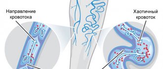
Venous blood flows through the body against gravity. To overcome it, the muscular-venous pump comes into operation, and the valves, having filled, do not allow the incoming fluid to return back along the bed of the vessel.
It is thanks to the valves that blood moves only towards the heart.
The valve is a fold that is formed from the inner layer consisting of collagen.
They resemble pockets in their structure, which, under the influence of the gravity of the blood, close, holding it in the desired area.
Valves can have from one to three leaflets, and they are located in small and medium-sized veins. Large vessels do not have such a mechanism.
Malfunction of the valves can lead to stagnation of blood in the veins and its erratic movement. This problem causes varicose veins, thrombosis and similar diseases.
Types of arteries
You should not think that all arteries are the same. There are different types of arteries in the body.
According to the diameter, all arteries are divided into:
- large arteries
- medium caliber arteries
- small arteries
According to the ratio of the number of elastic and muscle cells in the middle shell:
- elastic arteries
- arteries of mixed type (muscular-elastic)
- muscular arteries
The closer the artery is to the heart, the larger it is, and the greater its diameter (or caliber). The closer the artery is to the heart, the more dense elastic fibers there are in its middle shell.
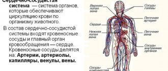
Human vascular system
Arterial vessels of the systemic circulation
Veins: structure and functions
Capillaries in the human body: structure and functions
Functions of arteries, veins and capillaries
All articles about blood and circulation
And this is easily explained: the closer the artery is located to the heart, the more pressure it must withstand, the stronger the shock wave acts on its wall. The stronger and more durable this wall should be.
The further away the artery is from the heart, the smaller its diameter and the more muscle fibers in its middle layer.
And this is understandable. After all, the further the artery is located from the heart, the more difficult it is for it to maintain the pressure inside the vessel and the speed of blood flow at the required level. The buoyant force of the heart does not help her much. Muscle fibers come to the rescue, regulating the width of the lumen of the vessel, thus maintaining the pressure and speed of blood flow at the proper level.
Main functions of the vein
The human venous system, the functions of which are practically invisible in everyday life unless you think about it, ensures the life of the body.
Blood, dispersed to all corners of the body, is quickly saturated with the products of all systems and carbon dioxide.
In order to remove all this and make room for blood rich in useful substances, the veins work.
In addition, hormones that are synthesized in the endocrine glands, as well as nutrients from the digestive system, are also distributed throughout the body through the veins.
And, of course, a vein is a blood vessel, so it is directly involved in regulating the process of blood circulation throughout the human body.
Thanks to it, there is a supply of blood to every part of the body, during paired work with the arteries.
How are vascular problems treated?
The venous circulatory system affects all areas of the body. Vascular diseases should be treated immediately. To avoid the formation of complicated stages of varicose veins or thrombosis, preventive measures are used. They try to remove dilated veins partially or completely. Blood clots are often cut out to prevent them from accidentally entering the bloodstream.
Common methods of treating veins help to eliminate further growth of the vessel, remove pathological areas, and reduce the risk of complications. Sclerotherapy is used in beauty salons and clinics. The procedure is safe and can be completed in a few minutes. A substance is injected into the affected vessel to glue the walls together.
The body gets rid of the glued vein on its own. It dissolves, and lightened tissues form in its place. There are no external defects. The procedure can be performed without pain relief. They try to use this method on small veins. On large vessels, abundant bluish areas appear.
The laser coagulation method is chosen when the affected veins are large. The procedure is painful and requires local anesthesia. After this, a light guide is introduced into the affected vessel, the radiation of which seals the liquid contents of the vein. If you follow the doctor's recommendations after surgery, the resulting area will resolve.
Structure and characteristics
The circulatory system has two circles, small and large, which have their own tasks and characteristics. The diagram of the human venous system is based precisely on this division.
Pulmonary circulation
The lesser circle is also called the pulmonary circle. Its task is to carry blood from the lungs to the left atrium.
The capillaries of the lungs have a transition to venules, which then unite into large vessels.
These veins go to the bronchi and parts of the lungs, and already at the entrances to the lungs (gates), they unite into large channels, of which two come out of each lung.
They do not have valves, but go, respectively, from the right lung to the right atrium, and from the left to the left.
Systemic circulation
The large circle is responsible for supplying blood to every organ and tissue area in a living organism.
The upper part of the body is attached to the superior vena cava, which at the level of the third rib flows into the right atrium.
Veins such as the jugular, subclavian, brachiocephalic and other adjacent veins supply blood here.
From the lower part of the body, blood flows into the iliac veins. Here the blood converges through the external and internal veins, which converge into the inferior vena cava at the level of the fourth lumbar vertebra.
For all organs that do not have a pair (except for the liver), blood flows through the portal vein first to the liver, and from here to the inferior vena cava.
Deviations in health status
The venous system of the lower extremities is vulnerable due to high loads during walking, running and even in a normal position - standing. Diseases of the venous system appear for many reasons, not only physical. This refers, for example, to poor nutrition. Excessive consumption of fried, salty, and sweet foods leads to the formation of plaques in the blood that stick into huge clots. Thrombosis is dangerous for anyone.
First, blockages occur in small veins. But as they grow, clots can get into the main vessels leading to the heart. Severe cases of the disease lead to its stop. Blood clots should be removed in a timely manner to prevent dangerous complications.
The most common are varicose veins. This disease affects more than half of the female population. With age, the elasticity of the veins decreases, but the load remains the same. Often excess weight leads to the formation of stretched vessel walls. The size of the heart does not change, but the volume of blood transferred increases with the acquisition of additional kilograms.
An additional negative factor is a sedentary lifestyle. Stagnation of blood provokes not only the appearance of vascular diseases, but also complications in other parts of the body. Oxygen starvation affects the appearance of the skin of the face, hands, and neck.
Features of blood movement through the veins
At some stages of movement, for example, from the lower extremities, the blood in the venous canals is forced to overcome gravity, rising almost one and a half meters on average.
This occurs due to the phases of breathing when negative pressure occurs in the chest during inhalation.
Initially, the pressure in the veins located near the chest is close to atmospheric.
In addition, blood is pushed through contracting muscles, indirectly participating in the blood circulation process, raising the blood upward.
Structure
The venous-vascular system experiences tissue pressure from blood circulation; it has several layers:
- Collagenous: tissues resist the internal pressure of blood flow.
- Muscle protection tissues: muscle contraction and stretching helps blood flow and at the same time protects blood vessels from external influences (temperature, pressure, mechanical damage).
- Longitudinal fibers have elasticity and work constantly when the body moves: flexion and extension of the torso, arms or legs, when tilting the head.
When the veins are stretched, outflow is difficult, but when the muscles contract, additional force is provided to push the blood. The speed of movement through the vessels is higher due to a set of the following factors: heartbeat, movement of the chest during breathing, flexion of limbs, changes in body position in space, blood thinning due to digestion or the action of drugs. Blood flow also increases due to an increase in the temperature surrounding the body: in a sauna, hot bath.
The main veins have a significant diameter. The movement of fluid inside the vessels occurs in a certain direction due to the presence of numerous valves. They consist of fabrics of increased elasticity and strength. They can withstand a huge number of compression cycles throughout a person’s life.
The venous system cannot function effectively without valves. During the period of their weakening, pathological conditions called varicose veins can form. The most common site of its occurrence is the lower extremities.